Review
. 2018 Jun 22;8(7):457.
doi: 10.3390/nano8070457.

Protein-Based Fiber Materials in Medicine: A Review

Affiliations
Review

Protein-Based Fiber Materials in Medicine: A Review

Kelsey G DeFrates et al. Nanomaterials (Basel). .

Abstract

Fibrous materials have garnered much interest in the field of biomedical engineering due to their high surface-area-to-volume ratio, porosity, and tunability. Specifically, in the field of tissue engineering, fiber meshes have been used to create biomimetic nanostructures that allow for cell attachment, migration, and proliferation, to promote tissue regeneration and wound healing, as well as controllable drug delivery. In addition to the properties of conventional, synthetic polymer fibers, fibers made from natural polymers, such as proteins, can exhibit enhanced biocompatibility, bioactivity, and biodegradability. Of these proteins, keratin, collagen, silk, elastin, zein, and soy are some the most common used in fiber fabrication. The specific capabilities of these materials have been shown to vary based on their physical properties, as well as their fabrication method. To date, such fabrication methods include electrospinning, wet/dry jet spinning, dry spinning, centrifugal spinning, solution blowing, self-assembly, phase separation, and drawing. This review serves to provide a basic knowledge of these commonly utilized proteins and methods, as well as the fabricated fibers’ applications in biomedical research.

Keywords: biomaterials fabrication; drug delivery; medicine; nanofibers; protein; tissue engineering; wound healing.


Conflict of interest statement

The authors declare no conflict of interest.

Figures

Figure 1
Figure 1
Protein-based biomaterials can be made from a variety of sources. The origin and structures of some of the most commonly used proteins are shown. These include collagen or gelatin, silk, keratin, elastin, soy, and corn zein. These proteins can then be processed into fibers with unique physical properties through a variety of methods.
Figure 2
Figure 2
Schematic showing the set-up of a (A) vertical electrospinning system and (B) horizontal electrospinning system.
Figure 3
Figure 3
Scanning electron microscope images of (A) pure silk nanofibers, (B) PCL-gelatin nanofibers, (C) silk-PEO nanofibers, and (D) type I collagen nanofibers fabricated using the electrospinning technique (reproduced with permission from [19,78,136,139], copyright Elsevier, 2017 (A); Elsevier, 2007 (B); John Wiley and Sons, Inc., 2010 (C); Elsevier, 2006 (D)).
Figure 4
Figure 4
Common systems of (A) wet spinning, and (B) dry-jet wet spinning. In dry-jet wet spinning, the polymer solution is extruded through an air gap before the coagulation bath, resulting in higher molecular alignment compared to conventional wet spinning [124,141,142].
Figure 5
Figure 5
(A) Scheme of a typical dry spinning system. (B) Representation of drawing mechanism for polymer-based fiber fabrication (reproduced with permission from [126,146], copyright Elsevier, 2011 (A); AIP Publishing, 2006 (B)).
Figure 6
Figure 6
Generalized model of potential tissue engineering and medicine applications for various organ systems. (Reproduced with permission from [158], Copyright John Wiley and Sons, Inc., 1998).
Figure 7
Figure 7
(A,B) Aortoiliac bypass using a PCL-collagen scaffold; (CE) Effects of mulberry B. mori silk and non-mulberry A. mylitta silk on bone regeneration (reproduced with permission from [168,170], Copyright Elsevier, 2009 (A,B); Elsevier 2016 (CE)).
Figure 8
Figure 8
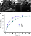
Corn zein nanofibers containing ferulic acid (FA) can be fabricated through single-fluid electrospinning (A) and modified coaxial electrospinning (B). Release of FA from both fiber types (F1 and F2) was then monitored over time (C) (reproduced with permission from [173], Copyright Elsevier, 2013).
Figure 9
Figure 9
(A) Hematoxylin and eosin staining for histological analysis of subcutaneously implanted mats made of varying PVA-silk nanofiber composite fibers. Mats containing non-mulberry silk showed greater cell infiltration and proliferation compared to those made of mulberry silk or those containing only PVA. Green arrows indicate infiltrating cells at the interface of the implant, and yellow arrows indicate the development of new blood vessels. (B) Images of wounds on rabbits treated with PVAA-silk mats. The percentage of wound closure is quantified in (C) (reproduced with permission from [25], Copyright Elsevier, 2017).

Similar articles

Cited by

References

    1. Sang Q., Williams G.R., Wu H., Liu K., Li H., Zhu L.-M. Electrospun gelatin/sodium bicarbonate and poly(lactide-co-ε-caprolactone)/sodium bicarbonate nanofibers as drug delivery systems. Mater. Sci. Eng. C. 2017;81:359–365. doi: 10.1016/j.msec.2017.08.007. - DOI - PubMed
    1. Löbmann K., Svagan A.J. Cellulose nanofibers as excipient for the delivery of poorly soluble drugs. Int. J. Pharm. 2017;533:285–297. doi: 10.1016/j.ijpharm.2017.09.064. - DOI - PubMed
    1. Lv Y., Pan Q., Bligh S.W.A., Li H., Wu H., Sang Q., Zhu L.-M. Core-Sheath Nanofibers as Drug Delivery System for Thermoresponsive Controlled Release. J. Pharm. Sci. 2017;106:1258–1265. doi: 10.1016/j.xphs.2016.12.031. - DOI - PubMed
    1. Vashisth P., Raghuwanshi N., Srivastava A.K., Singh H., Nagar H., Pruthi V. Ofloxacin loaded gellan/PVA nanofibers—Synthesis, characterization and evaluation of their gastroretentive/mucoadhesive drug delivery potential. Mater. Sci. Eng. C. 2017;71:611–619. doi: 10.1016/j.msec.2016.10.051. - DOI - PubMed
    1. Mutlu G., Calamak S., Ulubayram K., Guven E. Curcumin-loaded electrospun PHBV nanofibers as potential wound-dressing material. J. Drug Deliv. Sci. Technol. 2018;43:185–193. doi: 10.1016/j.jddst.2017.09.017. - DOI