. 2018 Jun 22;6(3):73.
doi: 10.3390/biomedicines6030073.

siRNAs Targeting Growth Factor Receptor and Anti-Apoptotic Genes Synergistically Kill Breast Cancer Cells through Inhibition of MAPK and PI-3 Kinase Pathways

Affiliations

siRNAs Targeting Growth Factor Receptor and Anti-Apoptotic Genes Synergistically Kill Breast Cancer Cells through Inhibition of MAPK and PI-3 Kinase Pathways

Nur Izyani Kamaruzman et al. Biomedicines. .

Abstract

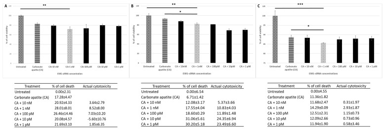
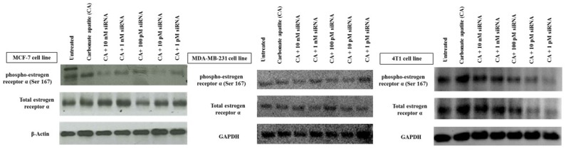
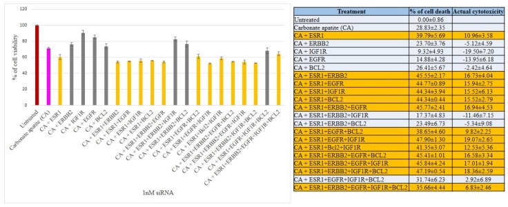
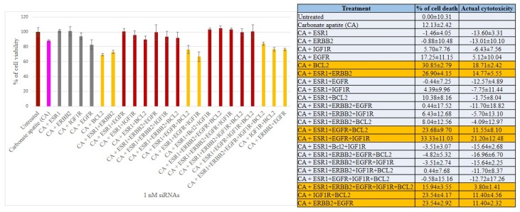
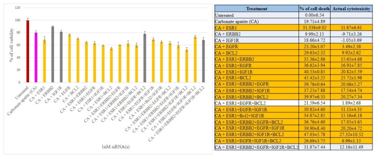
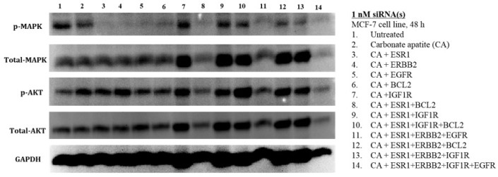
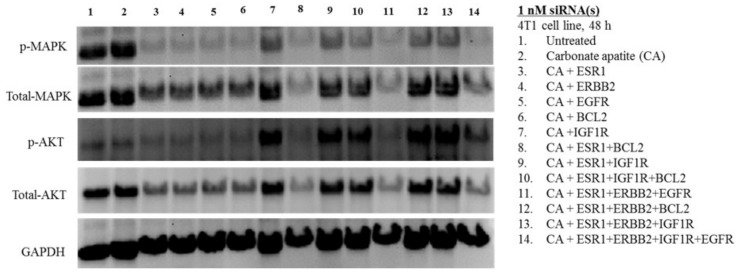
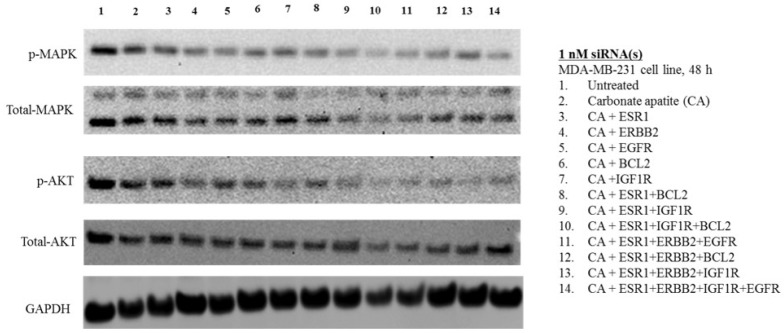
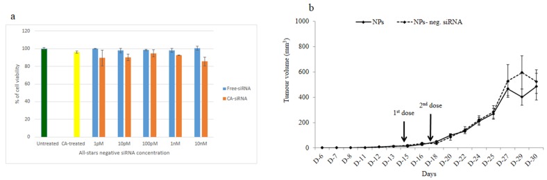
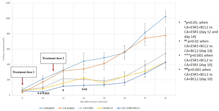
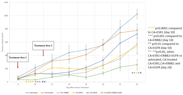
Breast cancer, the second leading cause of female deaths worldwide, is usually treated with cytotoxic drugs, accompanied by adverse side-effects, development of chemoresistance and relapse of disease condition. Survival and proliferation of the cancer cells are greatly empowered by over-expression or over-activation of growth factor receptors and anti-apoptotic factors. Identification of these key players that cross-talk to each other, and subsequently, knockdown with their respective siRNAs in a synchronous manner could be a promising approach to precisely treat the cancer. Since siRNAs demonstrate limited cell permeability and unfavorable pharmacokinetic behaviors, pH-sensitive nanoparticles of carbonate apatite were employed to efficiently carry the siRNAs in vitro and in vivo. By delivering selective siRNAs against the mRNA transcripts of the growth factor receptors, such as ER, ERBB2 (HER2), EGFR and IGFR, and anti-apoptotic protein, such as BCL2 in human (MCF-7 and MDA-MB-231) and murine (4T1) breast cancer cell lines, we found that ESR1 along with BCL-2, or with ERBB2 and EGFR critically contributes to the growth/survival of the cancer cells by activating the MAPK and PI-3 kinase pathways. Furthermore, intravenous delivery of the selected siRNAs aiming to suppress the expression of ER/BCL2 and ER/ERBB2/EGFR groups of proteins led to a significant retardation in tumor growth in a 4T1-induced syngeneic mouse model.

Keywords: breast cancer; carbonate apatite nanoparticle; estrogen receptor (ER); mitogen-activated protein kinase (MAPK); protein kinase B (AKT); siRNA.


Conflict of interest statement

The authors declare no conflict of interest.

Figures

Figure 1
Figure 1
Cell viability and cytotoxicity assessments of MCF-7 cells (A), MDA-MB-231 (B) and 4T1 (C) cells treated with CA-ESR1 siRNA complexes for 48 h. Preparation of CA-ESR1 siRNA complexes involved introduction of different concentrations of ESR1 siRNA (0 to 10 nM) and 3.5 mM of CaCl2 to 1 mL of bicarbonate-buffered DMEM (pH 7.4) medium. The mixture was allowed incubation at 37 °C for 30 min prior to addition of 10% FBS. The cells were incubated for the next 48 h with the prepared complexes. MTT assay was performed and absorbance reading was taken at 595 nm with 630 nm as references wavelength. Data was presented as mean ± SD of triplicates. * p < 0.05, ** p < 0.01 and *** p < 0.001.
Figure 2
Figure 2
Effects of intracellular delivery of CA-ESR1 siRNA complexes on expression and activation of estrogen receptor α in MCF-7, MDA-MB-231 and 4T1 cell lines. Cells were incubated with CA-ESR1 complexes for 48 h prior to cell lysis for protein extraction and Western blot analysis. Proteins were loaded on SDA-PAGE and transferred onto nitrocellulose membrane for detection of phosphorylated estrogen receptor (ER) α, total ER α and GAPDH (housekeeping protein) expression.
Figure 3
Figure 3
Cell viability and cytotoxicity assessments of MCF-7 cells treated with various CA-siRNA complexes involving ESR1, ERBB2, IGF1R, EGFR and BCL2 siRNAs (1 nM) individually or in combination, for a period of 48 h. Preparation of the CA-siRNA(s) complexes involved introduction of ESR1, ERBB2, IGF1R, EGFR and BCL2 siRNAs individually or in combination with 3.5 mM of CaCl2 to 1 mL of bicarbonate-buffered DMEM (pH 7.4). The mixture was allowed incubation at 37 °C for 30 min before the addition of 10% FBS. The cells were incubated for the next 48 h with the prepared complexes. MTT assay was performed and absorbance reading was taken at 595 nm with 630 nm as references wavelength. Data was presented as mean ± SD of triplicates.
Figure 4
Figure 4
Cell viability and cytotoxicity assessments of MDA-MB-231 cells treated with various CA-siRNA complexes involving ESR1, ERBB2, IGF1R, EGFR and BCL2 siRNAs (1 nM) individually or in combination, for a period of 48 h. Preparation of the CA-siRNA(s) complexes involved introduction of ESR1, ERBB2, IGF1R, EGFR and BCL2 siRNAs individually or in combination with 3.5 mM of CaCl2 to 1 mL of bicarbonate-buffered DMEM (pH 7.4). The mixture was allowed incubation at 37 °C for 30 min before the addition of 10% FBS. The cells were incubated for the next 48 h with the prepared complexes. MTT assay was performed and absorbance reading was taken at 595 nm with 630 nm as references wavelength. Data was presented as mean ± SD of triplicates.
Figure 5
Figure 5
Cell viability and cytotoxicity assessments of 4T1 cells treated with various CA-siRNA complexes involving ESR1, ERBB2, IGF1R, EGFR and BCL2 siRNAs (1 nM) individually or in combination, for a period of 48 h. Preparation of the CA-siRNA(s) complexes involved introduction of ESR1, ERBB2, IGF1R, EGFR and BCL2 siRNAs individually or in combination with 3.5 mM of CaCl2 to 1 mL of bicarbonate-buffered DMEM (pH 7.4). The mixture was allowed incubation at 37 °C for 30 min before the addition of 10% FBS. The cells were incubated for the next 48 h with the prepared complexes. MTT assay was performed and absorbance reading was taken at 595 nm with 630 nm as references wavelength. Data was presented as mean ± SD of triplicates.
Figure 6
Figure 6
Effects of intracellular delivery of various CA-siRNA complexes on expression and activation of AKT and MAPK proteins in MCF-7 cell line. Preparation of the CA-siRNA(s) complexes involved introduction of ESR1, ERBB2, IGF1R, EGFR and BCL2 siRNAs individually or in combination with 3.5 mM of CaCl2 to 1 mL of bicarbonate-buffered DMEM (pH 7.4). The mixture was allowed incubation at 37 °C for 30 min before the addition of 10% FBS. The cells were incubated for a consecutive period of 48 h with the prepared complexes, prior to cell lysis for protein extraction and Western blot analysis. Proteins were loaded on SDS-PAGE and transferred onto nitrocellulose membrane for detection of p-MAPK, p-AKT, total MAPK, total AKT and GAPDH (housekeeping protein) expressions.
Figure 7
Figure 7
Effects of intracellular delivery of various CA-siRNA complexes on expression and activation of AKT and MAPK proteins in 4T1 cell line. Preparation of the CA-siRNA(s) complexes involved introduction of ESR1, ERBB2, IGF1R, EGFR and BCL2 siRNAs individually or in combination with 3.5 mM of CaCl2 to 1 mL of bicarbonate-buffered DMEM (pH 7.4). The mixture was allowed incubation at 37 °C for 30 min before the addition of 10% FBS. The cells were incubated for a consecutive period of 48 h with the prepared complexes, prior to cell lysis for protein extraction and Western blot analysis. Proteins were loaded on SDS-PAGE and transferred onto nitrocellulose membrane for detection of p-MAPK, p-AKT, total MAPK, total AKT and GAPDH (housekeeping protein) expressions.
Figure 8
Figure 8
Effects of intracellular delivery of various CA-siRNA complexes on expression and activation of AKT and MAPK proteins in MDA-MB-231 cell line. Preparation of the CA-siRNA(s) complexes involved introduction of ESR1, ERBB2, IGF1R, EGFR and BCL2 siRNAs individually or in combination with 3.5 mM of CaCl2 to 1 mL of bicarbonate-buffered DMEM (pH 7.4). The mixture was allowed incubation at 37 °C for 30 min before the addition of 10% FBS. The cells were incubated for a consecutive period of 48 h with the prepared complexes, prior to cell lysis for protein extraction and Western blot analysis. Proteins were loaded on SDS-PAGE and transferred onto nitrocellulose membrane for detection of p-MAPK, p-AKT, total MAPK, total AKT and GAPDH (housekeeping protein) expressions.
Figure 9
Figure 9
Effect of nanoparticles with loaded negative control siRNA on tumor outgrowth in 4T1-cells induced mouse model. (a) After formulation of CA nanoformulation of a negative control siRNA, MCF-7 cells were incubated with it for a consecutive period of 48 h and subsequently, MTT assay was carried out. Mice were treated intravenously through tail-vein injection with 100 µL of either nanoparticles or nanoparticles with electrostatically associated negative control siRNA (50 nM). (b) Six mice/group were used and data were represented as mean ± SD.
Figure 10
Figure 10
Tumor outgrowth volume of mice intravenously treated with CA+ESR1 siRNA, CA+BCL-2 siRNA and CA+ESR1+BCL-2 siRNA complexes on a 4T1 induced breast tumor mouse model. Mice were administered twice (three days apart) with 100 µL of CA, CA+ESR1 siRNA, CA+BCL-2 siRNA and CA+ESR1+BCL-2 siRNA complexes. CA+siRNA complexes were formed by mixing 50 mM of a particular siRNA along with 4 μL of 1 M CaCl2 in 100 μL of freshly prepared bicarbonated (44 mM) DMEM media and incubating at 37 °C for 30 min. Six mice per group were used and data was represented as mean±SD. Values were significant with * p < 0.05 compare to the control group.
Figure 11
Figure 11
Tumor outgrowth volume of mice intravenously treated with CA+ERBB2 siRNA, CA+ESR1 siRNA, CA+EGFR siRNA and CA+ESR1+ERBB2+EGFR siRNAs complexes on a 4T1 induced breast tumor mouse model. Mice were administered twice (three days apart) with 100 µL of CA, CA+ERBB2 siRNA, CA+ESR1 siRNA, CA+EGFR siRNA and CA+ESR1+ERBB2+EGFR siRNAs complexes. CA+siRNA complexes were formed by mixing 50 mM of a particular siRNA along with 4 μL of 1 M CaCl2 in 100 μL of freshly prepared bicarbonated (44 mM) DMEM media and incubating at 37 °C for 30 min. Six mice per group were used and data was represented as mean ± SD. Values were significant with * p < 0.05 compare to the control group.

Similar articles

Cited by

References

    1. Ferlay J., Soerjomataram I., Dikshit R., Eser S., Mathers C., Rebelo M., Parkin D.M., Forman D., Bray F. Cancer incidence and mortality worldwide: Sources, methods and major patterns in GLOBOCAN 2012. Int. J. Cancer. 2015;136:359–386. doi: 10.1002/ijc.29210. - DOI - PubMed
    1. De Moraes G.N., Delbue D., Silva K.L., Robaina M.C., Khongkow P., Gomes A.R., Zona S., Crocamo S., Mencalha A.L., Magalhães L.M., et al. FOXM1 targets XIAP and survivin to modulate breast cancer survival and chemoresistance. Cell Signal. 2015;27:2496–2505. doi: 10.1016/j.cellsig.2015.09.013. - DOI - PubMed
    1. Fatemian T., Chowdhury E.H. Targeting oncogenes and tumor suppressors genes to mitigate chemoresistance. Curr. Cancer Drug Targets. 2014;14:599–609. doi: 10.2174/156800961407140926104458. - DOI - PubMed
    1. Masood S. Estrogen and progesteron receptors in cytology: A comprehensive review. Diagn. Cytopathol. 1992;8:475–491. doi: 10.1002/dc.2840080508. - DOI - PubMed
    1. Levin E.R. Cellular functions of plasma membrane estrogen receptors. Steroids. 2002;67:471–475. doi: 10.1016/S0039-128X(01)00179-9. - DOI - PubMed

LinkOut - more resources