Review
. 2018 Aug 15;35(8):792-837.
doi: 10.1039/c7np00067g.

Recent examples of α-ketoglutarate-dependent mononuclear non-haem iron enzymes in natural product biosyntheses

Affiliations
Review

Recent examples of α-ketoglutarate-dependent mononuclear non-haem iron enzymes in natural product biosyntheses

Shu-Shan Gao et al. Nat Prod Rep. .

Abstract

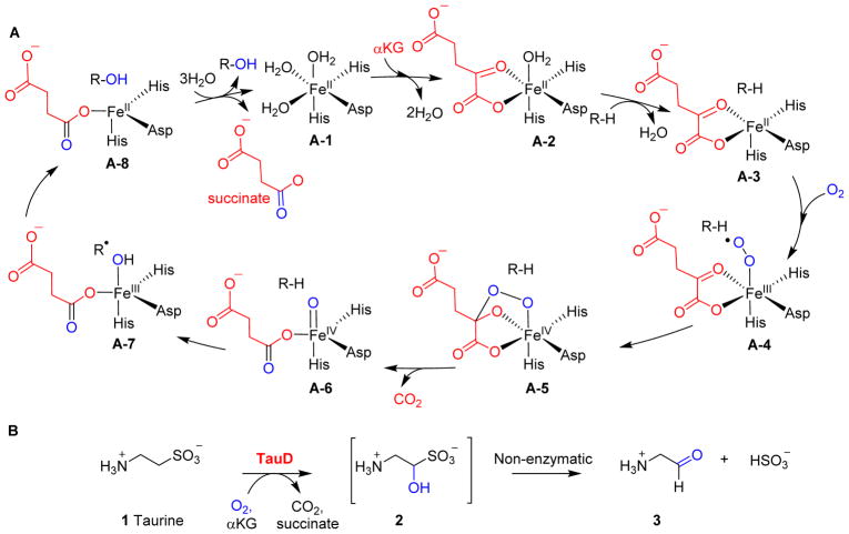
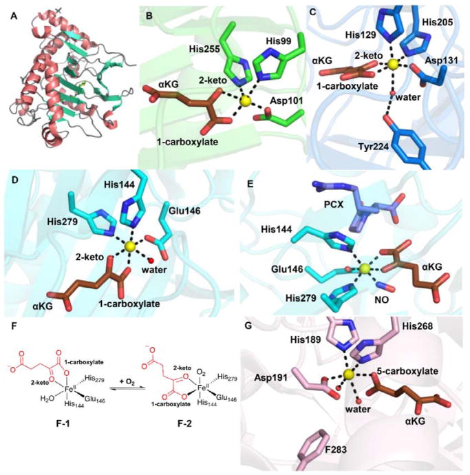
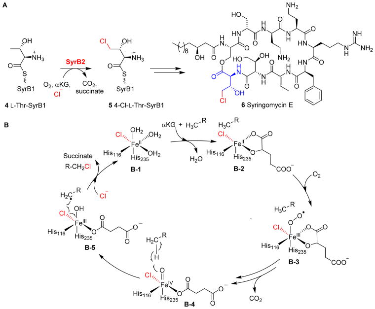
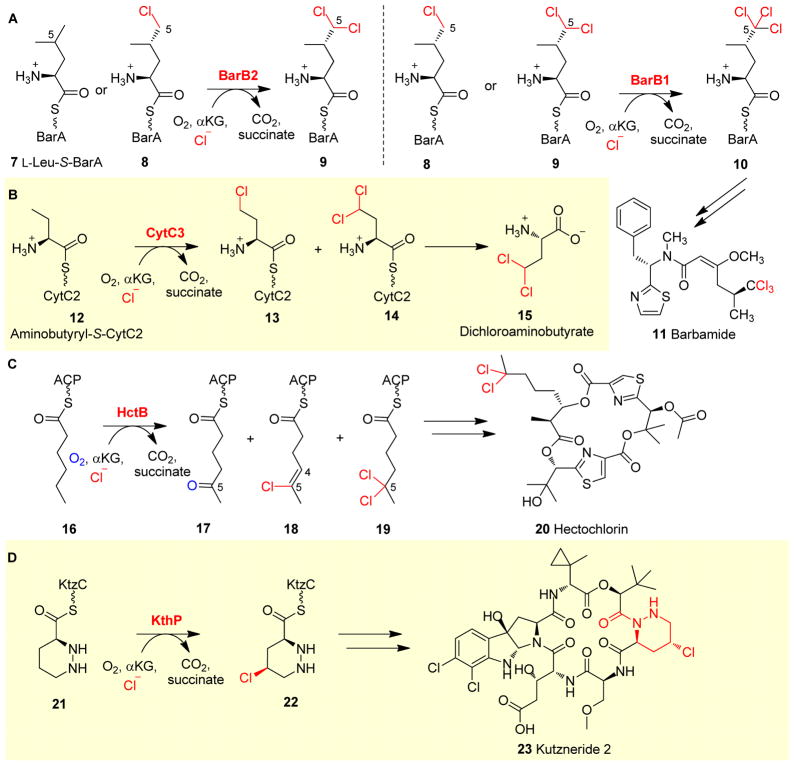
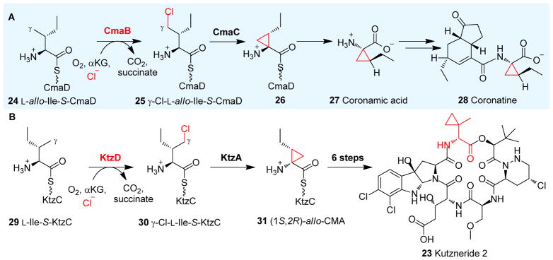
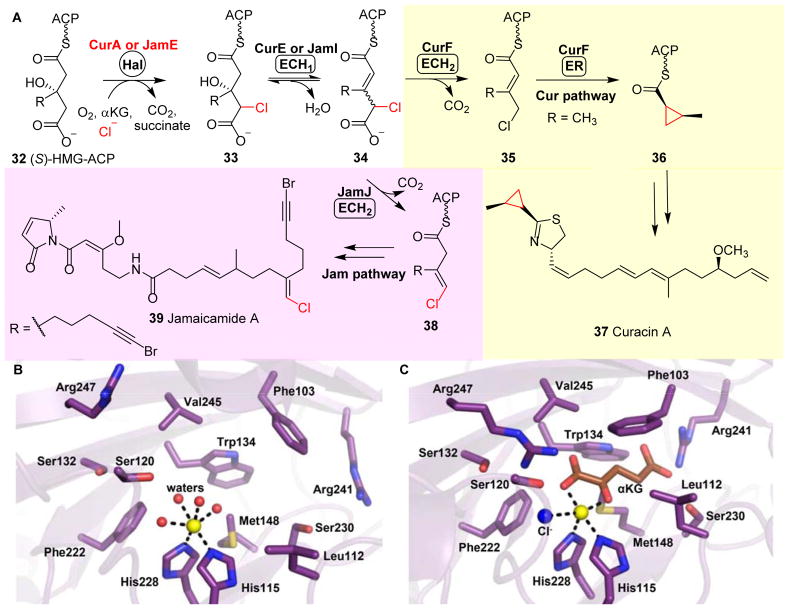
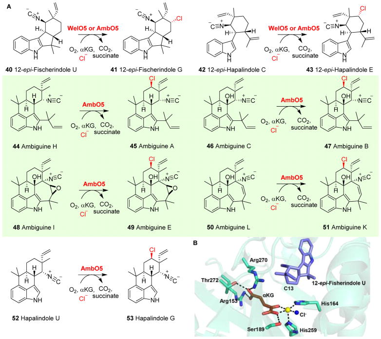
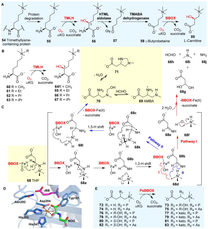
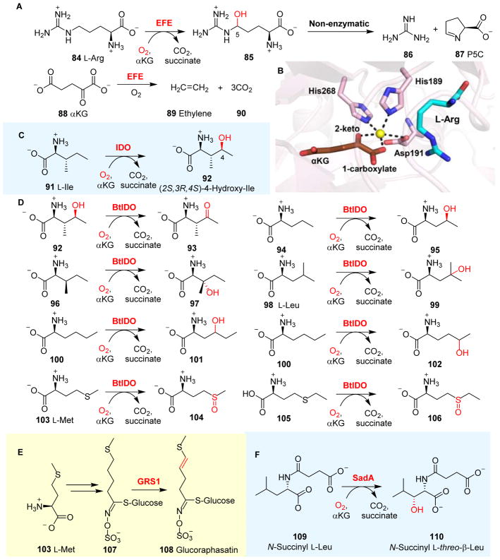
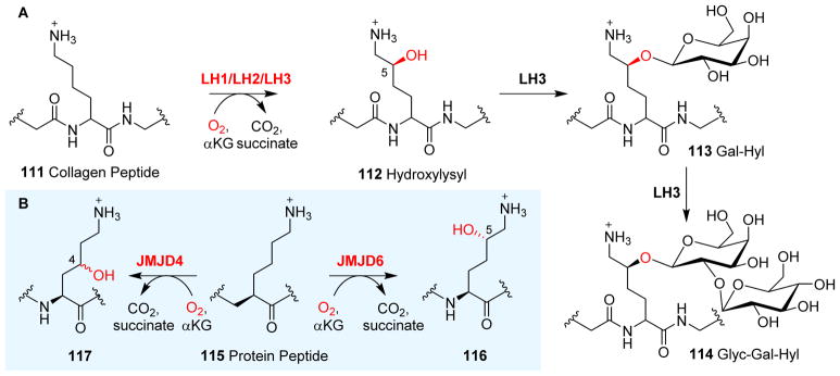
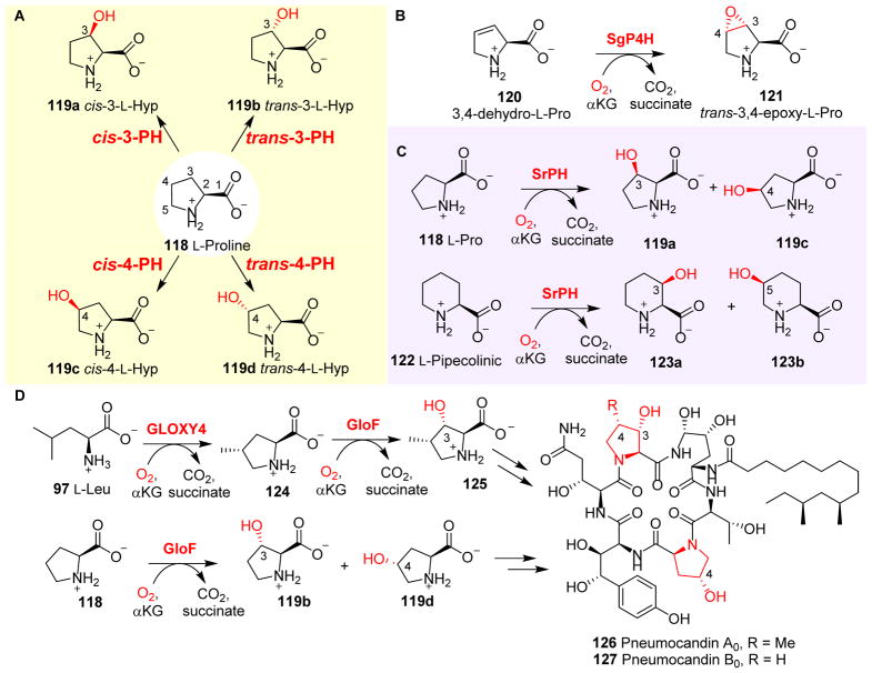
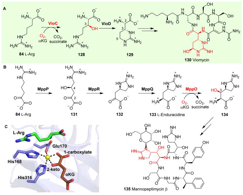
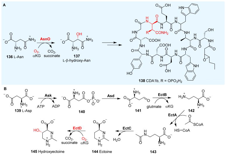
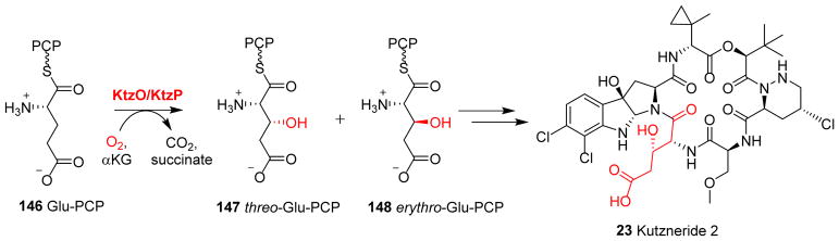
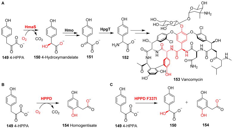
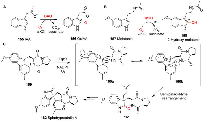
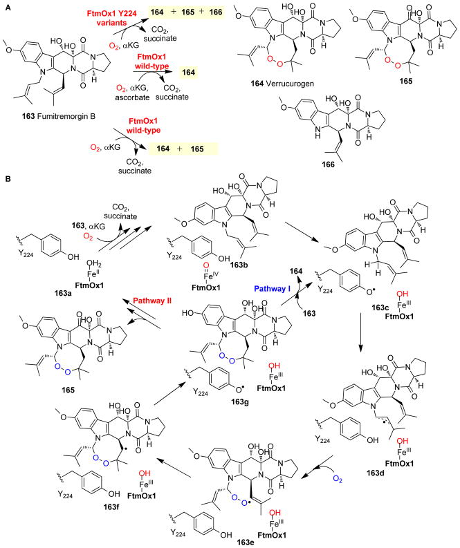
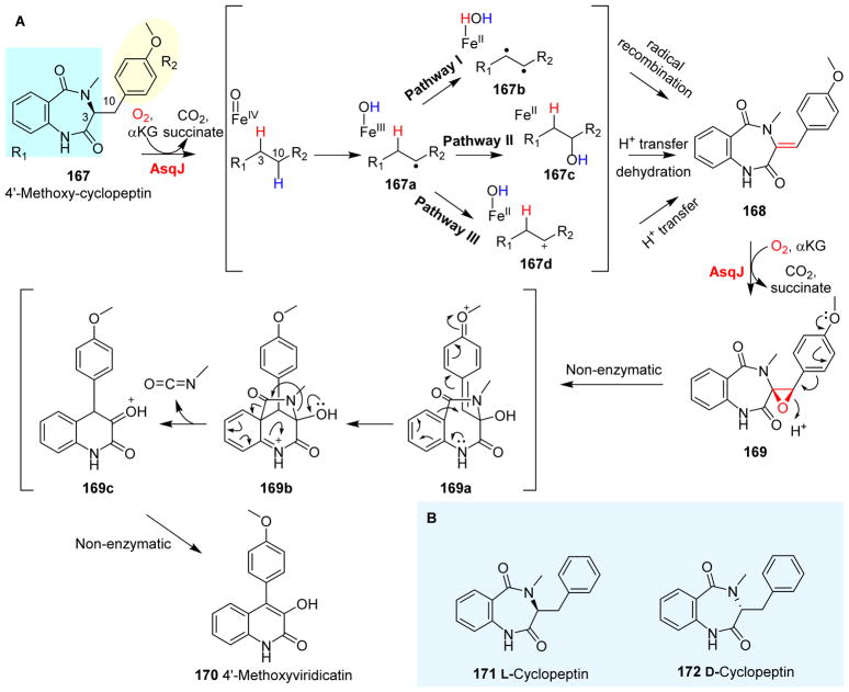
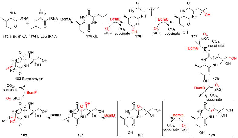
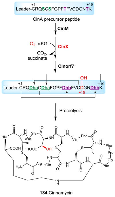
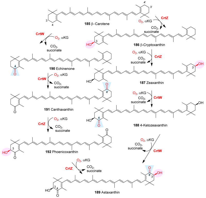
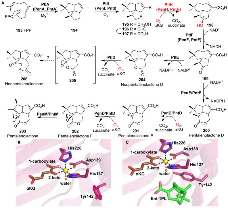
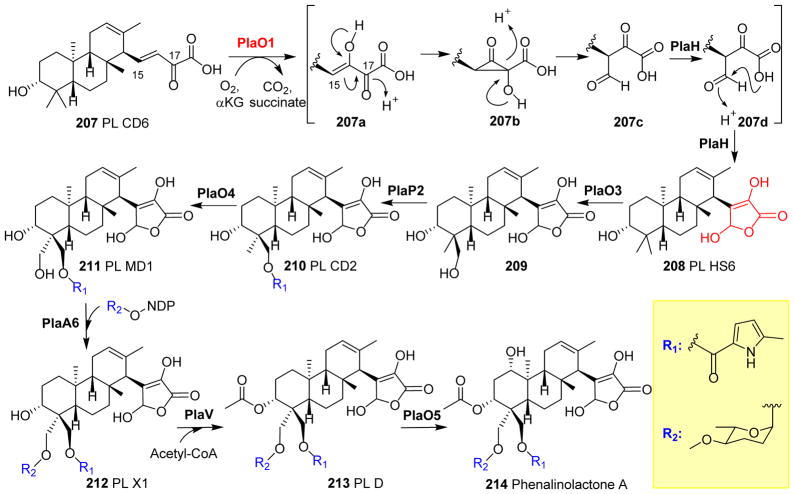
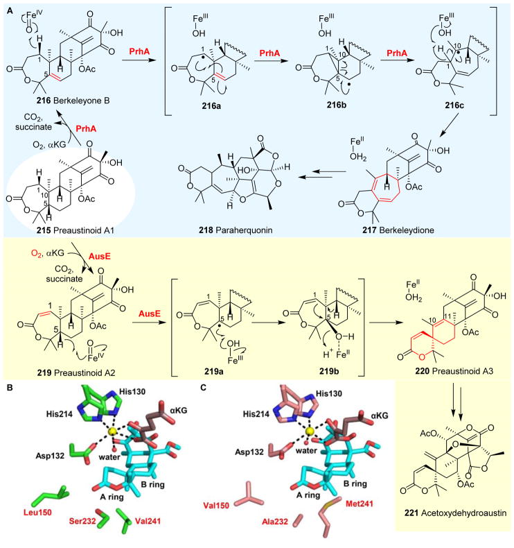
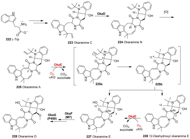
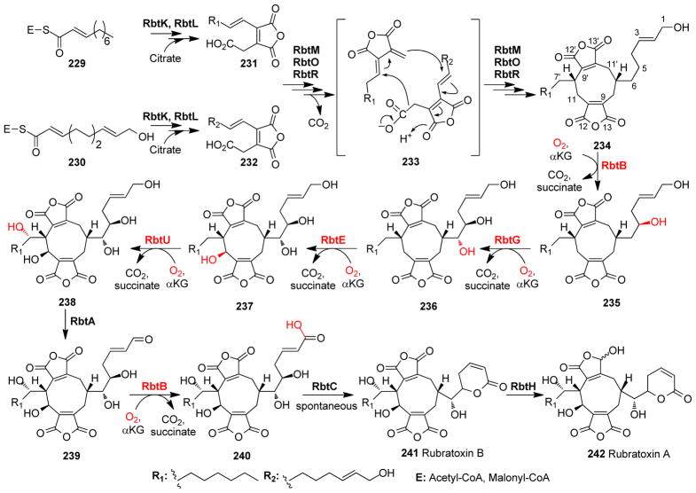
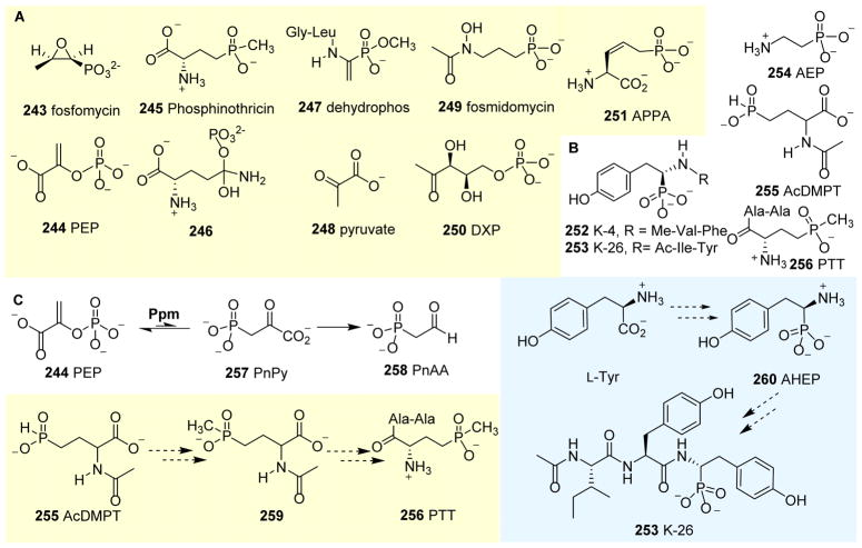
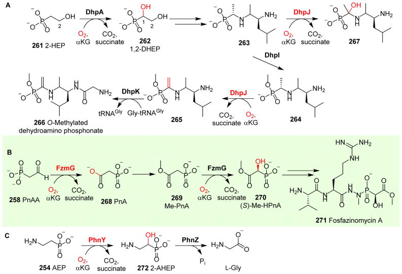
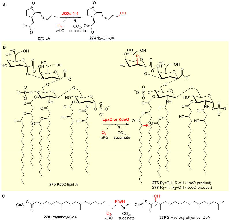
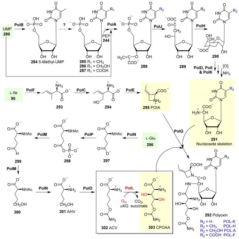
Covering: up to 2018 α-Ketoglutarate (αKG, also known as 2-oxoglutarate)-dependent mononuclear non-haem iron (αKG-NHFe) enzymes catalyze a wide range of biochemical reactions, including hydroxylation, ring fragmentation, C-C bond cleavage, epimerization, desaturation, endoperoxidation and heterocycle formation. These enzymes utilize iron(ii) as the metallo-cofactor and αKG as the co-substrate. Herein, we summarize several novel αKG-NHFe enzymes involved in natural product biosyntheses discovered in recent years, including halogenation reactions, amino acid modifications and tailoring reactions in the biosynthesis of terpenes, lipids, fatty acids and phosphonates. We also conducted a survey of the currently available structures of αKG-NHFe enzymes, in which αKG binds to the metallo-centre bidentately through either a proximal- or distal-type binding mode. Future structure-function and structure-reactivity relationship investigations will provide crucial information regarding how activities in this large class of enzymes have been fine-tuned in nature.


Conflict of interest statement

9 Conflicts of Interest

There are no conflicts to declare.

Figures

Fig. 1
Fig. 1
Hydroxylation mediated by αKG-dependent NHFe enzymes. (A) The generic mechanism of the αKG-NHFe enzyme-mediated hydroxylation reaction, involving an Fe(IV)=O species. (B) Hydroxylation of taurine catalysed by TauD.
Fig. 2
Fig. 2
αKG-NHFe enzyme structural information. (A) The double-stranded helical fold (DSBH fold) first observed in IPNS structure. (B) A typical proximal αKG binding conformation represented by the TauD•Fe•αKG complex. (C) Distal-type αKG binding conformation represented by the FtmOx1•Fe•αKG complex. The proposed αKG conformational switch from the proximal (D) to the distal mode upon exposing the CAS•Fe•αKG•substrate complex to NO (E). (F) Schematic representation of αKG conformational switch between a proximal (F1)- and distal (F2)-type of conformation. (G) Another type of αKG binding conformation observed in the EFE•Fe•αKG binary complex where αKG binds to the Fe(II) centre monodentately using its C5 carboxylate. The iron centre is shown as yellow sphere, αKG is shown as brown sticks, water is shown as a red sphere, and NO is shown as sticks.
Fig. 3
Fig. 3
Halogenation on protein-tethered substrates. (A) Halogenation reaction catalyzed by SyrB2 on L-Thr tethered on SyrB1 4 in Syringomycin E biosynthesis. (B) The proposed mechanism for SyrB2 reaction begins with oxygen activation, similarly to other enzymes in this family, to form the Fe(IV)=O species. Notably, the carboxylate ligand is replaced with a chloride ligand at the iron centre allowing the halogen atom to react with a substrate-based radical to give the halogenated product.
Fig. 4
Fig. 4
Halogenation on protein-tethered substrates. (A) In barbamide 11 biosynthesis, BarB2 works along with BarB1 to yield a trichloro-Leu 10, which is further incorporated into the final product 11. (B) A similar chlorination strategy is observed in CytC3 reaction in the biosynthesis of dichloroaminobutyrate 15. (C) HtcB-mediated chlorination reactions in hectochlorin 20 biosynthesis. (D) Chlorination reaction using a piperazyl-group tethered to a carrier protein as the substrate has been observed in KthP catalysis in the biosynthesis of kutzneride 2.
Fig. 5
Fig. 5
Halogenation-initiated formation of cyclopropane. (A) Halogenation reactions catalyzed by CmaB on CmaD-tethered Ile 24 in the coronatine 28 biosynthetic pathway. (B) KtzD chlorinates KtzC-tethered L-Ile 29 and the chlorinated product is further cyclized by KtzA-catalysis.
Fig. 6
Fig. 6
Halogenation-initiated formation of cyclopropane. (A) In the biosynthesis of curacin 37 and jamaicamide 39, two homologous megasynthases, namely CurA and JamE, catalyze the chlorination of (S)-3-hydroxy-3-methylglutaryl-ACP 32. In the curacin pathway, the ECH2 domain catalyzes the decarboxylation to give an α,β-enoyl thioester 35, while the ECH2 domain in the jamaicamide pathway catalyzes the formation of the vinyl chloride moiety 38. (B) Structure of CurA halogenase in a ligand-free open form. (C) CurA halogenase•Fe•αKG• structural complex showing that αKG (brown stick) and chloride ion (blue sphere) binding trigger a conformation change to a closed form, which allows substrate binding.
Fig. 7
Fig. 7
Halogenation versatility of WelO5 and AmbO5. (A) WelO5 chlorinates hapalindole-type molecules, while AmbO5 exhibits a higher substrate tolerance chlorinating ambiguine, fisherindole and hapalindole alkaloids. (B) Structure of WelO5•Fe•αKG•substrate shows a chloride ligand (blue sphere) at the iron centre (yellow sphere). A second-coordination shell Ser189 was proposed to be involved in controlling the rearrangement of αKG (brown sticks) binding conformation to re-orient the chloride group in the halo-oxo-iron(IV) intermediate towards the substrate for the chlorination reaction.
Fig. 8
Fig. 8
Carnitine biosynthesis. (A) L-carnitine 59 biosynthesis involves two αKG-NHFes: TMLH and BBOX. (B) The biocatalytic versatility of TMLH-mediated hydroxylation on trimethyl-Lys analogues. (C) BBOX also oxidizes THP 68 as the substrate through a Stevens-type rearrangement reaction. (D) Structure of BBOX in complex with zinc (orange sphere), N-oxalylglycine (NOG, green sticks) andγBB substrate (magenta sticks) showing a distal-type αKG binding mode. (E) The studies of PsBBOX show that the positively charged trimethylammonium group on the substrate is crucial for recognition.
Fig. 9
Fig. 9
Amino acid modifications by αKG-NHFe enzymes. (A) In EFE catalysis, L-Arg 84 hydroxylation and αKG fragmentation to ethylene 89 are the two reactions. (B) EFE•Fe•αKG•L-Arg complex shows that αKG (brown stick) binds to the Fe(II) centre bidentately. (C) Hydroxylation on L-Ile mediated by IDO. (D) BtIOD not only catalyzes hydroxylation on a wide range of substrates, but also catalyzes reactions other than hydroxylations. (E) In the biosynthesis of glucoraphasatin, GRS1 catalyzes the desaturation of the side chain of compound 107 to form the aliphatic glucoraphasatin 108. (F) SadA-mediated β-hydroxylation of N-succinyl-L-Ile 109.
Fig. 10
Fig. 10
Hydroxylations of lysyl residues. (A) Collagen polypeptide lysyl 111 hydroxylation mediated by LH1/LH2/LH3. In contrast to LH1 and LH2, LH3 can further modify hydroxylysyl 112 to galactosyl hydroxylysyl 113 and glucosylgalactosyl hydroxylysyl 114. (B) Reactions catalyzed by JMJD6/JMJD4 on lysyl residue in post-translational modifications of proteins.,
Fig. 11
Fig. 11
L-Pro modifications catalyzed by PHs. (A) Four different isomers of monohydroxyl-L- Pro (119a–d) produced by stereo- and regio-specific PHs. (B) Stereospecific epoxidation reactions catalyzed by SgP4H. (C) SrPH can hydroxylate both L-Pro 118 and L-pipcolinic acid 122. (D) In pneumocandin (126 and 127) biosynthesis, Gloxy4 catalyzes the oxidative cyclization of L-Leu 97 to methyl-Pro 124, which is further hydroxylated by GloF to produce hydroxyproline as one of the building blocks for pneumocandin biosynthesis.
Fig. 12
Fig. 12
L-Arg hydroxylation reactions catalyzed by αKG-NHFe oxygenases. (A) VioC hydroxylates L-Arg 84 and VioD further hydroxylates the product 128 to (2S,3R)-capreomycidine 129, which serves as a building block for viomycin biosynthesis. (B) In mannopeptimycin β 135, L-Arg 84 hydroxylation is mediated by MppO to produce a hydroxyenduracididine 134 moiety in the final product. (C) Crystal structure of VioC•Fe•αKG complex.
Fig. 13
Fig. 13
Asparagine and aspartate hydroxylation reactions mediated by αKG-NHFe enzymes. (A) L-Asn 136 hydroxylation is catalyzed by AsnO, to generate the building block 137 for CDA 138 biosynthesis. (B) Biosynthetic pathway of ectoine 144. The hydroxylation of ectoine 144 to hydroxylectoine 145 is mediated by an αKG-dependent EctD.
Fig. 14
Fig. 14
Hydroxylation of glutamate tethered to a carrier protein mediated by KtzO/KtzP in kutzneride 2 biosynthesis, which results in the production of threo 147 and erytho 148.
Fig. 15
Fig. 15
Reactions of 4-hydroxyphenylpyruvate oxygenases. (A) In vancomycin biosynthesis, HmaS-mediated hydroxylation affords L-4-hydroxymandelate 150. (B) HPPD hydroxylates aromatic carbon to yield homogentisate. (C) In the reaction catalyzed by the HPPD F337I variant, both 150 and 154 are produced.
Fig. 16
Fig. 16
Hydroxylation of tryptophan derivatives. (A) DAO-catalyzed production of the natural plant auxin IAA 155. (B) M2H-catalyzed melatonin 157 hydroxylation. (C) FqzB-catalyzed rearrangement in the biosynthesis of spirotryprostatin A 162.
Fig. 17
Fig. 17
Endoperoxide formation in verroculogen biosynthesis. (A) FtmOx1 reaction with ascorbate affords verruculogen 164 as the dominant product, while in the absence of ascorbate, compound 165 is the dominant product. The reactions of FtmOx1 Y224 variants produce the N-1 deprenylation 166 as the major product. (B) Proposed FtmOx1 catalysis involves a tyrosyl radical species, which is key to the endoperoxidation reaction.
Fig. 18
Fig. 18
AsqJ catalysis. (A) AsqJ-catalyzed dehydrogenation and epoxidation. (B) Probes used in the AsqJ-dehydrogenation reaction mechanistic study.
Fig. 19
Fig. 19
The biosynthetic pathway of bicyclomycin involves five αKG-NHFe enzymes in the tailoring reactions to produce the final product bicyclomycin 183.
Fig. 20
Fig. 20
β-Hydroxylation of Asp15 in the precursor peptide CinA catalyzed by an αKG-NHFe enzyme CinX in the cinnamycin 184 biosynthetic pathway.
Fig. 21
Fig. 21
Multiple oxidative modifications in the astaxanthin 189 biosynthetic pathway involving two αKG-dependent NHFe enzymes: CtrZ and CrtW. CrtZ hydroxylates either the 3 or 3′ position of the β-ionone ring, while CrtW oxidizes methylene to keto groups at the 4 or 4′ position of the β-ionone ring.
Fig. 22
Fig. 22
Pentalenolactone and neopentalenolactone biosynthesis. (A) The biosynthetic pathways of pentalenolactone 203 and neopentalenolactone 206. PenH/PntH/PtlH catalyzes the hydroxylation of 1-deoxypentalenic acid to 11-β-hydroxy-1-deoxypentalenic acid 198. (B) Crystal structure of PtlH•Fe•αKG showing a proximal-type αKG (brown sticks) coordination to the iron centre (yellow sphere). (C) Notably, the structure of PtlH with the substrate analogue ent-1-deoxypentalenic acid (green sticks) reveals a conformation change of an active site Y142 relative to that in (B), where there is no substrate.
Fig. 23
Fig. 23
Modification reactions in terpene biosynthesis. In the biosynthesis of phenalinolactone A, PlaO1 is responsible for converting PL CD6 207 to a γ-butyrolactone moiety formation of PL HS6 208 through a proposed cyclopropanone intermediate.
Fig. 24
Fig. 24
Multiple chemical transformations catalyzed by αKG-NHFe enzymes in the paraherquonin and acetoxydehydroaustin pathways. (A) PrhA mediates the dehydrogenation of 215, followed by oxidation to yield paraherquonin as the final product. AusE acts on the same substrate 215. Unlike PrhA, AusE-catalysis goes through a dehydrogenation reaction followed by rearrangements to form the spiro-ring. (B) Crystal structure of AusE•Mn•αKG with substrate 215 modelled into the active site. (C) Crystal structure of PrhA•Fe•αKG•substrate preaustinoid A1 215 (cyan sticks). The residues involved in the structure–function studies are labelled in red.
Fig. 25
Fig. 25
The biosynthetic pathway of okaramine D. Okaramine A 225 can be converted to 12-deshydroxyl okaramine E 226 and okaramine 227 by OkaE-catalysis via radical intermediates.
Fig. 26
Fig. 26
Proposed biosynthetic pathway of rubratoxin A. Four αKG-NHFe enzymes RbtB, RbtG, RbtE and RbtU catalyze the sequential hydroxylations, converting 234 to 238.
Fig. 27
Fig. 27
Natural phosphonates. (A) Phosphonates and their corresponding enzymatic substrates or transition state analogues. (B) Four categories of phosphonates are represented by K-26: 253, 2-aminoethylphosphonic acid (AEP) 254, N-acetyl demethylphos-phinothricin (AcDMPT) 255 and PTT 256, based on how their C-P bonds are constructed. (C) Key steps of the biosynthesis of PnAA 258, PTT 256 and K26 253.
Fig. 28
Fig. 28
Phosphonate modifications mediated by αKG-NHFe enzymes. (A) Reactions catalyzed by DhpA and DhpJ in the O-methylated dehydroamino phosphonate 266 biosynthetic pathway. (B) FzmG mediates multiple hydroxylation reactions in the biosynthesis of fosfazinomycin A 271. (C) The organophosphate metabolism involves the PhnY-mediated hydroxylation of 254 to yield 2-amino-1-hydroxyethylphosphonic acid 272.
Fig. 29
Fig. 29
Lipid and fatty acid modification reactions. (A) Hydroxylation of jasmonic acid 273 mediated by JOXs 1–4. (B) LpxO and KdoO-catalyzed hydroxylations of Kdo2-lipid A 275., (C) Hydroxylation of phytanoyl-CoA 278 catalyzed by PhyH in the phytanic acid metabolism.
Fig. 30
Fig. 30
Biosynthesis of capuramycin 283. The αKG-NHFe Cpr19 catalyzes the conversion of UMP 280 to uridine-5-aldehyde 281 through a germinal hydroxyl-phosphoester intermediate 280a, followed by phosphate elimination.
Fig. 31
Fig. 31
The biosynthetic pathway of polyoxin 292. The reactions catalyzed by PolL introduce two hydroxyl groups onto the structure of CPOAA 303, which is one of the modules used in the biosynthesis of polyoxin.

References

    1. Hausinger RP. Crit Rev Biochem Mol Biol. 2004;39:21–68. - PubMed
    1. Simmons JM, Müller TA, Hausinger RP. Dalton Trans. 2008:5132–5142. - PMC - PubMed
    1. Purpero V, Moran GR. J Biol Inorg Chem. 2007;12:587–601. - PubMed
    1. Loenarz C, Schofield CJ. Nat Chem Biol. 2008;4:152–156. - PubMed
    1. Loenarz C, Schofield CJ. Trends Biochem Sci. 2011;36:7–18. - PubMed

Publication types