Review
. 2019 Feb;145(Pt B):160-176.
doi: 10.1016/j.neuropharm.2018.06.021. Epub 2018 Jun 20.

Novel therapies for combating chronic neuropathological sequelae of TBI

Affiliations
Review

Novel therapies for combating chronic neuropathological sequelae of TBI

Milos D Ikonomovic et al. Neuropharmacology. 2019 Feb.

Abstract

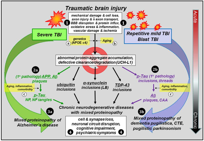
Traumatic brain injury (TBI) is a risk factor for development of chronic neurodegenerative disorders later in life. This review summarizes the current knowledge and concepts regarding the connection between long-term consequences of TBI and aging-associated neurodegenerative disorders including Alzheimer's disease (AD), chronic traumatic encephalopathy (CTE), and Parkinsonism, with implications for novel therapy targets. Several aggregation-prone proteins such as the amyloid-beta (Aβ) peptides, tau proteins, and α-synuclein protein are involved in secondary pathogenic cascades initiated by a TBI and are also major building blocks of the hallmark pathological lesions in chronic human neurodegenerative diseases with dementia. Impaired metabolism and degradation pathways of aggregation-prone proteins are discussed as potentially critical links between the long-term aftermath of TBI and chronic neurodegeneration. Utility and limitations of previous and current preclinical TBI models designed to study the link between TBI and chronic neurodegeneration, and promising intervention pharmacotherapies and non-pharmacologic strategies to break this link, are also summarized. Complexity of long-term neuropathological consequences of TBI is discussed, with a goal of guiding future preclinical studies and accelerating implementation of promising therapeutics into clinical trials. This article is part of the Special Issue entitled "Novel Treatments for Traumatic Brain Injury".

Keywords: Alpha synuclein; Alzheimer's disease; Amyloid; Brain trauma; Chronic traumatic encephalopathy; Tau.


Figures

Figure 1.
Figure 1.. Consequences of single severe TBI and repetitive mild TBI: abnormal protein aggregation and risk for chronic neurodegenerative diseases.
TBI results in multiple pathological cascades involving acute cell loss, axonal injury, blood brain barrier (BBB) disruption, oxidative stress, inflammation, vascular damage and ischemia (1). There is considerable overlap in acute pathology in severe, rmTBI, and blast TBI despite the different injury mechanisms. All are characterized in part by accumulation of aggregation-prone molecules, however, the primary pathology of each depends on activation and propagation of different aggregated molecules chronically after TBI. Specifically, severe TBI is more closely associated with the activation of amyloidogenic pathway due to altered amyloid-β (Aβ) precursor protein (APP) metabolism and accumulation of Aβ peptides (2a), with secondary development of over-phosphorylated tau (p-tau) pathology, while the pathology of rmTBI and blast TBI is driven primarily by p-tau protein (2b) and secondary development of amyloidosis at more advanced age. In addition, aggregates of other proteins (ubiquitin; α-synuclein; transactive response DNA binding protein 43, TDP-43) can be initiated by all forms of TBI and contribute to the mixed proteinopathy of AD (3a) or dementia pugilistica, chronic traumatic encephalopathy (CTE), and parkinsonism (3b). Development and progression of these pathologies can be influenced by genetic factors (a) and aging (b,c), inflammation and comorbidities (c; vascular disease, depression, substance abuse), as well as defective clearance mechanisms involving dysregulation of ubiquitin carboxyl-terminal hydrolase isozyme L1 (UCH-L1) and the proteasome. Chronic neuronal injury combined with toxic intracellular inclusions ultimately results in cell death, loss of synapses and circuits, cognitive impairment, and psychiatric symptoms (4). Early intervention to prevent the initiation and/or propagation of pathological protein aggregates may be the key to breaking the link between TBI and chronic neurodegenerative diseases. Abbreviations: APOE, apolipoprotein E; CAA, cerebral amyloid angiopathy; LB, Lewy bodies; NP, neuritic plaques; NF, neurofibrillary.
Figure 2.
Figure 2.. Amyloid-β (Aβ) deposition and intracellular amyloid precursor protein (APP) accumulation in the temporal cortex after severe TBI and in AD.
Diffuse extracellular deposits of Aβ are scattered throughout the gray matter in a biopsy sample from an acute severe TBI patient (A), contrasting the more compact and numerous Aβ plaques in AD (B). The arrow in (B) marks an Aβ-immunoreactive blood vessel commonly observed in AD but not acutely after TBI. Intracellular APP immunoreactivity is present in clusters of dystrophic neurites in both severe TBI (C) and in AD (D). APP is also detected in damaged axons in severe TBI (E, arrows point at axonal bulbs) and in neuronal cell bodies in both TBI and AD (F, arrows point at corkscrew-appearing dendrites in AD). Illustrations are representative of results obtained in biopsy samples of temporal lobe from 20 subjects admitted to the University of Pittsburgh emergency department with severe closed head injury (GCS < 9). Brain specimens were obtained under the approval of the University of Pittsburgh Institutional Review Board for the Clinical Core of the University of Pittsburgh’s Brain Trauma Research Center and for the University of Pittsburgh ADRC. All TBI patients underwent temporal lobe resection for decompressive craniectomy, either to relieve intractable cerebral swelling or for removal of severely traumatized brain tissue as described in (DeKosky et al., 2007; Ikonomovic et al., 2004). Time interval from injury to surgery averaged 11.25 hr ± 12.5 (range 2–72 hr). Aβ plaques were observed in eight (40%) subjects, confirming prior reports, while 16 subjects had APP aggregates in neuronal cell bodies and processes. Autopsy samples examined were middle temporal gyrus tissue from 10 cases of severe AD obtained from brain bank of the University of Pittsburgh Alzheimer’s Disease Research Center (ADRC). Neuropathological confirmation of AD diagnosis was made according to established criteria, with all cases categorized by a certified ADRC neuropathologist as Braak stage 6, with high frequency of neuritic plaques by CERAD criteria (definite AD) and high likelihood of AD by the NIA-Reagan Institute criteria. TBI patients/AD cases illustrated: biopsy sample of left temporal cortex obtained from a 39 y.o. male, 10 hours after TBI. AD autopsy sample obtained from a 77 y.o. female with a post mortem interval of 3 hr. All samples were fixed in paraformaldehyde, sectioned at 40 µm, and processed using chromogen-based immunohistochemistry using antibodies clone 4G8 (A,B; BioLegend #SIG-39220; Aβ) and clone anti-6 (C-F; gift from Athena Neurosciences; APP). Scale bar = 200 µm (A,B); 50 µm (C,D,F); 25 µm (E).
Figure 3.
Figure 3.. Complexity of acute pathological sequelae of acute severe TBI: potential seeds of chronic neurodegeneration.
Accumulation of amyloid-β (A; Aβ; diffuse extracellular plaques), tau (B; tau miss-sorting into dendrites and cell soma), and alpha-synuclein (C; α-syn; immunoreactive cells are marked by arrows), activated microglia expressing cluster differentiation factor 68 antigen (D; CD68; immunoreactive cells bodies) and increased astrocytosis (E; glial fibrillary acidic protein, GFAP; immunoreactive cell bodies and processes), and evidence of neurofilament disruption (F; NF-H; arrows mark dystrophic axons). See Figure 2 legend for details of sample acquisition. Patient illustrated: biopsy sample of left temporal cortex obtained from a 45 y.o. female, 8 hours after TBI. Samples were formalin-fixed and paraffin-embedded, sectioned at 8 µm, and processed using chromogen-based immunohistochemistry using antibodies clone 4G8 (BioLegend, Aβ), a polyclonal antibody against tau (Dako #0024), antibody clone LB509 (Abcam #ab27766, α-syn), antibody clone KP1 (Thermo #MA5-13324, CD68), a polyclonal antibody against GFAP (Dako #Z0334), and antibody clone SMI-32 (Millipore # NE1023, NF-H). Chromogen signal is brown and sections were counterstained with hematoxylin (blue) to mark nuclei. Scale bar = 25 µm.
Figure 4.
Figure 4.. Diverse p-tau lesions characteristic of CTE in relation to AD pathology.
A-D: p-tau pathology (antibody clone PHF-1, gift from Dr. P. Davies, Albert Einstein College of Medicine) in inferior temporal cortex (IT) and hippocampal CA2 from a 59 y.o. male with a history of rmTBI (PMI = 5 hr). Samples were formalin-fixed and paraffin-embedded, sectioned at 8 µm, and processed using chromogen-based immunohistochemistry. Neuropathological evidence of CTE is detected as p-tau immunoreactive glial processes at subpial (A) and perivascular (empty arrow in C and D) locations, and p-tau immunoreactive neurons in hippocampus CA2 (B) and inferior temporal cortex (IT, arrows in C and D). No Aβ pathology was found in the same brain regions. E-F: Samples of IT cortex from a 62-year old case with AD (Braak stage VI; PMI = 4 hr) and a history of TBI were formalin-fixed and paraffin-embedded, sectioned at 8 µm, and processed using chromogen-based immunohistochemistry. Extensive p-tau pathology is detected at deep sulcal locations (E, asterisk) as dense neuropil threads, neuritic plaques (E, NP), and tangles (E, small arrows). Numerous Aβ plaques (F; antibody clone 4G8, Biolegend) are seen in a directly adjacent section (F). This illustrates the challenge faced in postmortem analyses when attempting to distinguish pathology specifically related to previous TBI in aged brains affected with advanced AD, due to overwhelming density of p-tau and Aβ pathology at time of death. Scale bar = 150 µm (A-C); 75 µm (D-F).
Figure 5.
Figure 5.. Multiple beneficial effects of simvastatin therapy after TBI in humanized Aβ mice.
Human amyloid-β (Aβ) mice (see text for description of mouse model) were exposed to vertically directed CCI injury and administered 3 mg/kg simvastatin (Merck) or vehicle (3% methylcellulose) daily by oral gavage. After a 14-day survival interval (A-F) whole brains from CCI-injured mice were preserved in 4% paraformaldehyde and assessed histopathologically in relation to non-surgically manipulated (Naïve) mice. Microglia activation (A-C; rat monoclonal antibody clone A3–1 against mouse macrophage glycoprotein F4/80; abcam #ab6640) induced in CA1 hippocampus after CCI injury (B; compare to naïve mice, A) is suppressed in mice receiving simvastatin. Simvastatin therapy also resulted in preservation of synaptic densities assessed using mouse monoclonal antibody clone SVP-38 (Sigma #S5768) in CA3 hippocampus after CCI injury (F) compared to a marked decrease in CCI-injured, vehicle-treated mice (E) relative to naïve mice (D). Simvastatin therapy also resulted in greater tissue preservation assessed in vivo using magnetic resonance imaging (MRI; G) and ex vivo (Nissl histology; H). Arterial spin labeling MRI was used to assess cerebral blood flow (CBF) in CCI-injured relative to naïve mice 21 days after surgery; in this study, CCI-injured, simvastatin-treated mice had markedly higher regional CBF rates compared to CCI-injured, vehicle-treated and naïve mice (I). Enzyme-linked immunosorbent assay (ELISA) quantification of Aβ showed that CCI-induced elevations in Aβ concentration (relative to naïve) are suppressed by simvastatin treatment (*p<0.05) at 21 days after CCI injury. Collectively, these examples demonstrate how a single drug with pleiotropic actions can have beneficial effects on a wide range of pathology after brain injury, including effects on neuroinflammation, synaptic preservation, neuropil preservation, preserved/enhanced CBF, and suppression of aggregation prone Aβ peptides. Scale bar = 40 µm (A-F); 2 mm (G-I).

Similar articles

Cited by

References

    1. Abisambra JF, Scheff S, 2014. Brain injury in the context of tauopathies. J Alzheimers Dis 40, 495–518. - PubMed
    1. Abrahamson EE, Foley LM, Dekosky ST, Hitchens TK, Ho C, Kochanek PM, Ikonomovic MD, 2013. Cerebral blood flow changes after brain injury in human amyloid-beta knock-in mice. J Cereb Blood Flow Metab 33, 826–833. - PMC - PubMed
    1. Abrahamson EE, Ikonomovic MD, Ciallella JR, Hope CE, Paljug WR, Isanski BA, Flood DG, Clark RSB, DeKosky ST, 2006. Caspase inhibition therapy abolishes brain trauma-induced increases in Abeta peptide: Implications for clinical outcome. Exp Neurol 197, 437–450. - PubMed
    1. Abrahamson EE, Ikonomovic MD, Dixon CE, DeKosky ST, 2009. Simvastatin therapy prevents brain trauma-induced increases in beta-amyloid peptide levels. Ann Neurol 66, 407–414. - PubMed
    1. Acosta SA, Tajiri N, de la Pena I, Bastawrous M, Sanberg PR, Kaneko Y, Borlongan CV, 2015. Alpha-synuclein as a pathological link between chronic traumatic brain injury and Parkinson’s disease. J Cell Physiol 230, 1024–1032. - PMC - PubMed

Publication types

MeSH terms