. 2018 Oct;13(10):1496-1507.
doi: 10.1016/j.jtho.2018.05.039. Epub 2018 Jun 19.

Innate Genetic Evolution of Lung Cancers and Spatial Heterogeneity: Analysis of Treatment-Naïve Lesions

Affiliations

Innate Genetic Evolution of Lung Cancers and Spatial Heterogeneity: Analysis of Treatment-Naïve Lesions

Kenichi Suda et al. J Thorac Oncol. 2018 Oct.

Abstract

Introduction: Data regarding the pre-treatment intertumor heterogeneity of potential biomarkers in advanced-stage lung cancers is limited. A finding of such heterogeneity between primary and metastatic lesions would prove valuable to determine if a metastatic lesion can be a surrogate for the primary tumor, as more biomarkers will likely be used in the future to inform treatment decisions.

Methods: We performed RNA sequencing to analyze intertumor heterogeneity in 30 specimens (primary tumors, intrathoracic, and extrathoracic metastatic lesions) obtained from five treatment-naïve lung cancer patients.

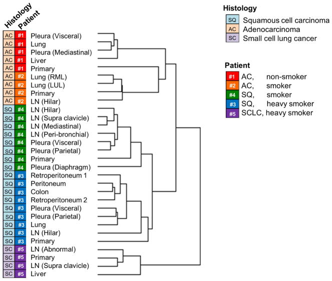
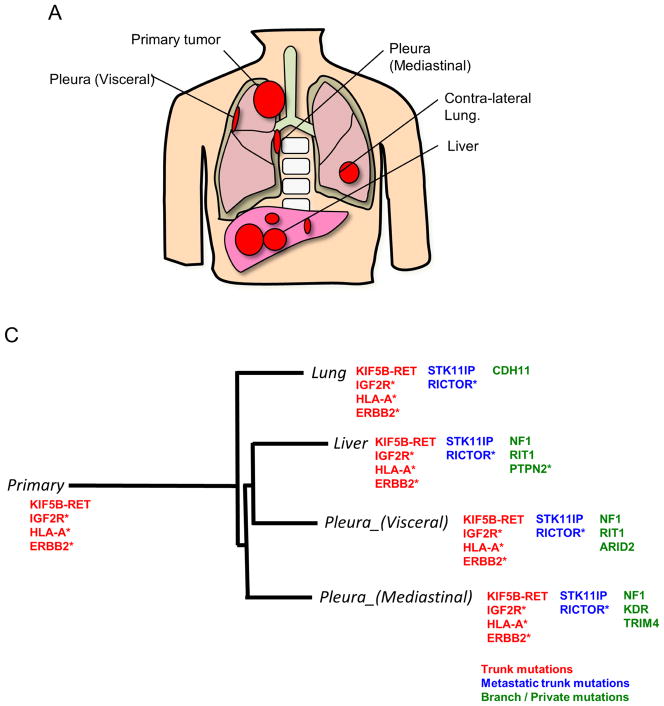
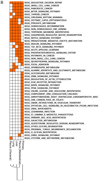
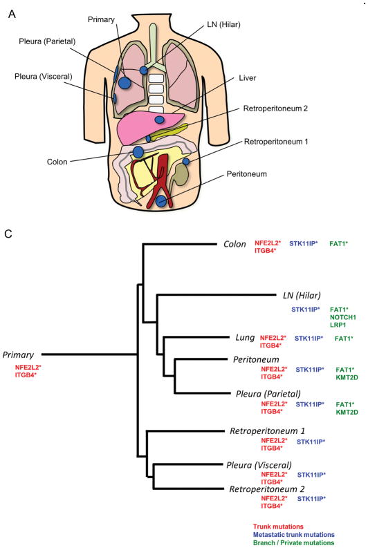
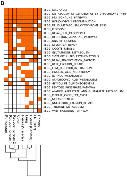
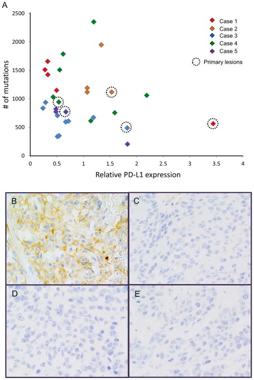
Results: The global unsupervised clustering analysis showed that the lesions clustered at the individual patient level rather than on the metastatic sites, suggesting that the characteristics of specific tumor cells have a greater impact on the gene expression signature than the microenvironment in which the metastasis develops. The mutational and transcriptional data highlight the presence of intertumor heterogeneity showing that the primary tumors are usually distinct from metastatic lesions. Through a comparison between metastatic lesions and the primary tumors, we observed that pathways related to cell proliferation were upregulated, whereas immune-related pathways were downregulated in metastatic lesions.

Conclusion: These data not only provide insight into the evolution of lung cancers, but also imply possibilities and limitations of biomarker-based treatment in lung cancers.

Keywords: Autopsy; Biomarkers; Immune-related markers; RET fusion; RNA sequencing; Tumor heterogeneity.


Conflict of interest statement

Conflicts of Interest and Source of Funding

T. Mitsudomi has received honoraria from AstraZeneca, Chugai, Boehringer-Ingelheim, Pfizer and Roche; has received compensation from AstraZeneca, Chugai, Boehringer-Ingelheim, Pfizer, Roche and Clovis Oncology for participating in advisory boards; and has received research funding (through Kindai University Faculty of Medicine) from AstraZeneca and Chugai. F. R. Hirsch has received compensation from Genentech/Roche, Pfizer, BMS, Lilly, Merck, Ventana/Roche, Novartis and Abbvie for participating in advisory boards; and has received research funding (through the University of Colorado) from Genetech/Roche, BMS, Lilly, Bayer, Amgen and Ventana/Roche. All other authors declare that they have no conflict of interest related to this study.

Our study was supported by an IASLC Young investigator Award (2015 – 2017) to K. Suda, JSPS KAKENHI Grant Number 18K07336 to K. Suda, the Pia and Fred Hirsch Chair in Lung Cancer at the University of Colorado Anschutz Medical Campus, the National Institutes of Health P50CA058187, P30CA046934, Cancer League of Colorado, and the David F. and Margaret T. Grohne Family Foundation.

Figures

Figure 1
Figure 1
The global unsupervised clustering analysis of expression data of all lesions analyzed. The left and right rows indicate the histology and the individual patient, respectively. LN, lymph node metastases; RML, right middle lobe of the lung; LUL, left upper lobe of the lung; AC, adenocarcinoma; SQ, squamous cell carcinoma; SCLC, small-cell lung cancer.
Figure 2
Figure 2
Mutational and pathway analyses of lesions obtained from a never-smoking lung adenocarcinoma patient (Case 1). A, Geographic locations of analyzed lesions are shown. B, Pathway analysis was performed with Gene Set Enrichment Analysis based on MSigDB C2 curated KEGG gene sets. Dysregulated pathways in all lesions are summarized in Supplementary Table 1, and are excluded from the figure. C, Representative mutations identified in Case 1. KIF5B-RET fusion was also identified in all lesions, but not in non-cancerous tissue, using TopHat-Fusion on the RNA-seq. IGF2R, HLA-A, and ERBB2 mutations were also identified as trunk mutations, while NF1 mutation was present only in some metastatic lesions. The phylogenetic tree was constructed using a Neighbor joining method implemented in the R phangom package, based on somatic and deleterious mutations in reliably expressed genes. The genes with asterisk indicate that somatic/deleterious mutations in those genes were identified in lesions obtained from the other lung adenocarcinoma patient (Case 2).
Figure 2
Figure 2
Mutational and pathway analyses of lesions obtained from a never-smoking lung adenocarcinoma patient (Case 1). A, Geographic locations of analyzed lesions are shown. B, Pathway analysis was performed with Gene Set Enrichment Analysis based on MSigDB C2 curated KEGG gene sets. Dysregulated pathways in all lesions are summarized in Supplementary Table 1, and are excluded from the figure. C, Representative mutations identified in Case 1. KIF5B-RET fusion was also identified in all lesions, but not in non-cancerous tissue, using TopHat-Fusion on the RNA-seq. IGF2R, HLA-A, and ERBB2 mutations were also identified as trunk mutations, while NF1 mutation was present only in some metastatic lesions. The phylogenetic tree was constructed using a Neighbor joining method implemented in the R phangom package, based on somatic and deleterious mutations in reliably expressed genes. The genes with asterisk indicate that somatic/deleterious mutations in those genes were identified in lesions obtained from the other lung adenocarcinoma patient (Case 2).
Figure 3
Figure 3
Mutational and pathway analyses of lesions obtained from a lung squamous cell carcinoma patient with heavy smoking history (Case 3). A, Geographic locations of analyzed lesions are shown. B, Pathway analysis was performed with Gene Set Enrichment Analysis based on MSigDB C2 curated KEGG gene sets. C, Representative mutations identified in Case 3. NFE2L2 and ITGB4 mutations were identified in all lesions except for the hilar lymph node metastasis. The phylogenetic tree was constructed using a Neighbor joining method implemented in the R phangom package, based on somatic and deleterious mutations in reliably expressed genes. The genes with asterisks indicate that somatic/deleterious mutations in those genes were identified in lesions obtained from the other lung squamous cell carcinoma patient (Case 4).
Figure 3
Figure 3
Mutational and pathway analyses of lesions obtained from a lung squamous cell carcinoma patient with heavy smoking history (Case 3). A, Geographic locations of analyzed lesions are shown. B, Pathway analysis was performed with Gene Set Enrichment Analysis based on MSigDB C2 curated KEGG gene sets. C, Representative mutations identified in Case 3. NFE2L2 and ITGB4 mutations were identified in all lesions except for the hilar lymph node metastasis. The phylogenetic tree was constructed using a Neighbor joining method implemented in the R phangom package, based on somatic and deleterious mutations in reliably expressed genes. The genes with asterisks indicate that somatic/deleterious mutations in those genes were identified in lesions obtained from the other lung squamous cell carcinoma patient (Case 4).
Figure 4
Figure 4
The inter-tumor heterogeneity of potential “predictive” biomarkers for PD-1/PD-L1 targeted immunotherapy. A, The relative expression of CD274 (PD-L1) and tumor mutation burden (the numbers of somatic/non-synonymous mutations detected) are shown. Primary lesion from each patient was marked by a dotted circle. B–D, Comparison of PD-L1 IHC staining in the primary tumor (B), visceral pleura (C), contra-lateral lung metastasis (D), and liver metastasis (E) in Case 1 is shown.

References

    1. Hirsch FR, Suda K, Wiens J, Bunn PA., Jr New and emerging targeted treatments in advanced non-small-cell lung cancer. Lancet. 2016;388(10048):1012–24. - PubMed
    1. Takano T, Fukui T, Ohe Y, et al. EGFR mutations predict survival benefit from gefitinib in patients with advanced lung adenocarcinoma: a historical comparison of patients treated before and after gefitinib approval in Japan. J Clin Oncol. 2008;26(34):5589–95. - PubMed
    1. Kris MG, Johnson BE, Berry LD, et al. Using multiplexed assays of oncogenic drivers in lung cancers to select targeted drugs. JAMA. 2014;311(19):1998–2006. - PMC - PubMed
    1. Yatabe Y, Matsuo K, Mitsudomi T. Heterogeneous distribution of EGFR mutations is extremely rare in lung adenocarcinoma. J Clin Oncol. 2011;29(22):2972–7. - PubMed
    1. de Bruin EC, McGranahan N, Mitter R, et al. Spatial and temporal diversity in genomic instability processes defines lung cancer evolution. Science. 2014;346(6206):251–6. - PMC - PubMed

Publication types