

The Bacterial Protein CNF1 as a Potential Therapeutic Strategy against Mitochondrial Diseases: A Pilot Study
- PMID: 29933571
- PMCID: PMC6073533
- DOI: 10.3390/ijms19071825
The Bacterial Protein CNF1 as a Potential Therapeutic Strategy against Mitochondrial Diseases: A Pilot Study
Abstract
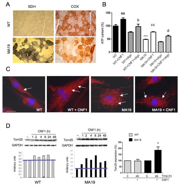
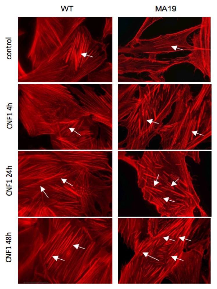
The Escherichia coli protein toxin cytotoxic necrotizing factor 1 (CNF1), which acts on the Rho GTPases that are key regulators of the actin cytoskeleton, is emerging as a potential therapeutic tool against certain neurological diseases characterized by cellular energy homeostasis impairment. In this brief communication, we show explorative results on the toxin’s effect on fibroblasts derived from a patient affected by myoclonic epilepsy with ragged-red fibers (MERRF) that carries a mutation in the m.8344A>G gene of mitochondrial DNA. We found that, in the patient’s cells, besides rescuing the wild-type-like mitochondrial morphology, CNF1 administration is able to trigger a significant increase in cellular content of ATP and of the mitochondrial outer membrane marker Tom20. These results were accompanied by a profound F-actin reorganization in MERRF fibroblasts, which is a typical CNF1-induced effect on cell cytoskeleton. These results point at a possible role of the actin organization in preventing or limiting the cell damage due to mitochondrial impairment and at CNF1 treatment as a possible novel strategy against mitochondrial diseases still without cure.
Keywords: actin cytoskeleton; adenosine triphosphate; cytotoxic necrotizing factor 1; mitochondrial diseases; myoclonic epilepsy with ragged red fibers syndrome.
Conflict of interest statement
The authors declare no conflict of interest.
Figures
Similar articles
-
Parkin-mediated mitophagy and autophagy flux disruption in cellular models of MERRF syndrome.Biochim Biophys Acta Mol Basis Dis. 2020 Jun 1;1866(6):165726. doi: 10.1016/j.bbadis.2020.165726. Epub 2020 Feb 13. Biochim Biophys Acta Mol Basis Dis. 2020. PMID: 32061767
-
CNF1 Enhances Brain Energy Content and Counteracts Spontaneous Epileptiform Phenomena in Aged DBA/2J Mice.PLoS One. 2015 Oct 12;10(10):e0140495. doi: 10.1371/journal.pone.0140495. eCollection 2015. PLoS One. 2015. PMID: 26457896 Free PMC article.
-
Enhancement of mitochondrial ATP production by the Escherichia coli cytotoxic necrotizing factor 1.FEBS J. 2014 Aug;281(15):3473-88. doi: 10.1111/febs.12874. Epub 2014 Jul 1. FEBS J. 2014. PMID: 24925215
-
Mitochondrial DNA mutation-elicited oxidative stress, oxidative damage, and altered gene expression in cultured cells of patients with MERRF syndrome.Mol Neurobiol. 2010 Jun;41(2-3):256-66. doi: 10.1007/s12035-010-8123-7. Epub 2010 Apr 23. Mol Neurobiol. 2010. PMID: 20411357 Review.
-
The E. coli CNF1 as a pioneering therapy for the central nervous system diseases.Toxins (Basel). 2014 Jan 7;6(1):270-82. doi: 10.3390/toxins6010270. Toxins (Basel). 2014. PMID: 24402235 Free PMC article. Review.
Cited by
-
Crystal structure of bacterial cytotoxic necrotizing factor CNFY reveals molecular building blocks for intoxication.EMBO J. 2021 Feb 15;40(4):e105202. doi: 10.15252/embj.2020105202. Epub 2021 Jan 7. EMBO J. 2021. PMID: 33410511 Free PMC article.
-
Cnf1 Variants Endowed with the Ability to Cross the Blood-Brain Barrier: A New Potential Therapeutic Strategy for Glioblastoma.Toxins (Basel). 2020 May 4;12(5):291. doi: 10.3390/toxins12050291. Toxins (Basel). 2020. PMID: 32375387 Free PMC article.
-
Treatment with the Bacterial Toxin CNF1 Selectively Rescues Cognitive and Brain Mitochondrial Deficits in a Female Mouse Model of Rett Syndrome Carrying a MeCP2-Null Mutation.Int J Mol Sci. 2021 Jun 23;22(13):6739. doi: 10.3390/ijms22136739. Int J Mol Sci. 2021. PMID: 34201747 Free PMC article.
-
Effects of the Escherichia coli Bacterial Toxin Cytotoxic Necrotizing Factor 1 on Different Human and Animal Cells: A Systematic Review.Int J Mol Sci. 2021 Nov 22;22(22):12610. doi: 10.3390/ijms222212610. Int J Mol Sci. 2021. PMID: 34830494 Free PMC article.
-
The best evidence for progressive myoclonic epilepsy: A pathway to precision therapy.Seizure. 2019 Oct;71:247-257. doi: 10.1016/j.seizure.2019.08.012. Epub 2019 Aug 23. Seizure. 2019. PMID: 31476531 Free PMC article. Review.
References
-
- Fukuhara N., Tokiguchi S., Shirakawa K., Tsubaki T. Myoclonus Epilepsy Associated with Ragged-Red Fibres (Mitochondrial Abnormalities): Disease Entity or a Syndrome? Light-and Electron-Microscopic Studies of Two Cases and Review of Literature. J. Neurol. Sci. 1980;47:117–133. doi: 10.1016/0022-510X(80)90031-3. - DOI - PubMed
Publication types
MeSH terms
Substances
LinkOut - more resources
Full Text Sources
Other Literature Sources
