Review
. 2018 Jun 21;6(3):29.
doi: 10.3390/proteomes6030029.

Proteomic Analysis of Histone Variants and Their PTMs: Strategies and Pitfalls

Affiliations
Review

Proteomic Analysis of Histone Variants and Their PTMs: Strategies and Pitfalls

Sara El Kennani et al. Proteomes. .

Abstract

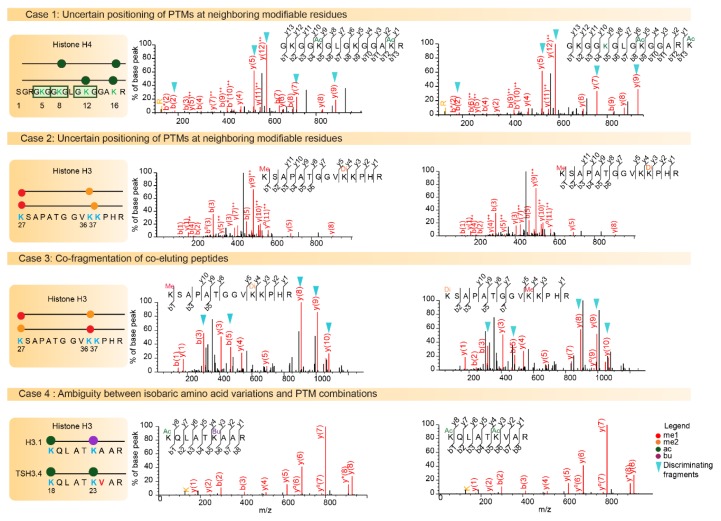
Epigenetic modifications contribute to the determination of cell fate and differentiation. The molecular mechanisms underlying histone variants and post-translational modifications (PTMs) have been studied in the contexts of development, differentiation, and disease. Antibody-based assays have classically been used to target PTMs, but these approaches fail to reveal combinatorial patterns of modifications. In addition, some histone variants are so similar to canonical histones that antibodies have difficulty distinguishing between these isoforms. Mass spectrometry (MS) has progressively developed as a powerful technology for the study of histone variants and their PTMs. Indeed, MS analyses highlighted exquisitely complex combinations of PTMs, suggesting “crosstalk” between them, and also revealed that PTM patterns are often variant-specific. Even though the sensitivity and acquisition speed of MS instruments have considerably increased alongside the development of computational tools for the study of multiple PTMs, it remains challenging to correctly describe the landscape of histone PTMs, and in particular to confidently assign modifications to specific amino acids. Here, we provide an inventory of MS-based strategies and of the pitfalls inherent to histone PTM and variant characterization, while stressing the complex interplay between PTMs and histone sequence variations. We will particularly illustrate the roles played by MS-based analyses in identifying and quantifying histone variants and modifications.

Keywords: bottom-up analysis; computational tools; crosstalk; histone variants; mass spectrometry; post-translational modifications.


Conflict of interest statement

None of the authors have any conflict of interest to declare.

Figures

Figure 1
Figure 1
Sequence similarity between histone variants in human and mouse. Core histones H2A (yellow), H2B (red), H3 (blue), and histone linker H1 (purple) are illustrated for mouse (A) and human (B). Sequence data were obtained from and treated as published in Reference [17].
Figure 2
Figure 2
Representation of bottom-up, middle-down, and top-down mass spectrometry experiments.
Figure 3
Figure 3
Challenges of histone post-translational modifications (PTM) assignments. The Mass Spectrometry fragmentation (MS/MS) spectra of higher energy collisional dissociation (HCD) fragmented histone peptides are shown. Case 1 illustrates positional isomers for the di-acetylated N-terminal tail of histone H4 containing the four Lysine residues K5, K8, K12, and K16. The left spectrum illustrates the identification of the first positional isomer indicated in the diagram, with a Mascot identification score of 65. On the right, the same spectrum is interpreted with fragments of the second possible modified sequence matched with a score of 56. Fragment ions b2, y5, y7, and y9 and y12++ can be used to assign acetylation on different lysine residues. Case 2 illustrates the difficulty associated with confidently assigning a Lys36 or Lys37 di-methylation to peptide K27SAPSTGGVK36K37PHR, as it relies only on the weak-intensity y4 fragment to discriminate between these two modifiable residues. Case 3 shows the probable co-fragmentation of the co-eluting peptides K27me1-K36me2 and K27me2-K36me1. The two positional isomers are matched to this MS/MS spectrum with Mascot scores 31 and 29, respectively. Discriminating b3, b5, y8, y9 and y10 fragment ions are detectable at high intensity in the spectrum. Case 4 illustrates ambiguity between amino acid variation and combination of PTMs. The masses of peptides K18QLATK23acVAR from TS H3.4 and K18QLATK23buAAR from H3.1/H3.2 are strictly equal. A diagnostic ion at m/z 126.091 indicates the presence of an acetylated Lys residue in the fragmented peptide, and thus orients toward the identification of acetylated TS H3.4.
Figure 4
Figure 4
Examples of isobaric PTM combinations. Differentiating structures in the Lys side chain are highlighted in yellow with an indication of their corresponding masses.
Figure 5
Figure 5
Sequence alignment for mouse histone H3 and its variants.

Similar articles

Cited by

References

    1. Luger K., Mäder A.W., Richmond R.K., Sargent D.F., Richmond T.J. Crystal structure of the nucleosome core particle at 2.8 A resolution. Nature. 1997;389:251–260. doi: 10.1038/38444. - DOI - PubMed
    1. Kowalski A., Pałyga J. Linker histone subtypes and their allelic variants. Cell Biol. Int. 2012;36:981–996. doi: 10.1042/CBI20120133. - DOI - PubMed
    1. Hergeth S.P., Schneider R. The H1 linker histones: Multifunctional proteins beyond the nucleosomal core particle. EMBO Rep. 2015;16:1439–1453. doi: 10.15252/embr.201540749. - DOI - PMC - PubMed
    1. Bednar J., Garcia-Saez I., Boopathi R., Cutter A.R., Papai G., Reymer A., Syed S.H., Lone I.N., Tonchev O., Crucifix C., et al. Structure and Dynamics of a 197 bp Nucleosome in Complex with Linker Histone H1. Mol. Cell. 2017;66:384–397. doi: 10.1016/j.molcel.2017.04.012. - DOI - PMC - PubMed
    1. Zhao Y., Garcia B.A. Comprehensive catalog of currently documented histone modifications. Cold Spring Harb. Perspect. Biol. 2015;7 doi: 10.1101/cshperspect.a025064. - DOI - PMC - PubMed

LinkOut - more resources