

The Contribution of EDF1 to PPARγ Transcriptional Activation in VEGF-Treated Human Endothelial Cells
- PMID: 29933613
- PMCID: PMC6073190
- DOI: 10.3390/ijms19071830
The Contribution of EDF1 to PPARγ Transcriptional Activation in VEGF-Treated Human Endothelial Cells
Abstract
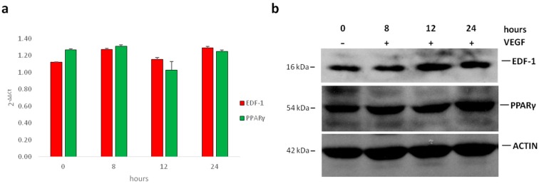
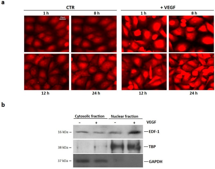
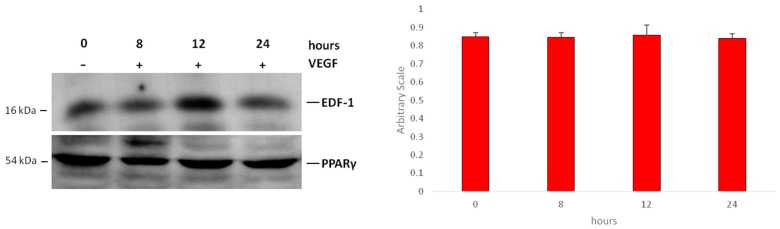
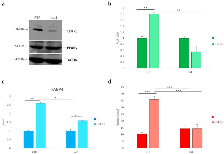
Vascular endothelial growth factor (VEGF) is important for maintaining healthy endothelium, which is crucial for vascular integrity. In this paper, we show that VEGF stimulates the nuclear translocation of endothelial differentiation-related factor 1 (EDF1), a highly conserved intracellular protein implicated in molecular events that are pivotal to endothelial function. In the nucleus, EDF1 serves as a transcriptional coactivator of peroxisome proliferator-activated receptor gamma (PPARγ), which has a protective role in the vasculature. Indeed, silencing EDF1 prevents VEGF induction of PPARγ activity as detected by gene reporter assay. Accordingly, silencing EDF1 markedly inhibits the stimulatory effect of VEGF on the expression of FABP4, a PPARγ-inducible gene. As nitric oxide is a marker of endothelial function, it is noteworthy that we report a link between EDF1 silencing, decreased levels of FABP4, and nitric oxide production. We conclude that EDF1 is required for VEGF-induced activation of the transcriptional activity of PPARγ.
Keywords: Endothelial Differentiation-related factor 1; Peroxisome proliferator-activated receptor γ; endothelial cells; vascular endothelial growth factor.
Conflict of interest statement
The authors declare no conflict of interest.
Figures
Similar articles
-
Rosiglitazone antagonizes vascular endothelial growth factor signaling and nuclear factor of activated T cells activation in cardiac valve endothelium.Endothelium. 2006 May-Jun;13(3):181-90. doi: 10.1080/10623320600760308. Endothelium. 2006. PMID: 16840174
-
Fatty acid activated PPARγ promotes tumorigenicity of prostate cancer cells by up regulating VEGF via PPAR responsive elements of the promoter.Oncotarget. 2016 Feb 23;7(8):9322-39. doi: 10.18632/oncotarget.6975. Oncotarget. 2016. PMID: 26814431 Free PMC article.
-
Peroxisome proliferator-activated receptor gamma ligands stimulate endothelial nitric oxide production through distinct peroxisome proliferator-activated receptor gamma-dependent mechanisms.Arterioscler Thromb Vasc Biol. 2005 Sep;25(9):1810-6. doi: 10.1161/01.ATV.0000177805.65864.d4. Epub 2005 Jul 14. Arterioscler Thromb Vasc Biol. 2005. PMID: 16020752
-
Peroxisome proliferator-activated receptor-γ in capillary endothelia promotes fatty acid uptake by heart during long-term fasting.J Am Heart Assoc. 2013 Jan 18;2(1):e004861. doi: 10.1161/JAHA.112.004861. J Am Heart Assoc. 2013. PMID: 23525438 Free PMC article.
-
Sphingosine 1-phosphate is a ligand for peroxisome proliferator-activated receptor-γ that regulates neoangiogenesis.FASEB J. 2015 Sep;29(9):3638-53. doi: 10.1096/fj.14-261289. Epub 2015 May 18. FASEB J. 2015. PMID: 25985799 Free PMC article.
Cited by
-
Synthesis, Regulatory Factors, and Signaling Pathways of Estrogen in the Ovary.Reprod Sci. 2023 Feb;30(2):350-360. doi: 10.1007/s43032-022-00932-z. Epub 2022 Apr 6. Reprod Sci. 2023. PMID: 35384637 Review.
-
System-Level Analysis of Alzheimer's Disease Prioritizes Candidate Genes for Neurodegeneration.Front Genet. 2021 Apr 6;12:625246. doi: 10.3389/fgene.2021.625246. eCollection 2021. Front Genet. 2021. PMID: 33889174 Free PMC article.
-
LncPrep + 96kb 2.2 kb Inhibits Estradiol Secretion From Granulosa Cells by Inducing EDF1 Translocation.Front Cell Dev Biol. 2020 Jun 30;8:481. doi: 10.3389/fcell.2020.00481. eCollection 2020. Front Cell Dev Biol. 2020. PMID: 32695776 Free PMC article.
-
Magnesium Deficiency Induces Lipid Accumulation in Vascular Endothelial Cells via Oxidative Stress-The Potential Contribution of EDF-1 and PPARγ.Int J Mol Sci. 2021 Jan 21;22(3):1050. doi: 10.3390/ijms22031050. Int J Mol Sci. 2021. PMID: 33494333 Free PMC article.
-
Prenatal diagnosis and mRNA profiles of fetal tetralogy of Fallot.BMC Pregnancy Childbirth. 2022 Nov 19;22(1):853. doi: 10.1186/s12884-022-05190-0. BMC Pregnancy Childbirth. 2022. PMID: 36402964 Free PMC article.
References
-
- Imai T., Takakuwa R., Marchand S., Dentz E., Bornert J.M., Messaddeq N., Wendling O., Mark M., Desvergne B., Wahli W., et al. Peroxisome proliferator-activated receptor γ is required in mature white and brown adipocytes for their survival in the mouse. Proc. Natl. Acad. Sci. USA. 2004;101:4543–4547. doi: 10.1073/pnas.0400356101. - DOI - PMC - PubMed
-
- Tomaru T., Steger D.J., Lefterova M.I., Schupp M., Lazar M.A. Adipocyte specific expression of murine resistin is mediated by synergism between peroxisome proliferator-activated receptor γ and CCAAT/enhancer-binding proteins. J. Biol. Chem. 2009;284:6116–6125. doi: 10.1074/jbc.M808407200. - DOI - PMC - PubMed
MeSH terms
Substances
LinkOut - more resources
Full Text Sources
Other Literature Sources
Research Materials
