Circulating cell-free DNA as a biomarker of tissue injury: Assessment in a cardiac xenotransplantation model
- PMID: 29933912
- PMCID: PMC6707066
- DOI: 10.1016/j.healun.2018.04.009
Circulating cell-free DNA as a biomarker of tissue injury: Assessment in a cardiac xenotransplantation model
Abstract
Background: Observational studies suggest that cell-free DNA (cfDNA) is a biomarker of tissue injury in a range of conditions including organ transplantation. However, the lack of model systems to study cfDNA and its relevance to tissue injury has limited the advancements in this field. We hypothesized that the predictable course of acute humoral xenograft rejection (AHXR) in organ transplants from genetically engineered donors provides an ideal system for assessing circulating cfDNA as a marker of tissue injury.
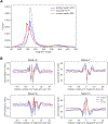
Methods: Genetically modified pig donor hearts were heterotopically transplanted into baboons (n = 7). Cell-free DNA was extracted from pre-transplant and post-transplant baboon plasma samples for shotgun sequencing. After alignment of sequence reads to pig and baboon reference sequences, we computed the percentage of xenograft-derived cfDNA (xdcfDNA) relative to recipient by counting uniquely aligned pig and baboon sequence reads.
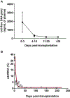
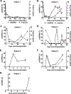
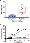
Results: The xdcfDNA percentage was high early post-transplantation and decayed exponentially to low stable levels (baseline); the decay half-life was 3.0 days. Post-transplantation baseline xdcfDNA levels were higher for transplant recipients that subsequently developed graft loss than in the 1 animal that did not reject the graft (3.2% vs 0.5%). Elevations in xdcfDNA percentage coincided with increased troponin and clinical evidence of rejection. Importantly, elevations in xdcfDNA percentage preceded clinical signs of rejection or increases in troponin levels.
Conclusion: Cross-species xdcfDNA kinetics in relation to acute rejection are similar to the patterns in human allografts. These observations in a xenotransplantation model support the body of evidence suggesting that circulating cfDNA is a marker of tissue injury.
Trial registration: ClinicalTrials.gov NCT02423070.
Keywords: acute humoral xenograft rejection; biomarker; cell-free DNA; cell-free DNA models; genetically engineered donors; tissue injury; xenotransplantation.
Copyright © 2018. Published by Elsevier Inc.
Conflict of interest statement
CONFLICT OF INTEREST AND FUNDING SOURCES DISCLOSURE
GRAfT study () was supported by the NHLBI intramural research program. DLA is an employee of Revivicor, Inc. None of the remaining authors have any conflicts of interest or relevant financial relationships with an external or commercial entity to disclose. Xenotransplantation work at the NHLBI was funded through contract HHSN268201300001C and gift funds from Revivicor, Inc. No external or commercial entities were involved in this study’s design, collection, analysis, or interpretation of data. The authors had access to all of the data in this study and take complete responsibility for the integrity of the data and the accuracy of the data analysis. The decision to write this report and to submit it for publication was made independently by the authors.
Figures


Comment in
-
Extracellular DNA in plasma: From marking to dissecting the cell biology of cardiac transplants.J Heart Lung Transplant. 2018 Aug;37(8):945-947. doi: 10.1016/j.healun.2018.05.006. Epub 2018 May 25. J Heart Lung Transplant. 2018. PMID: 29937215 No abstract available.
References
-
- Lo YM, et al., Presence of donor-specific DNA in plasma of kidney and liver-transplant recipients. Lancet, 1998. 351(9112): p. 1329–30. - PubMed
-
- Fournie G, et al., Plasma DNA as a marker of cancerous cell death. Investigations in patients suffering from lung cancer and in nude mice bearing human tumours. Cancer Lett, 1995. 91. - PubMed
-
- Margraf S, et al., Neutrophil-derived circulating free DNA (CF-DNA/NETS): a potential prognostic marker for posttraumatic development of inflammatory second hit and sepsis. Shock, 2008. 30. - PubMed
Publication types
MeSH terms
Substances
Associated data
Grants and funding
LinkOut - more resources
Full Text Sources
Other Literature Sources
Medical

