Review
. 2018 Nov;26(11):913-928.
doi: 10.1016/j.tim.2018.05.012. Epub 2018 Jun 19.

Yellow Fever Virus: Knowledge Gaps Impeding the Fight Against an Old Foe

Affiliations
Review

Yellow Fever Virus: Knowledge Gaps Impeding the Fight Against an Old Foe

Florian Douam et al. Trends Microbiol. 2018 Nov.

Abstract

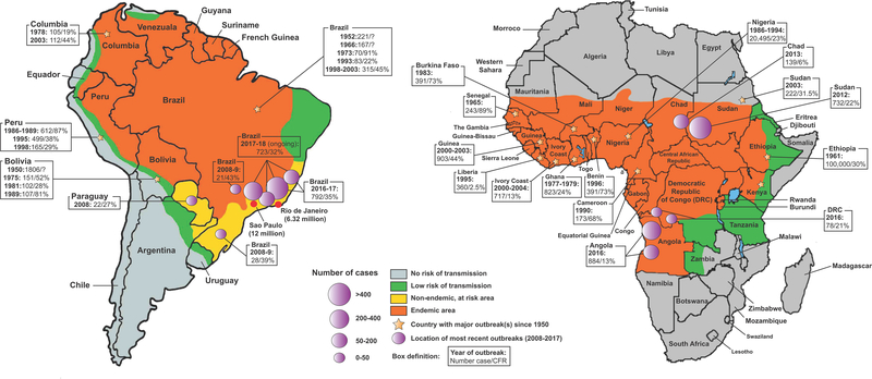
Yellow fever (YF) was one of the most dangerous infectious diseases of the 18th and 19th centuries, resulting in mass casualties in Africa and the Americas. The etiologic agent is yellow fever virus (YFV), and its live-attenuated form, YFV-17D, remains one of the most potent vaccines ever developed. During the first half of the 20th century, vaccination combined with mosquito control eradicated YFV transmission in urban areas. However, the recent 2016-2018 outbreaks in areas with historically low or no YFV activity have raised serious concerns for an estimated 400-500 million unvaccinated people who now live in at-risk areas. Once a forgotten disease, we highlight here that YF still represents a very real threat to human health and economies. As many gaps remain in our understanding of how YFV interacts with the human host and causes disease, there is an urgent need to address these knowledge gaps and propel YFV research forward.

Keywords: flaviviruses; viral pathogenesis; yellow fever; yellow fever vaccine; yellow fever virus.


Figures

Figure 1.
Figure 1.. YFV endemic areas and epidemiological distribution of YF outbreaks since 1950.
YFV endemic areas (orange) in South America (left) and Africa (right) are shown. Non-endemic, at risk areas (yellow), areas with low risk of transmission (green) and no risk of transmission (grey) are also displayed. On each continent, countries where major YF outbreaks occurred between 1950 and 2018 are marked by a yellow star, and epidemiological information (number of cases/CFR) for some of these outbreaks are shown. Precise locations of the most recent YF outbreaks (2008–2018) are represented by purple circles, sized in proportion to the intensity of the reported outbreak. Data were extracted from the WHO Global Yellow Fever Data Base (i) and from the periodic WHO “Yellow Fever Situation Reports” (viii). Epidemiological information related to the ongoing 2017–2018 outbreak in Brazil shows the number of confirmed cases and related CFRs reported between July 1, 2017 and February 27, 2018 (vi).
Figure 2.
Figure 2.. YFV genome organization.
A. Cryo-EM representation of an immature YFV particle (PDB 1NA4). B. Schematic representation of YFV viral RNA and polyprotein. Each viral protein is represented using a distinct color. Arrows indicate cleavage sites in the polyprotein that are processed by proteases of cellular (red or black arrow) or viral (blue arrow) origin. C. Schematic representation of the YFV polyprotein anchored into the endoplasmic reticulum (ER) membrane following translation.
Figure 3.
Figure 3.. Schematic representation of YFV life cycle.
Key steps of the YFV replication life cycle are displayed from 1 to 11. The few identified host factors regulating some of these steps are shown in red circles (see section 3 for description). The viral RNA replication step and the PrM-E maturation step are enlarged in white boxes. Major gaps in our understanding of specific steps of the life cycle are highlighted by red question marks.
Figure 4.
Figure 4.. Model of YF pathogenesis.
Schematic model of the YF-induced pathogenesis process, starting from a mosquito bite and leading to hepatic apoptosis and cytokine storm. The pathogenesis process is divided into five distinct steps (noted from 1 to 5), and each tissue compartment into which YFV circulates is displayed in a distinctly colored box. Intense viral replication in the liver is thought to trigger a molecular chain reaction inducing severe cytokine imbalance and pro-inflammatory cytokine secretion, leading to severe vasculopathy and multi-organ dysfunction. DC, dendritic cells; pDCs, plasmacytoid dendritic cells.
Figure 5.
Figure 5.. Molecular and phenotypic differences between YFV-17D and YFV-Asibi infection.
A. Location in the polyprotein of the amino acid differences between YFV-17D and YFV-Asibi. 32 amino-acid mutations between the two strains are shown, as well as three additional mutations (either M/M,I or S/S,P) due to Asibi clonal differences. B. Major differences between YFV-Asibi (red capsid) and YFV-17D (yellow capsid) infection directly or indirectly suggested by the literature and that could play a critical role in regulating YFV virulence or attenuation. Important biological questions related to each of the listed differences are highlighted. At the bottom of the figure are three more general outstanding questions that encompass our currently limited understanding of the viral and host determinants regulating YFV course of infection.

References

    1. Monath TP and Vasconcelos PF (2015) Yellow fever. Journal of clinical virology : the official publication of the Pan American Society for Clinical Virology 64, 160–173 - PubMed
    1. Quaresma JA, et al. (2013) Immunity and immune response, pathology and pathologic changes: progress and challenges in the immunopathology of yellow fever. Reviews in medical virology 23, 305–318 - PubMed
    1. Garske T, et al. (2014) Yellow Fever in Africa: estimating the burden of disease and impact of mass vaccination from outbreak and serological data. PLoS medicine 11, e1001638. - PMC - PubMed
    1. Shearer FM, et al. (2017) Global yellow fever vaccination coverage from 1970 to 2016: an adjusted retrospective analysis. The Lancet. Infectious diseases - PMC - PubMed
    1. Paules CI and Fauci AS (2017) Yellow Fever - Once Again on the Radar Screen in the Americas. The New England journal of medicine 376, 1397–1399 - PubMed

Resource section

    1. WHO Global Yellow Fever Data Base; http://apps.who.int/globalatlas/default.asp
    1. WHO (2016) Situation Report Yellow Fever - 28 October 2016; http://www.who.int/emergencies/yellow-fever/situation-reports/28-october...
    1. WHO/PAHO (2017) Epidemiological Update Yellow Fever - 10 July 2017. http://www.paho.org/hq/index.php?option=com_docman&task=doc_view&Itemid=...
    1. WHO – Disease outbreak news of January 22nd 2018; http://www.who.int/csr/don/22-january-2018-yellow-fever-brazil/en/
    1. WHO – Disease outbreak news of February 27th 2018; http://www.who.int/csr/don/27-february-2018-yellow-fever-brazil/en/

Publication types

MeSH terms