. 2018 Aug 17;293(33):12663-12680.
doi: 10.1074/jbc.RA117.000871. Epub 2018 Jun 22.

A potent, proteolysis-resistant inhibitor of kallikrein-related peptidase 6 (KLK6) for cancer therapy, developed by combinatorial engineering

Affiliations

A potent, proteolysis-resistant inhibitor of kallikrein-related peptidase 6 (KLK6) for cancer therapy, developed by combinatorial engineering

Amiram Sananes et al. J Biol Chem. .

Abstract

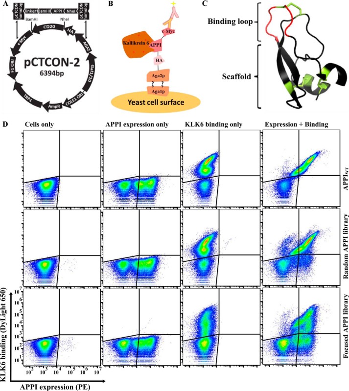
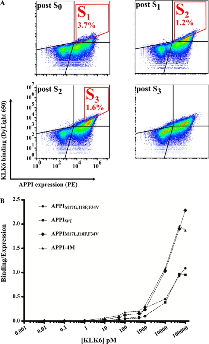
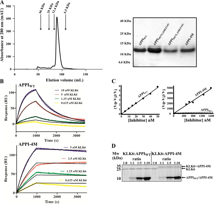
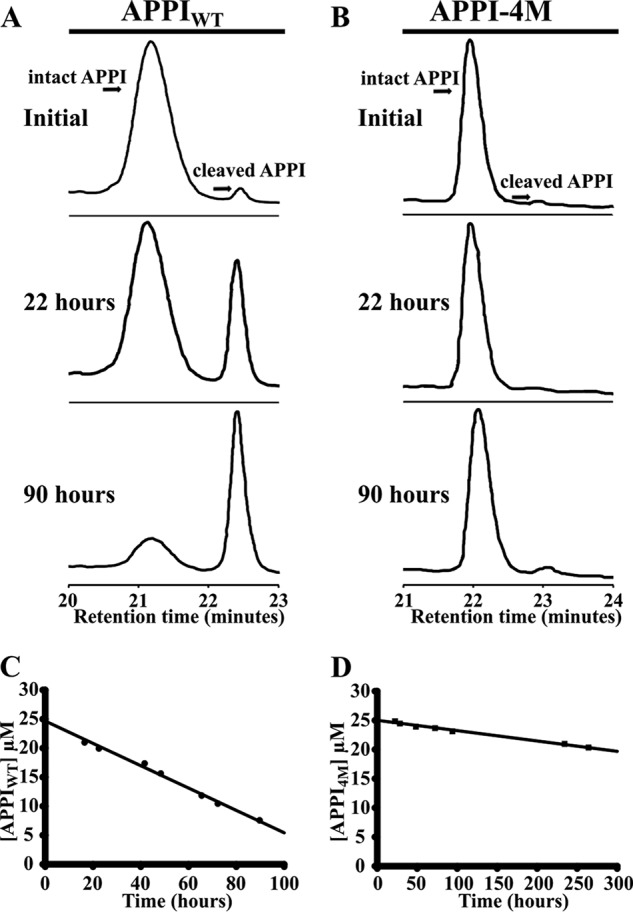
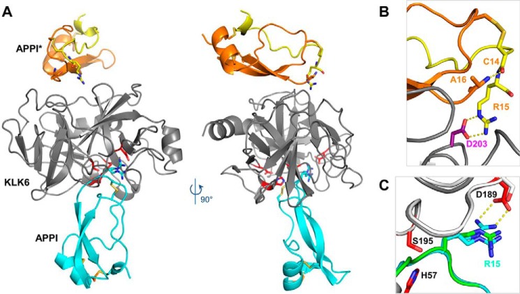
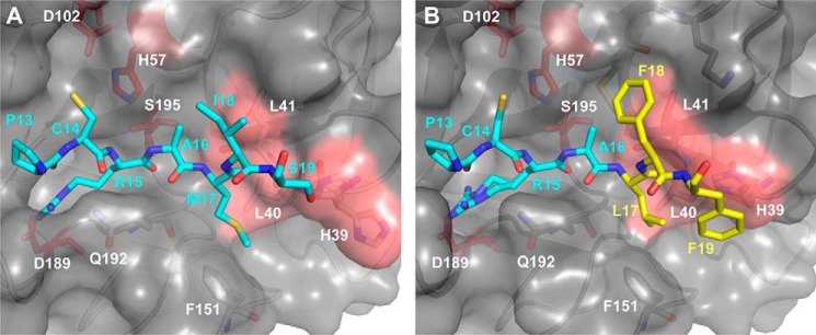
Human tissue kallikrein (KLK) proteases are hormone-like signaling molecules with important functions in cancer pathophysiology. KLK-related peptidase 6 (KLK6), specifically, is highly up-regulated in several types of cancer, where its increased activity promotes cancer invasion and metastasis. This characteristic suggests KLK6 as an attractive target for therapeutic interventions. However, inhibitors that specifically target KLK6 have not yet been reported, possibly because KLK6 shares a high sequence homology and structural similarity with other serine proteases and resists inhibition by many polypeptide inhibitors. Here, we present an innovative combinatorial approach to engineering KLK6 inhibitors via flow cytometry-based screening of a yeast-displayed mutant library of the human amyloid precursor protein Kunitz protease inhibitor domain (APPI), an inhibitor of other serine proteases, such as anionic and cationic trypsins. On the basis of this screening, we generated APPIM17L,I18F,S19F,F34V (APPI-4M), an APPI variant with a KLK6 inhibition constant (Ki ) of 160 pm and a turnover time of 10 days. To the best of our knowledge, APPI-4M is the most potent KLK6 inhibitor reported to date, displaying 146-fold improved affinity and 13-fold improved proteolytic stability compared with WT APPI (APPIWT). We further demonstrate that APPI-4M acts as a functional inhibitor in a cell-based model of KLK6-dependent breast cancer invasion. Finally, the crystal structures of the APPIWT/KLK6 and APPI-4M/KLK6 complexes revealed the structural and mechanistic bases for the improved KLK6 binding and proteolytic resistance of APPI-4M. We anticipate that APPI-4M will have substantial translational potential as both imaging agent and therapeutic.

Keywords: X-ray crystallography; cell invasion; directed evolution; enzyme inhibition; protease inhibitor; protein engineering; proteolysis; selective binding; yeast surface display.


Conflict of interest statement

The authors declare that they have no conflicts of interest with the contents of this article

Figures

Figure 1.
Figure 1.
FACS analysis of the different YSD APPI libraries, showing expression and KLK6 binding. A, YSD vector (pCTCON-2) is aligned with the general scheme of the insert: the insert gene consists of the APPI gene, which is flanked by two restriction sites (BamHI and NheI), followed by a linker sequence (LPDKPLAFQDPS) on the 3′ end, along with two pCTCON homologous sequences. B, APPI is displayed on the yeast cell surface as a translational fusion to Aga2p, which is linked to Aga1p by two disulfide bonds. Surface expression is detected by using fluorescence-labeled antibodies binding to the C terminus of the c-Myc epitope tag, whereas target binding is detected using fluorescence-labeled KLK6, via FACS. C, 3D structure of APPI (PDB code 1ZJD). D, expression of APPI and the binding of 10 nm labeled KLK6 were determined in a YSD system for an APPIWT clone, for a library of mutants based on APPIWT, and for a library of mutants based on APPIG17M,I18F,F34V.
Figure 2.
Figure 2.
Affinity maturation of the APPI library. A, to enrich the APPI library for high-affinity KLK6 binders, four sequential rounds of FACS sorting were conducted (S1–S3 marked in red). The results of the analysis after each sorting round are presented. S0 represents an initial sorting round intended only for selecting a cell population with high APPI expression, regardless of KLK6 binding. In S1, 500 pm DyLight-650–labeled KLK6 was used to select cells with a high APPI expression and a high KLK6 binding, which comprised 3.7% of the entire cell population. In S2, 50 pm DyLight-650–labeled KLK6 was used to select cells with a high APPI expression and a high KLK6 binding (gated in red), which comprised 1.2% of the entire cell population. In S3, 50 pm DyLight-650–labeled KLK6 was used to select cells with a high APPI expression and a high KLK6 binding, which comprised 1.6% of the entire cell population. B, FACS binding titration curve of KLK6 to yeast cells expressing APPIWT or its three variants. A leftward shift in the curve indicates a higher affinity. The binding signal of KLK6 is normalized to the expression signal of the APPIs.
Figure 3.
Figure 3.
Binding and inhibition kinetics of KLK6 by APPIWT and APPI-4M. A, purification of soluble APPI variants. Left, size-exclusion chromatography for APPI-4M. Arrows indicate correlations between the elution volume and size, according to known standards. Right, SDS-PAGE analysis of the purified APPI variants on a 15% polyacrylamide gel under reducing conditions. B, surface plasmon resonance binding experiment, in which 2 μg of either APPIWT (top panel) or APPI-4M (lower panel) was mounted as the ligands on a GLC chip. Six KLK6 concentrations (0–10 nm; represented by different colors) were used as the analytes. C, slow tight–binding inhibition of KLK6 catalytic activity by APPIWT (left, 1 nm KLK6) and APPI-4M (right, 100 nm KLK6). The Ki value of the reaction was calculated by using Equation 1 (see under “Experimental procedures”). V0 represents the uninhibited rate, and Vi represents the rate in the presence of APPI. In the APPI-4M plot, the dashed regression line indicates the slope of the inhibition by APPIWT. D, formation of complexes between KLK6 and APPI-4M. The SDS-PAGE was performed under reducing conditions and with KLK6/APPI molar ratios of 1:0, 1:1, 1:5, and 1:10. Note the formation of stable, higher molecular weight complexes in lanes loaded with KLK6/APPI-4M but not with KLK6/APPIWT complexes.
Figure 4.
Figure 4.
Hydrolysis of APPIWT (A and C) and APPI-4M (B and D) by KLK6, as determined by HPLC. The depletion of intact APPI over time was quantified by integrating the HPLC peak, which represents the intact APPI variant, at eight different time points (examples shown in A and B), and then determining the rate of depletion by linear regression (C and D). APPI concentrations were 25 μm for each variant, and the molar ratio of KLK6 to APPI was 1:8.
Figure 5.
Figure 5.
APPI-4M decreases the invasiveness of BT-20 breast cancer cells, but not their viability. The invasive ability of BT-20 cells in the presence of APPI-4M was evaluated by using a Matrigel-coated Boyden chamber. The cells that successfully invaded the Matrigel 36 h after plating were stained and quantified. A, representative images of the invading cells treated with either buffer (vehicle), APPIWT, or APPI-4M (100 nm). siRNA-transfected cells were used as a control. B, reverse-transcriptase PCR (RT-PCR) analysis was conducted in BT-20 breast cancer cells that were transfected with KLK6 siRNA. After the cells were transfected with siKLK6 or with a small-interfering control (scrambled siRNA) for 48 h, whole RNA was extracted, and the RT-PCR was performed. GAPDH, glyceraldehyde-3-phosphate dehydrogenase. C, quantification of invading cells. C is control (scrambled siRNA); K is KLK6–siRNA. Bars represent means ± S.D. of three biological repetitions. D, quantification of cell viability using the XTT assay, 36 h after incubation with APPIWT or APPI-4M. Bars represent means ± S.D. of three biological repetitions. E, quantification of migrating cells (see F), normalized to untreated cells (control). Bars represents the mean (± S.D.) of triplicate experiments. *, p < 0.05; **, p < 0.01 (Student's t test, as compared with untreated control). F, BT-20 cells were scratched by removing a strip across the well. Then, the cells were treated with APPI-4M for 24 h. The area free from cells was counted.
Figure 6.
Figure 6.
Crystal structure reveals an inhibited KLK6/APPI-4M complex and a cleaved product APPI-4M*. A, co-crystal structure of KLK6 with the APPI-4M variant shows one molecule of KLK6 (gray) bound to a molecule of APPI-4M occupying the active site in the inhibitory mode (cyan). The crystal structure also reveals an additional APPI-4M molecule that has undergone proteolysis at Arg-15–Ala-16 to yield the product APPI-4M*, which is comprised of two protein chains (yellow and orange) connected by two disulfide bonds. B, cleaved APPI-4M* molecule reveals a large rotation of the Cys-14 ψ angle (relative to the intact APPI-4M), allowing the formation of a salt bridge between the Arg-15 side chain and the Asp-203 residue of KLK6, thus stabilizing the crystal lattice, whereas the Cys-14 carbonyl forms a hydrogen bond with the Ala-16 N terminus. C, Arg-15 of the intact APPI-4M was observed in both the expected “up” conformation, in which it forms a salt bridge with KLK6 Asp-189 (dotted yellow lines), and a “down” conformation, which is similar to that previously reported in the superposed structure of APPI (green) bound to chymotrypsin (white) (PDB code 1CA0).
Figure 7.
Figure 7.
Binding loop mutations of APPI-4M optimize interactions with the primed side subsites of KLK6. A, in the complex of KLK6 (gray surface) with APPIWT (cyan sticks), APPI residues Met-17, Ile-18, and Ser-19 interact with the S2′, S3′, and S4′ subsites of KLK6, respectively, which are shaped by His-39, Leu-40, and Leu-41 (salmon surface patch). B, in the APPI-4M/KLK6 complex, Phe-18 of APPI-4M makes more extensive contacts with the S3′ subsite of KLK6, whereas Phe-19 forms a ring-stacking interaction with His-39 of KLK6, thus locking this residue into a single side-chain conformation. Leu-17 and Phe-19 residues of APPI-4M also form intramolecular hydrophobic contacts, which may help to stabilize the conformation of the binding loop.
Figure 8.
Figure 8.
Four APPI mutations optimize intra- and intermolecular packing interactions with KLK6. A and B, side view of the primed side of the substrate-binding cleft between KLK6 residues 39–41 (salmon surfaces) and Phe-151 (lavender) portrays van der Waals packing interactions in complexes with APPIWT (cyan) (A) and APPI-4M (mutated residues shown in yellow) (B). The mutated residues Leu-17, Phe-19, and Val-34 of APPI-4M form a hydrophobic cluster that fills the crevice and form hydrophobic interactions with KLK6 residues Phe-151 and Leu-40, whereas Phe-19 also forms a ring-stacking interaction with KLK6 His-39. C and D, rotated view, revealing how the mutated residues Phe-18 and Phe-19 of APPI-4M wrap around the ridge formed by KLK6 residues 39–41 (D), thus forming a more extensive contact interface than in the APPIWT/KLK6 complex (C).

References

    1. Prassas I., Eissa A., Poda G., and Diamandis E. P. (2015) Unleashing the therapeutic potential of human kallikrein-related serine proteases. Nat. Rev. Drug Discov. 14, 183–202 10.1038/nrd4534 - DOI - PubMed
    1. Emami N., and Diamandis E. P. (2007) New insights into the functional mechanisms and clinical applications of the kallikrein-related peptidase family. Mol. Oncol. 1, 269–287 10.1016/j.molonc.2007.09.003 - DOI - PMC - PubMed
    1. Marceau F., and Regoli D. (2004) Bradykinin receptor ligands: therapeutic perspectives. Nat. Rev. Drug Discov. 3, 845–852 10.1038/nrd1522 - DOI - PubMed
    1. Oikonomopoulou K., Diamandis E. P., and Hollenberg M. D. (2010) Kallikrein-related peptidases: proteolysis and signaling in cancer, the new frontier. Biol. Chem. 391, 299–310 - PubMed
    1. Kontos C. K., and Scorilas A. (2012) Kallikrein-related peptidases (KLKs): a gene family of novel cancer biomarkers. Clin. Chem. Lab. Med. 50, 1877–1891 - PubMed

Publication types