. 2018 Aug;30(8):1745-1769.
doi: 10.1105/tpc.18.00121. Epub 2018 Jun 22.

Plastidial NAD-Dependent Malate Dehydrogenase: A Moonlighting Protein Involved in Early Chloroplast Development through Its Interaction with an FtsH12-FtsHi Protease Complex

Affiliations

Plastidial NAD-Dependent Malate Dehydrogenase: A Moonlighting Protein Involved in Early Chloroplast Development through Its Interaction with an FtsH12-FtsHi Protease Complex

Tina B Schreier et al. Plant Cell. 2018 Aug.

Abstract

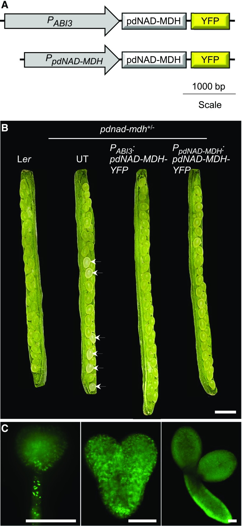
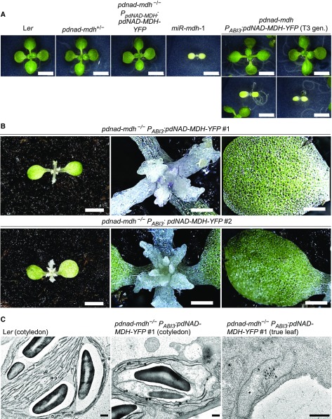
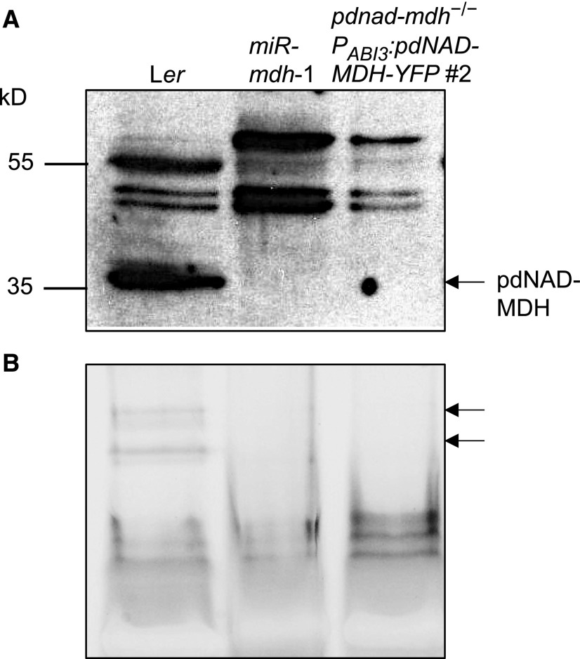
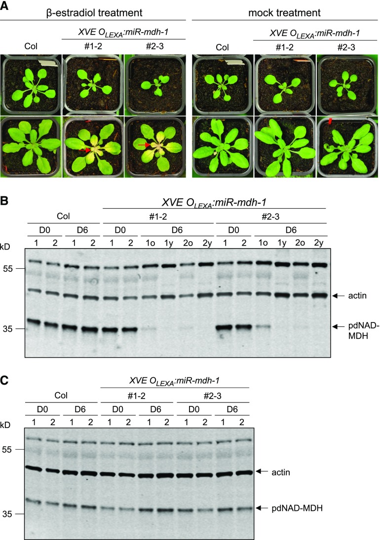
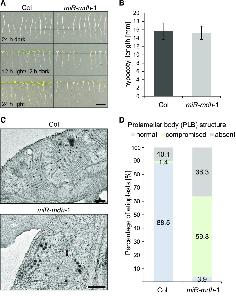
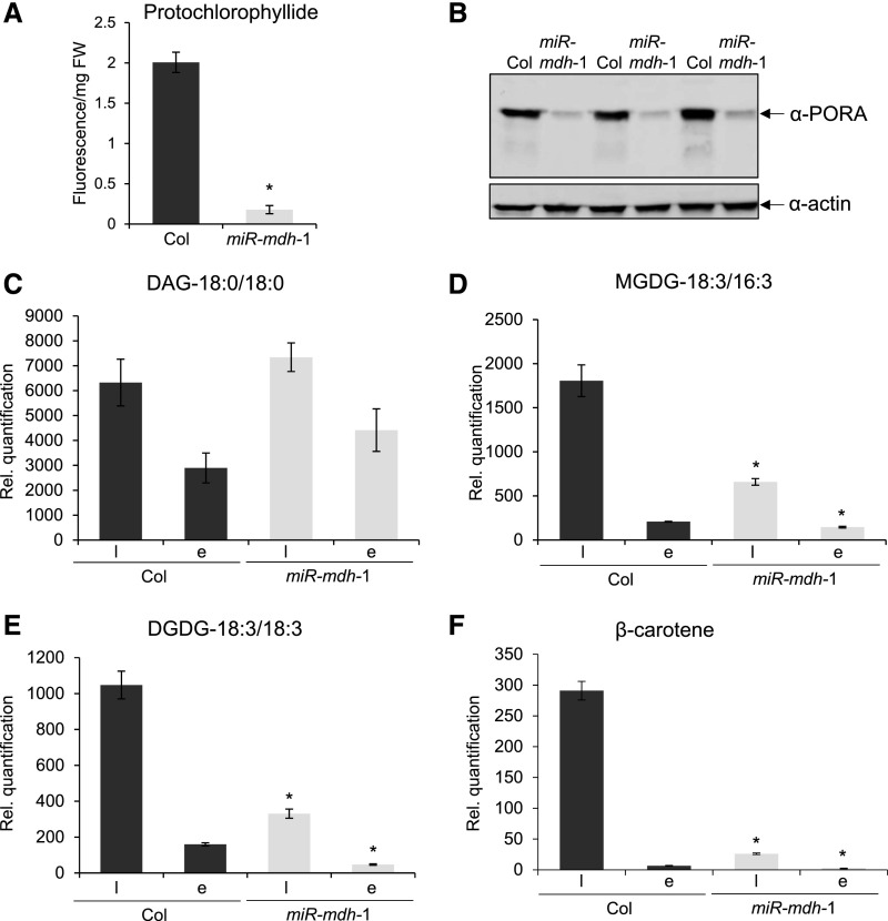
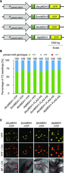
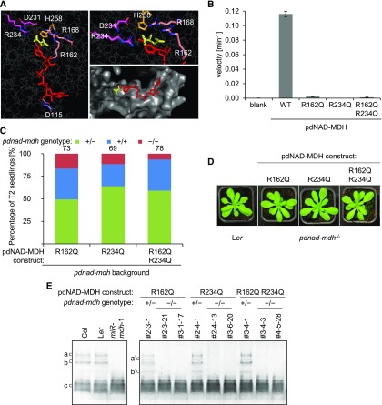
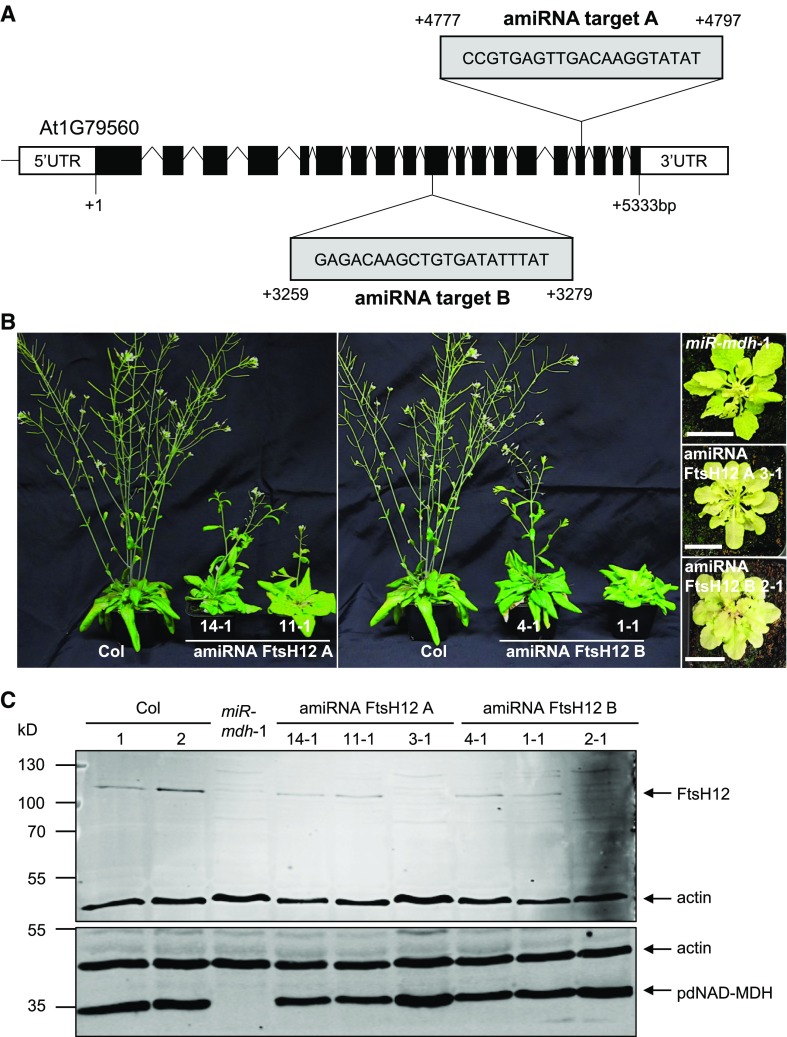
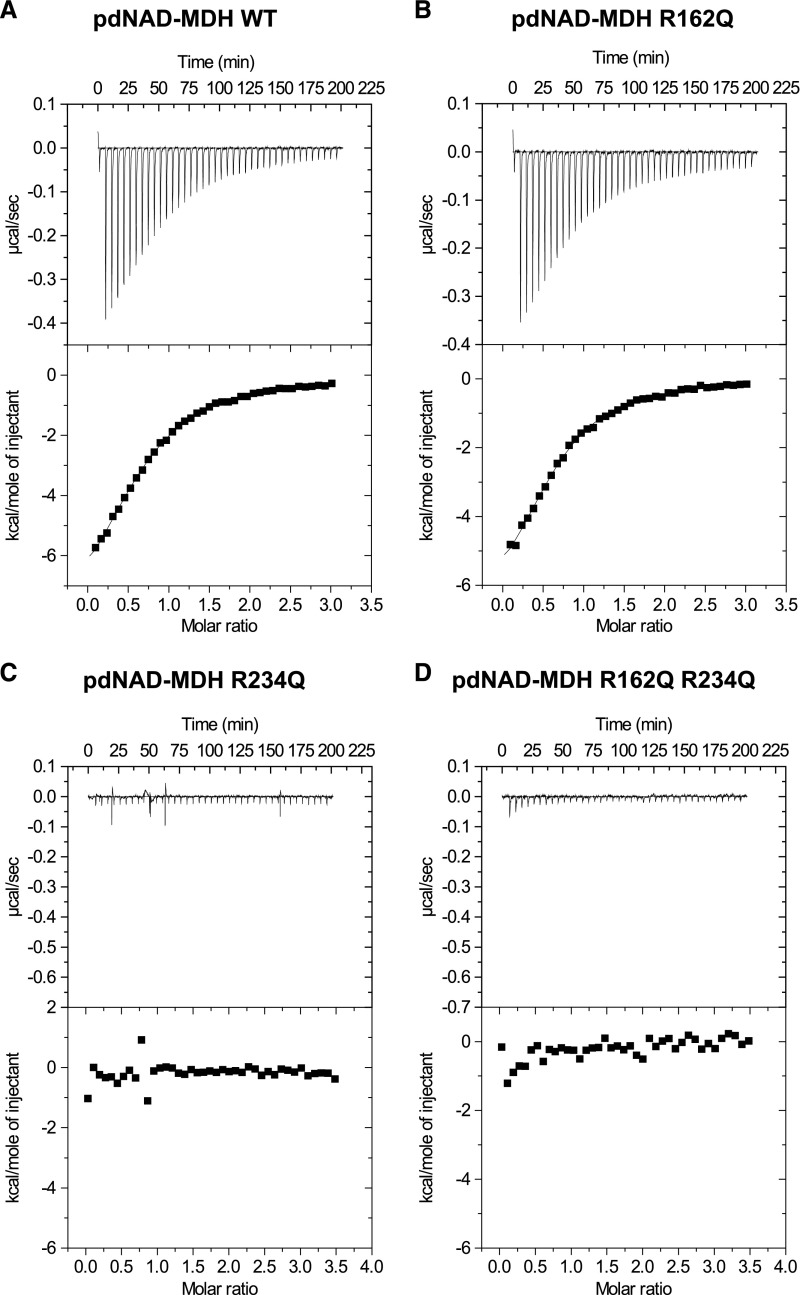
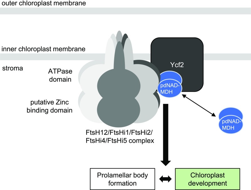
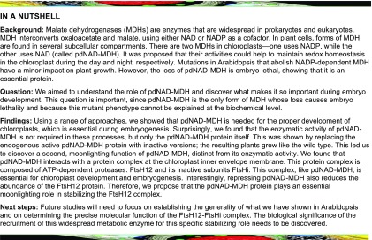
Malate dehydrogenases (MDHs) convert malate to oxaloacetate using NAD(H) or NADP(H) as a cofactor. Arabidopsis thaliana mutants lacking plastidial NAD-dependent MDH (pdnad-mdh) are embryo-lethal, and constitutive silencing (miR-mdh-1) causes a pale, dwarfed phenotype. The reason for these severe phenotypes is unknown. Here, we rescued the embryo lethality of pdnad-mdh via embryo-specific expression of pdNAD-MDH. Rescued seedlings developed white leaves with aberrant chloroplasts and failed to reproduce. Inducible silencing of pdNAD-MDH at the rosette stage also resulted in white newly emerging leaves. These data suggest that pdNAD-MDH is important for early plastid development, which is consistent with the reductions in major plastidial galactolipid, carotenoid, and protochlorophyllide levels in miR-mdh-1 seedlings. Surprisingly, the targeting of other NAD-dependent MDH isoforms to the plastid did not complement the embryo lethality of pdnad-mdh, while expression of enzymatically inactive pdNAD-MDH did. These complemented plants grew indistinguishably from the wild type. Both active and inactive forms of pdNAD-MDH interact with a heteromeric AAA-ATPase complex at the inner membrane of the chloroplast envelope. Silencing the expression of FtsH12, a key member of this complex, resulted in a phenotype that strongly resembles miR-mdh-1. We propose that pdNAD-MDH is essential for chloroplast development due to its moonlighting role in stabilizing FtsH12, distinct from its enzymatic function.


Figures

Figure 1.
Figure 1.
Embryo-Specific Expression of pdNAD-MDH to Rescue the Embryo Lethality of pdnad-mdh Plants. (A) Schematic diagram of the PABI3:pdNAD-MDH-YFP and PpdNAD-MDH:pdNAD-MDH-YFP constructs. For the former construct, the ABI3 promoter (PABI3) was placed upstream of the pdNAD-MDH coding sequence fused at its C terminus to YFP. The latter construct was described by Beeler et al. (2014). (B) Opened siliques of pdNAD-MDH+/− plants transformed with the PABI3:pdNAD-MDH-YFP construct. Siliques from the wild type (Ler), untransformed (UT) pdNAD-MDH+/− plants, and pdNAD-MDH+/− transformed with the PpdNAD-MDH:pdNAD-MDH-YFP construct are shown for comparison. White seeds are indicated with an arrow. Bar = 1 mm. (C) Expression of the PABI3:pdNAD-MDH-YFP construct in Arabidopsis embryos after transformation of pdNAD-MDH+/− plants. Expression was detected by fluorescence microscopy on embryos at three different developmental stages (globular, heart, and torpedo). Bar = 50 µm.
Figure 2.
Figure 2.
Phenotype of pdnad-mdh Mutants Rescued by the Embryo-Specific Expression of pdNAD-MDH. (A) Photographs of 14-d-old seedlings of pdnad-mdh plants expressing the PABI3:pdNAD-MDH-YFP construct, grown on 0.5× strength MS agar plates under a 12-h-light/12-h-dark regime. The PABI3:pdNAD-MDH-YFP construct was transformed into pdNAD-MDH+/− plants, and a T3 population that was segregating for the pdnad-mdh T-DNA insertion but not for the PABI3:pdNAD-MDH-YFP construct was obtained. Two examples of pale plants that resembled the constitutive silencing line, miR-mdh-1, are shown, while the other plants resembled the wild-type Ler or pdnad-mdh+/−. A pdnad-mdh−/− plant complemented with the PpdNAD-MDH:pdNAD-MDH-YFP construct is also shown. Bars = 500 µm. (B) Photographs of 4-week-old pdnad-mdh−/− seedlings rescued with the PABI3:pdNAD-MDH-YFP construct. Close-up images of their meristematic zones and their cotyledons are shown. Bars in the left-hand panels = 500 µm, and bars in the middle and right-hand panels = 100 µm. (C) Transmission electron micrographs of plastids in pdnad-mdh−/− seedlings rescued with the PABI3:pdNAD-MDH-YFP construct. Plastids from cotyledons are shown for the wild type (Ler) and pdnad-mdh−/−, and a plastid in a true leaf of pdnad-mdh−/− is shown on the right. Bars = 500 nm.
Figure 3.
Figure 3.
Immunoblot and Native-PAGE Detection of pdNAD-MDH in Ler, miR-mdh-1, and pdnad-mdh−/− Seedlings Rescued with the PABI3:pdNAD-MDH-YFP Construct. (A) Immunoblots were conducted with the pdNAD-MDH antibody. Equal amounts of soluble protein (5 µg) were loaded. The migration of molecular weight markers is indicated (left). (B) In-gel activity assay of the NAD-MDH. The two bands corresponding to pdNAD-MDH activity are indicated (arrows, right). Equal amounts of protein (15 µg) were loaded.
Figure 4.
Figure 4.
β-Estradiol-Inducible Silencing of pdNAD-MDH in Mature Plant Rosettes. XVE OLEXA:miR-mdh-1 plants photographed before and after a 6-d treatment with β-estradiol or a mock treatment. (A) Photographs in the top row were taken before β-estradiol treatment (D0) and those in the bottom row after 6 d (D6). β-Estradiol solution (20 µM) was sprayed every second day onto the entire rosette. β-Estradiol was applied to wild-type (Col) plants as a control. Mock-treated samples were sprayed with water containing the same amount of DMSO as the treatment solution (used to dissolve the β-estradiol). Red arrows indicate examples of white, newly emerging leaves. (B) Immunoblot detection of pdNAD-MDH in total protein extracts of wild-type and XVE OLEXA:miR-mdh-1 plants before (D0) and 6 d into β-estradiol treatment (D6). For the treated XVE OLEXA:miR-mdh-1 plants, proteins were extracted from the old, green leaves (D6o) and young, white leaves (D6y) separately. Gels were loaded on an equal leaf area basis. The migration of molecular mass markers is indicated on the left. pdNAD-MDH and actin (as a loading control) were detected concurrently on the same membrane using secondary antibodies conjugated to different infrared fluorescence dyes (800CW for pdNAD-MDH and 680RD for actin). (C) As for (B), but with old leaves from mock-treated samples.
Figure 5.
Figure 5.
Etiolated Growth of miR-mdh-1 and Etioplast Structure. (A) Wild-type (Col) and miR-mdh-1 seedlings were grown on 0.5× strength MS agar plates in different diel light conditions (24 h dark, 12 h light/12 h dark, 24 h light). Bar = 1 cm. (B) Quantification of the hypocotyl length of etiolated wild-type and miR-mdh-1 seedlings. Values represent the mean ± se of measurements conducted on n = 89 and n = 86 seedlings for the wild type and miR-mdh-1, respectively. (C) Etioplast ultrastructure in cotyledons of etiolated wild-type and miR-mdh-1 seedlings observed by transmission electron microscopy. Bars = 500 nm (D) Proportion of etioplasts showing normal, compromised, or absent prolamellar body structures in cotyledons of the wild type and miR-mdh-1. The first 139 and 256 etioplasts, observed in wild-type and miR-mdh-1 cotyledons, respectively, were categorized based on their prolamellar body structure.
Figure 6.
Figure 6.
Protochlorophylide, Galactolipid, and β-Carotene Contents of miR-mdh-1 Seedlings. (A) Protochlorophyllide levels in 5-d-old wild-type (Col) and miR-mdh-1 etiolated seedlings, measured by fluorescence after excitation at 440 nm. Values are the means ± se of three biological replicates, each measured with four technical replicates. Each biological replicate is a pool of 100 to 200 individual seedlings. (B) Immunoblot detection of PORA in total protein extracts from 6-d-old etiolated wild-type and miR-mdh-1 seedlings (upper panel). Immunoblots for actin was performed as a loading control (lower panel). Gels were loaded on an equal fresh weight basis. Three biological replicates are shown. Each biological replicate is a pool of 50 to 100 individual seedlings. (C) to (F) Lipids and carotenoids were extracted from 6-d-old wild-type and miR-mdh-1 seedlings, either etiolated (E) or light grown (l; grown under a 12-h-light/12-h-dark regime). Levels of DAG (C), MGDG 18:3/16:3 (D) DGDG 18:3/18:3 (E), and β-carotene (F) are shown. For the lipids, only the most common species (in terms of fatty acid chain composition) is shown; similar trends were seen in other detected species (Supplemental Table 1). All lipids and carotenoids were quantified relative to an internal standard, and values were corrected for differences in fresh weight (see Methods for details). Values are the mean ± se from four biological replicates. Each biological replicate is a pool of 50 to 100 individual seedlings. Significant differences (P < 0.05) within the respective light-grown and etiolated samples of miR-mdh-1 and the wild type, determined using a two-tailed t test, are indicated with an asterisk.
Figure 7.
Figure 7.
Complementation Test with Various NAD-MDH Isoforms Expressed in pdnad-mdh. (A) Constructs encoding NAD-MDH isoforms under the control of the pdNAD-MDH promoter. The plastidial transit peptide from Rubisco small subunit was fused to the N terminus of each isoform. The constructs shown are tagged at the C terminus with YFP. Similar constructs were cloned with the Flag-HA tag in place of YFP. (B) Genotyping results of the T2 progeny from T1 plants heterozygous for the pdnad-mdh mutation and expressing the different NAD-MDH isoforms. Numbers above the bars indicate the number of BASTA-resistant T2 plants that were genotyped. (C) Chloroplast localization of the NAD-MDH isoforms. Similar constructs to those shown in (A), except with the 35S promoter in place of the native promoter, were transiently expressed in Nicotiana benthamiana leaves and imaged using confocal microscopy. Bar = 5 µm.
Figure 8.
Figure 8.
Enzymatically Inactive pdNAD-MDH Proteins Can Complement the Embryo Lethality and Growth Defects of the pdnad-mdh Mutant. (A) Catalytic center of the Arabidopsis pdNAD-MDH structure. The pdNAD-MDH protein sequence (without cTP) was modeled using the human malate- and NADH-bound MMDH2 crystal structure (PDB: 2DFD) as a template. Left panel shows NADH (red) and malate (yellow), and the relative position of the conserved catalytic amino acid residues of pdNAD-MDH. A detailed view of the malate binding site is shown in the upper right panel. The surface of the catalytic pocket is shown in the lower right panel. (B) An in vitro activity assay was performed using purified recombinant proteins. The proteins (10 µg) were incubated with an excess of cofactor (NADH) at 22°C. The reaction was started by the addition of excess substrate (oxaloacetate). The velocity was determined by measuring the decrease in absorbance at 340 nm resulting from the conversion of NADH to NAD+. Error bars indicate mean ± se (n = 3). (C) Genotyping results of T2 progeny from T1 plants heterozygous for pdnad-mdh and expressing the enzymatically inactive pdNAD-MDH proteins. Numbers above the bars indicate the number of BASTA-resistant T2 plants that were genotyped. (D) Photographs showing 3-week-old rosettes of homozygous pdnad-mdh plants complemented with enzymatically-inactive pdNAD-MDH mutants. For comparison, wild-type (Ler) plants are shown. Plants were grown under long days (16 h light/8 h dark). (E) NAD-MDH activity observed by native-PAGE. Equal amounts of protein (15 µg) were loaded per lane, and all lanes were run on the same gel. pdNAD-MDH runs in distinct activity bands (a, b, and c). While the fastest migrating band (c) corresponding to the free dimer is masked by other NAD-MDH activity, the two slower migrating bands that correspond to NAD-MDH in protein complexes can be easily observed (a and b). Additional activity bands (a’ and b’) are observed for plants heterozygous for the pdnad-mdh T-DNA insertion expressing the catalytic-inactive pdNAD-MDH variants, possibly due to dimer formation between an inactive pdNAD-MDH with an endogenous form of pdNAD-MDH protein.
Figure 9.
Figure 9.
Constitutive and Inducible Silencing of the FtsH12 Protein. (A) In Arabidopsis, the FtsH12 gene consists of 19 exons and 18 introns. The sequence and position of the target for artificial microRNA silencing are indicated. Numbers represent nucleotide positions relative to the translational start site +1. (B) Constitutive silencing of FtsH12 resulted in plants with varying degrees of paleness and delayed growth phenotype in the T1 generation. Plants were grown under a 12-h-light/12-h-dark regime for 6 weeks. Both amiRNA constructs produced plants with comparable phenotypes. The identical wild type (Col) plant was used for both the left and right panels. Bar = 2 cm. (C) Immunoblot analysis of total protein extracts from the amiRNA FtsH12 lines, using the FtsH12 antibody (upper panel), pdNAD-MDH antibody (lower panel), and actin antibody as a loading control (both panels). FtsH12 and actin, as well as pdNAD-MDH and actin, were detected concurrently on the same membrane using secondary antibodies conjugated to different infrared fluorescence dyes (800CW for FtsH12 and pdNAD-MDH, and 680RD for actin). The migration of molecular mass markers is indicated (left). The gel was loaded on an equal protein (15 µg) basis.
Figure 10.
Figure 10.
ITC Thermograms of NADH Titrated into the pdNAD-MDH Wild-Type and Inactive Proteins. (A) ITC profile of NADH injected into a solution of recombinant His-pdNAD-MDH wild-type protein at 25°C. The upper panel shows the raw calorimetric data. The plot below shows the integrated enthalpy as a function of the NADH/pdNAD-MDH molar ratio. Reactions are exothermic. (B) to (D) Experiments were conducted as described for (A) with the His-pdNAD-MDH R162Q protein (B), the His-pdNAD-MDH R234Q protein (C), and the His-pdNAD-MDH R162Q R234Q protein (D).
Figure 11.
Figure 11.
Model for the Interaction of pdNAD-MDH with the Heteromeric FtsH12-FtsHi AAA-ATPase Complex at the Chloroplast Inner Envelope Membrane, Which Plays an Essential Role in Chloroplast Development.
None

Comment in

References

    1. Bahl J., Francke B., Monéger R. (1976). Lipid composition of envelopes, prolamellar bodies and other plastid membranes in etiolated, green and greening wheat leaves. Planta 129: 193–201. - PubMed
    1. Beeler S., Liu H.-C., Stadler M., Schreier T., Eicke S., Lue W.-L., Truernit E., Zeeman S.C., Chen J., Kötting O. (2014). Plastidial NAD-dependent malate dehydrogenase is critical for embryo development and heterotrophic metabolism in Arabidopsis. Plant Physiol. 164: 1175–1190. - PMC - PubMed
    1. Berkemeyer M., Scheibe R., Ocheretina O. (1998). A novel, non-redox-regulated NAD-dependent malate dehydrogenase from chloroplasts of Arabidopsis thaliana L. J. Biol. Chem. 273: 27927–27933. - PubMed
    1. Birktoft J.J., Banaszak L.J. (1983). The presence of a histidine-aspartic acid pair in the active site of 2-hydroxyacid dehydrogenases. X-ray refinement of cytoplasmic malate dehydrogenase. J. Biol. Chem. 258: 472–482. - PubMed
    1. Bodi Z., Zhong S., Mehra S., Song J., Graham N., Li H., May S., Fray R.G. (2012). Adenosine methylation in Arabidopsis mRNA is associated with the 3′ end and reduced levels cause developmental defects. Front. Plant Sci. 3: 48. - PMC - PubMed

Publication types

MeSH terms