Comparative Study
. 2019 Jun;68(6):1003-1013.
doi: 10.1136/gutjnl-2018-316226. Epub 2018 Jun 22.

Lactobacillus rhamnosus GG protects the intestinal epithelium from radiation injury through release of lipoteichoic acid, macrophage activation and the migration of mesenchymal stem cells

Affiliations
Comparative Study

Lactobacillus rhamnosus GG protects the intestinal epithelium from radiation injury through release of lipoteichoic acid, macrophage activation and the migration of mesenchymal stem cells

Terrence E Riehl et al. Gut. 2019 Jun.

Abstract

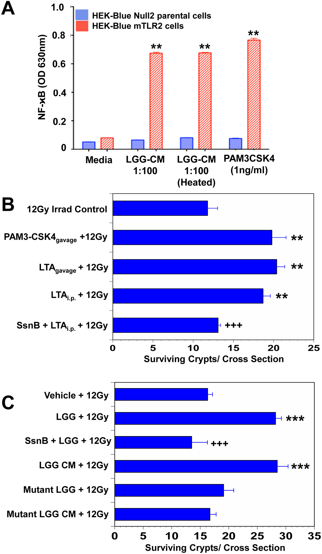
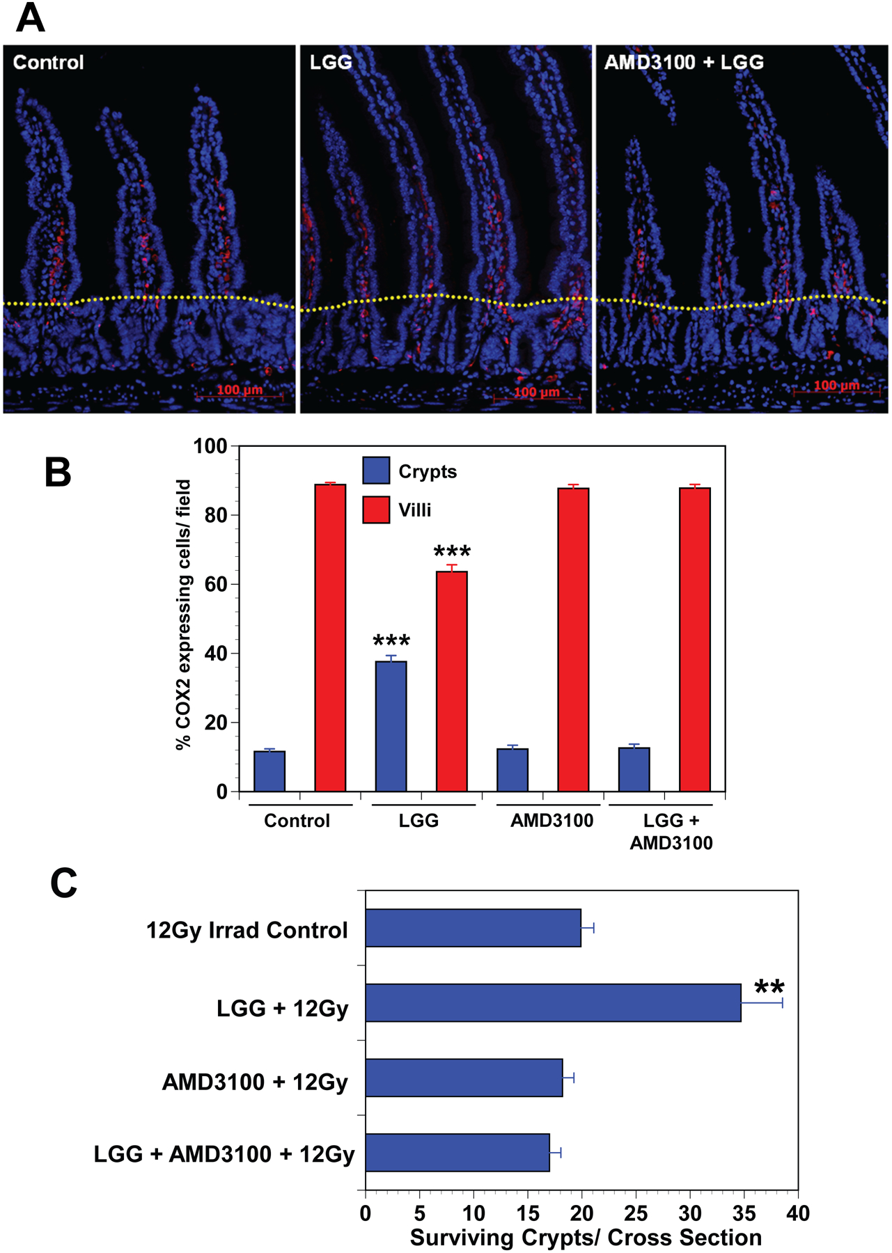
Objective: Lactobacillus rhamnosus GG (LGG), a probiotic, given by gavage is radioprotective of the mouse intestine. LGG-induced radioprotection is toll-like receptor 2 (TLR2) and cyclooxygenase-2 (COX-2)-dependent and is associated with the migration of COX-2+mesenchymal stem cells (MSCs) from the lamina propria of the villus to the lamina propria near the crypt epithelial stem cells. Our goals were to define the mechanism of LGG radioprotection including identification of the TLR2 agonist, and the mechanism of the MSC migration and to determine the safety and efficacy of this approach in models relevant to clinical radiation therapy.

Design: Intestinal radioprotection was modelled in vitro with cell lines and enteroids as well as in vivo by assaying clinical outcomes and crypt survival. Fractionated abdominal and single dose radiation were used along with syngeneic CT26 colon tumour grafts to assess tumour radioprotection.

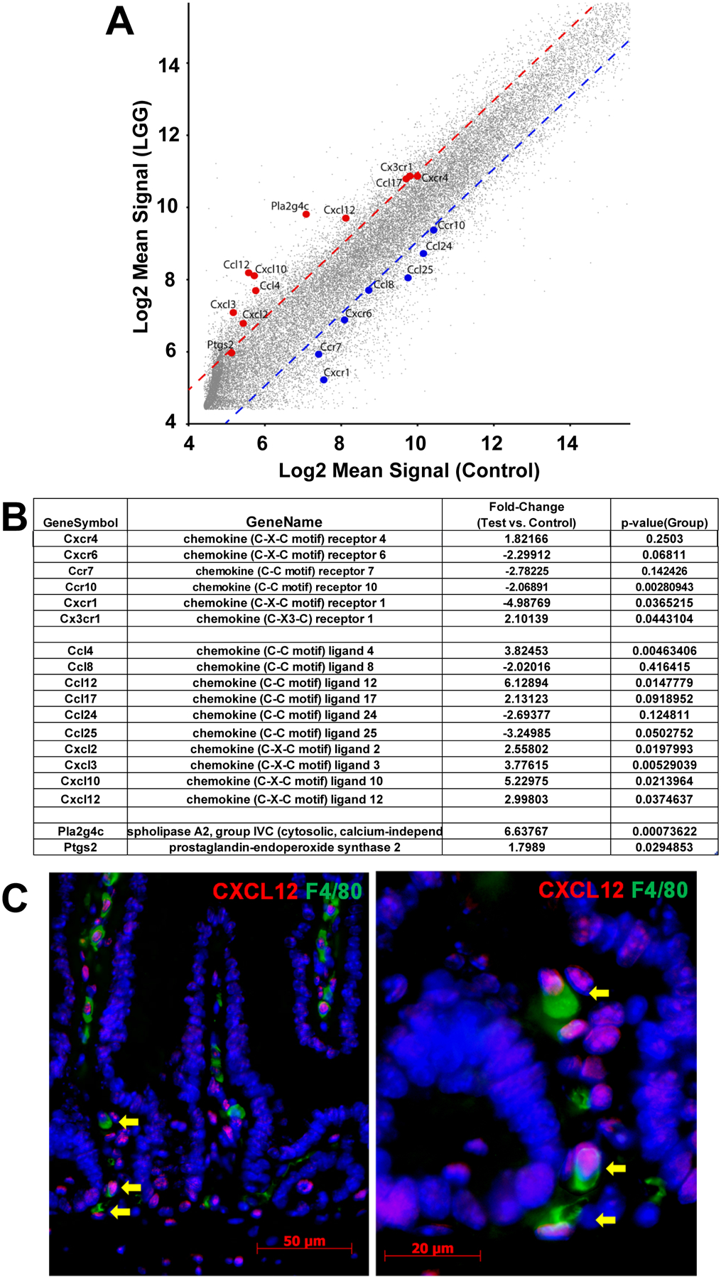
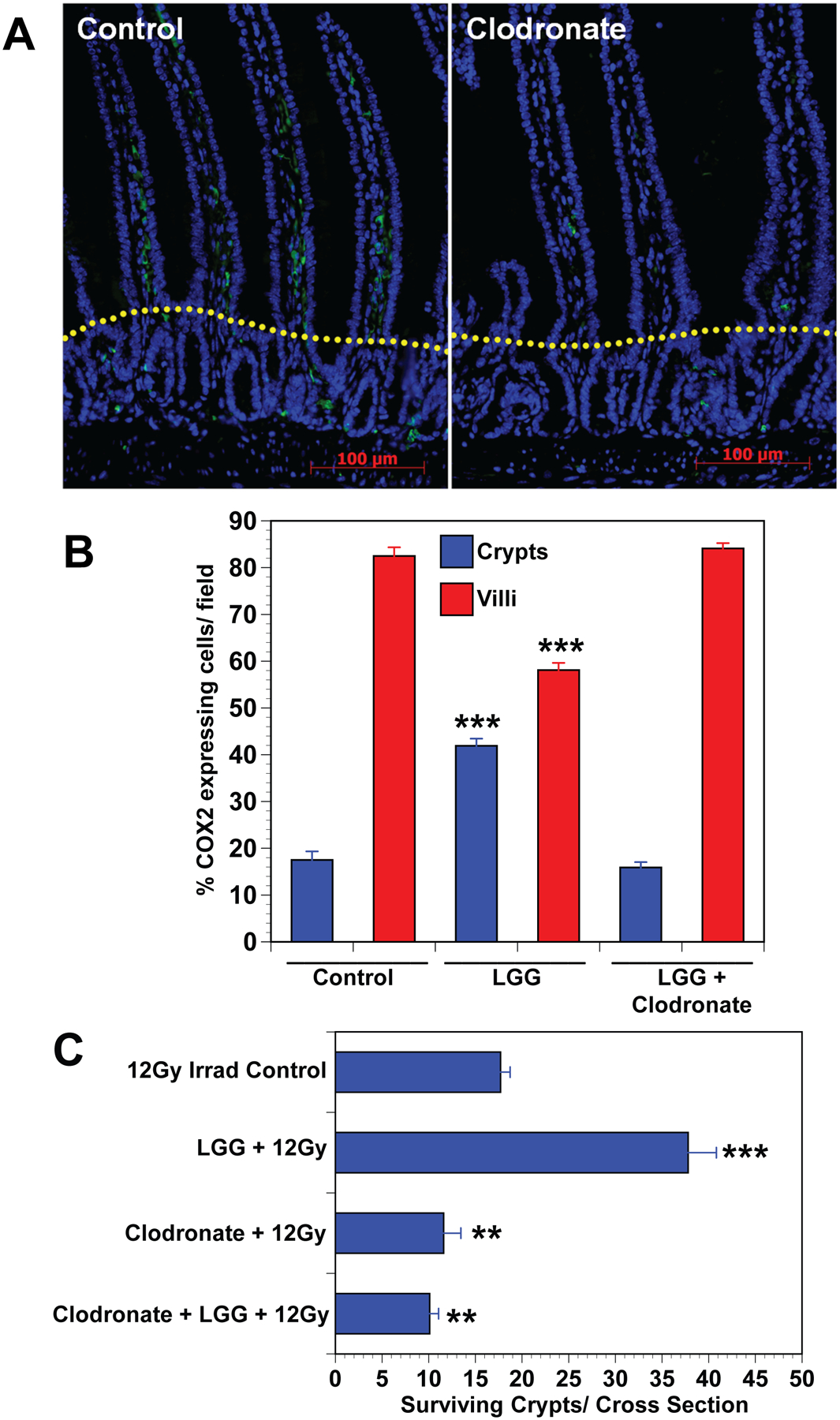
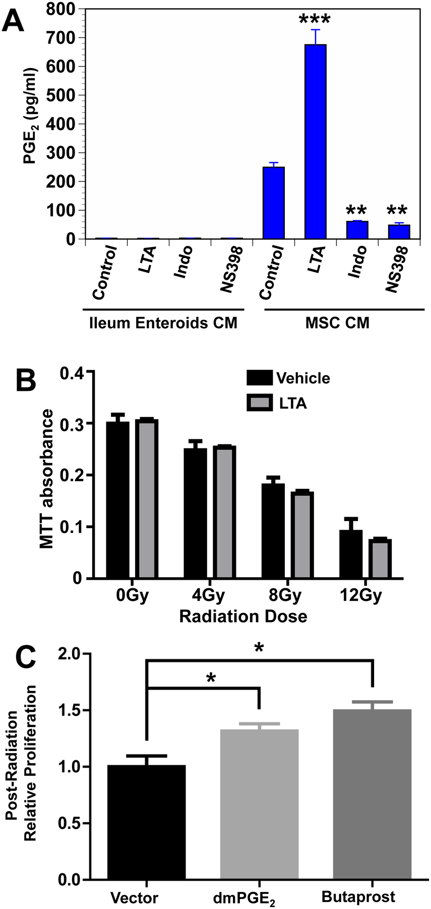
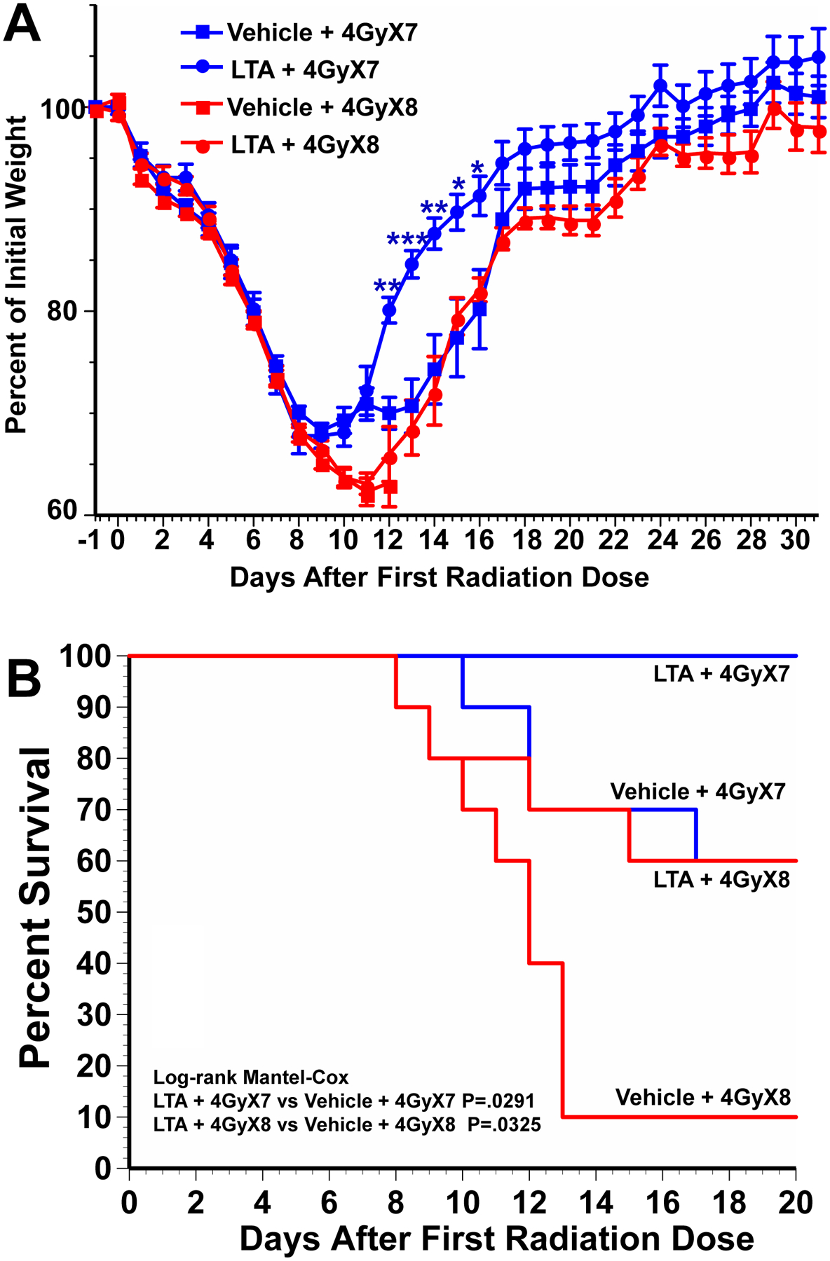
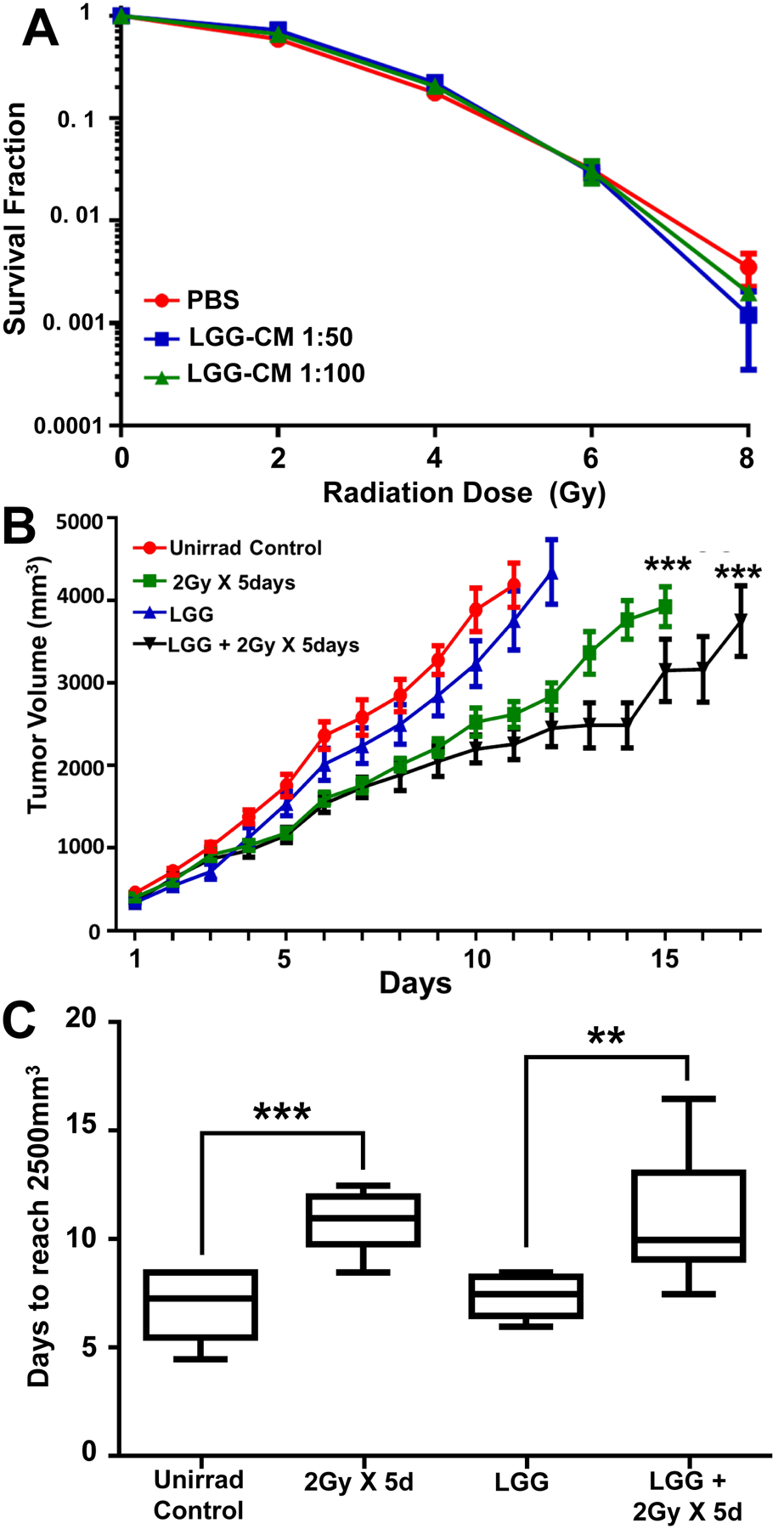
Results: LGG with a mutation in the processing of lipoteichoic acid (LTA), a TLR2 agonist, was not radioprotective, while LTA agonist and native LGG were. An agonist of CXCR4 blocked LGG-induced MSC migration and LGG-induced radioprotection. LGG given by gavage induced expression of CXCL12, a CXCR4 agonist, in pericryptal macrophages and depletion of macrophages by clodronate liposomes blocked LGG-induced MSC migration and radioprotection. LTA effectively protected the normal intestinal crypt, but not tumours in fractionated radiation regimens.

Conclusions: LGG acts as a 'time-release capsule' releasing radioprotective LTA. LTA then primes the epithelial stem cell niche to protect epithelial stem cells by triggering a multicellular, adaptive immune signalling cascade involving macrophages and PGE2 secreting MSCs.

Trial registration number: NCT01790035; Pre-results.

Keywords: intestinal epithelium; probiotics; prostaglandins; radiotherapy; stem cells.


Conflict of interest statement

Competing interests: None declared.

Figures

Fig. 1.
Fig. 1.. LTA is the radioprotective agent in LGG-CM.
(A) LGG-CM, and the TLR2 agonist PAM3-CSK4 activated NFκB and alkaline phosphatase in HEK-Blue mTLR2 reporter cells. Alkaline phosphatase activation is assessed spectrophotometrically. Data are means ± SEM for 6 samples per treatment group. **P<.001 compared with media controls. (B) Mice pretreated for 3 consecutive days with the TLR2 agonist LTA from Staphylococcus aureus by gavage or by intraperitoneal (i.p.) injection, or with the synthetic TLR2 agonist PAM3-CSK4 by gavage prior to receiving 12Gy TBI had significantly improved crypt survival. The TLR2 antagonist Sparstolonin B (SsmB) blocked LTA-induced radioprotection. **P<.001 compared with irradiated control. Data are means ± SEM for 10 mice per treatment group. (C) Mice had significantly improved crypt survival after radiation when pretreated with WT LGG or WT LGG-CM by gavage for 3 consecutive days. SsmBblocked LGG-induced radioprotection. There was no radioprotection in the small intestines of mice pretreated with mutant LGG dltD, which produces inactive modified LTA, or mutant LGG-CM. ***P<.0006 compared with irradiated control. +++P<.005 compared with LTA ip + 12 Gy or LGG+12Gy N=10 mice per treatment group.
Fig. 2.
Fig. 2.. Inhibition of CXCR4 by AMD3100 blocks LGG-induced migration of COX2 expressing MSCs and LGG-induced radioprotection in mice.
(A) Immunofluorescence for COX2 in the small intestine shows an increased percentage of MSCs in the crypt zone of LGG-treated mice compared with controls or mice treated with LGG and AMD3100 treated mice. Dotted yellow line separates crypt and villus zones. (B) Cell counts provide quantitative data indicating that LGG-induced migration of COX2 expressing MSCs from the villus to the crypt zone, and that AMD3100 blocks LGG induced migration. Data are means ± SEM for 5 to 7 mice per treatment group. ***P<.0001 compared with controls. (C) Crypt survival is significantly improved in LGG treated mice, and AMD3100 blocks LGG induced radioprotection. Data are means ± SEM for 5 to 7 mice per treatment group. **P<.005 compared with irradiated controls.
Fig. 3.
Fig. 3.. LGG induces CXCL12 in pericryptal macrophages.
(A,B) Mice were given LGG or vehicle by gavage and sacrificed 24 hours later. Laser Capture Microdissection (LCM) was performed and analyzed by gene chip. (A) Figure illustrates the relative expression of gene expression in the pericryptal mRNA from LGG treated and vehicle treated mice. Dotted lines represent 2 standard deviations from the means. Genes for chemokines, chemokine receptors and enzymes related to arachidonic acid metabolism are shown. (B) Table presents data for chemokine receptors, chemokines and arachidonic acid metabolism genes, comparing gene expression in pericryptal cells from vehicle treated and LGG treated mice. (C) Immunofluorescence for CXCL12(red) and F4/80(green) in mouse small intestines shows that some macrophages, including pericryptal macrophages (arrows), express CXCL12.
Fig. 4.
Fig. 4.. Depletion of macrophages with anionic liposomal clodronate blocks LGG-induced MSC migration and LGG radioprotection.
(A) Immunofluorescence for F4/80 expressing macrophages compares placebo treated mice having normal macrophage population with anionic clodronate liposome treated mice showing macrophage depletion. Dotted yellow line separates crypt and villus zones. (B) Cell counts provide quantitative data showing that treatment of mice with clodronate blocks LGG induced migration of COX2 expressing MSCs from the villus to the crypt zone in the small intestine. Data are means ± SEM for 5 to 7 mice per treatment group. ***P<.0001 compared with controls. (C) Crypt survival is significantly improved in LGG treated mice, and depletion of macrophages by treatment with clodronate blocks LGG induced radioprotection. Clodronate liposome (7mg/ml) were given i.p. at 4 days before (200μl/20g mouse), and again at 2 days before (100μl/20g mouse), and finally at eight hours before receiving 12Gy TBI. Data are means ± SEM for 5 to 7 mice per treatment group. **P<.001, ***P<.0001 compared with controls.
Fig. 5.
Fig. 5.. LTA induced PGE2 in MSCs protects epithelial stem cells from radiation.
(A) LTA induces PGE production in MSCs but not in enteroids. Ileum enteroids and small intestine MSCs were incubated in media alone, or in media containing LTA (10μ/mL) or indomethacin (50μM), or NS398 (5μM). After 24-hour incubation, PGE2 concentration was determined by ELISA. Data are means ± SEM for 4 replicates in each treatment group. **P<.0002, ***P<.0004. (B) Gamma radiation reduces primary enteroid cell proliferation in a dose dependent manner. Cells were pretreated with LTA (10μg/mL) or vehicle in medium for 2 hours, then irradiated with zero, 4, 8, or 12 Gy. LTA or vehicle was removed 2 hours post irradiation and cells were cultured for 72 hours. Proliferation was assessed by 3-(4,5-dimethylthiazol-2-yl)-2,5-diphenyltetrazolium bromide (MTT) incorporation. Using this pretreatment strategy, LTA did not protect against radiation injury. N.S., not significant. (C) dmPGE2 and the EP2 agonist butaprost are radioprotective of enteroids. WT enteroids were plated in 50% stem cell media (SCM)(11) for 18h, then grown in 5% SCM for 24h. Enteroids were then treated with dmPGE2 (10μM) or the EP2 agonist butaprost (10μM) for 1h in 5% SCM, followed by 6 Gy irradiation. Culture medium was immediately replaced with 50% SCM without drugs and enteroids were grown for 48h. Proliferation was measured by CCK-8 proliferation assay.
Fig. 6.
Fig. 6.. LTA improves post-irradiation weight recovery and mortality in mice receiving fractionated abdominal irradiation.
Mice received 4Gy total abdominal irradiation (TAI) on 7 or 8 consecutive days and intraperitoneal injections of LTA (5mg/kg) or vehicle at 1 hour before each radiation dose. Mice were followed for (A) weight change and (B) survival. N=10 mice per treatment group. For weight change time course, ***P<.0001, **P<.001, *P<.01 comparing LTA treated with vehicle treated controls. For survival Log-rank Mantel-COX: P=.0291 for LTA + 4GyX7 vs Vehicle + 4GyX7, P=.0325 for LTA + 4GyX8 vs Vehicle + 4GyX8.
Fig. 7.
Fig. 7.. LGG-CM is not radioprotective of CT26 colon cancer cells in vitro or in vivo.
(A) Cells were plated in 10% FBS RPMI media and left to attach overnight, Media was changed to LGG-CM at 1 hour before irradiation. Media was again changed at 1 hour after irradiation back to 10% FBS RPMI and cells allowed to grow for 5 days. Cells were fixed with 70% ethanol and stained with 1% methylene blue. (B) LGG does not provide radioprotection to CT26 cell tumors grown in mice. CT26 cells (7.50 × 10⁵) were injected into the flanks of mice and allowed to grow for 7 days, at which time mice were stratified into four treatment groups, each representing similar distributions of tumor size. Mice were treated once a day for 6 consecutive days with vehicle (groups 1 and 2) or LGG (groups 3 and 4) by gavage. Mice in groups 2 and 4 also received 2Gy of radiation to the tumor alone on days 2 through 6. Tumor volumes were measured with a digital caliper. ***P<.0001 compared with unirradiated controls using Tukey’s multiple comparisons test. (C) Data from B showing that tumors in irradiated mice take 40% longer (10.5 days) to reach 2500mm3 compared with tumors in unirradiated mice (7.5 days).Data are means ± SEM for 10 mice per treatment group. **P<.003, ***P<.0001 compared with unirradiated controls.

References

    1. Williams JP, Brown SL, Georges GE, Hauer-Jensen M, Hill RP, Huser AK, et al. Animal models for medical countermeasures to radiation exposure. Radiat Res 2010;173:557–78. - PMC - PubMed
    1. Potten CS. A comprehensive study of the radiobiological response of the murine (BDF1) small intestine. Int J Radiat Biol 1990;58:925–73. - PubMed
    1. Bismar MM, Sinicrope FA. Radiation enteritis. Current Gastroenterology Reports 2002;4:361–5. - PubMed
    1. Hauer-Jensen M, Denham JW, Andreyev HJ. Radiation enteropathy--pathogenesis, treatment and prevention. Nat Rev Gastroenterol Hepatol 2014;11:470–9. - PMC - PubMed
    1. Wardill HR, Van Sebille YZA, Ciorba MA, Bowen JM. Prophylactic probiotics for cancer therapy-induced diarrhoea: a meta-analysis. Current opinion in supportive and palliative care 2018;12:187–97. - PubMed

Publication types

MeSH terms

Associated data