. 2018 Jun 22;8(1):9494.
doi: 10.1038/s41598-018-27497-9.

Quantitative assessment of individual populations within polymicrobial biofilms

Affiliations

Quantitative assessment of individual populations within polymicrobial biofilms

Susana Patrícia Lopes et al. Sci Rep. .

Abstract

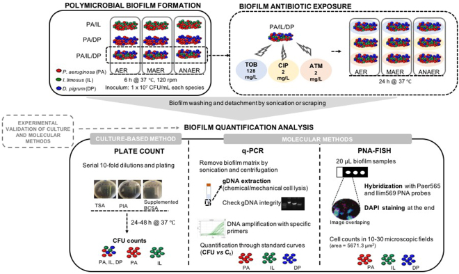
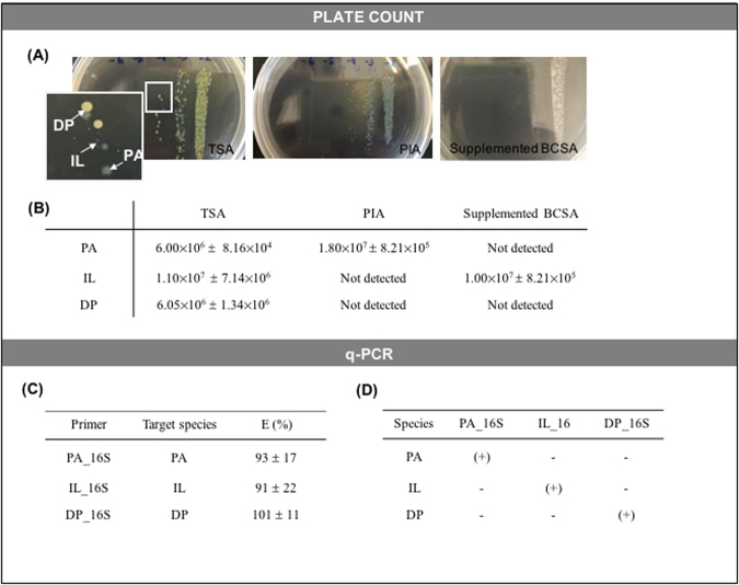
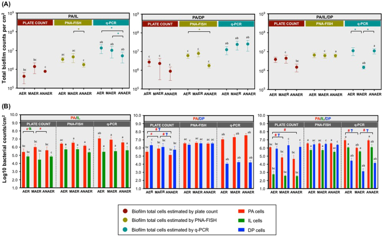
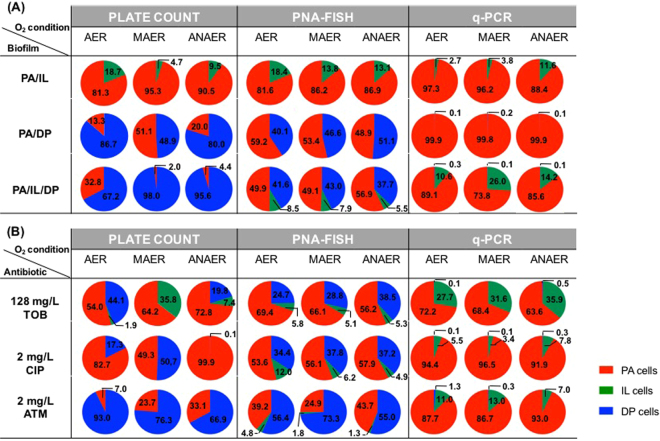
Selecting appropriate tools providing reliable quantitative measures of individual populations in biofilms is critical as we now recognize their true polymicrobial and heterogeneous nature. Here, plate count, quantitative real-time polymerase chain reaction (q-PCR) and peptide nucleic acid probe-fluorescence in situ hybridization (PNA-FISH) were employed to quantitate cystic fibrosis multispecies biofilms. Growth of Pseudomonas aeruginosa, Inquilinus limosus and Dolosigranulum pigrum was assessed in dual- and triple-species consortia under oxygen and antibiotic stress. Quantification methods, that were previously optimized and validated in planktonic consortia, were not always in agreement when applied in multispecies biofilms. Discrepancies in culture and molecular outcomes were observed, particularly for triple-species consortia and antibiotic-stressed biofilms. Some differences were observed, such as the higher bacterial counts obtained by q-PCR and/or PNA-FISH (≤4 log10 cells/cm2) compared to culture. But the discrepancies between PNA-FISH and q-PCR data (eg D. pigrum limited assessment by q-PCR) demonstrate the effect of biofilm heterogeneity in method's reliability. As the heterogeneity in biofilms is a reflection of a myriad of variables, tailoring an accurate picture of communities´ changes is crucial. This work demonstrates that at least two, but preferentially three, quantification techniques are required to obtain reliable measures and take comprehensive analysis of polymicrobial biofilm-associated infections.


Conflict of interest statement

The authors declare no competing interests.

Figures

Figure 1
Figure 1
Experimental design and workflow. Two- and triple-species biofilms involving P. aeruginosa, I. limosus, and D. pigrum developed under aerobic, microaerophilic, and anaerobic environments and the triple consortia exposed to antibiotics were assessed through culture (plate count) and molecular (q-PCR and PNA-FISH) methods. In culture-based method, individual biofilm populations were quantified by unspecific and selective growth media. Regarding q-PCR, DNA extracted from the biofilm-cells was amplified by using specific designed oligonucleotide primers and individual biofilm populations quantified by previous established standard curves (plotting CFU/mL vs Ct for pure cultures). A multiplex PNA-FISH assay with an additional staining step with DAPI was performed ex situ, using specific PNA probes - Paer565 and Ilim569 - previously developed and optimized and bacteria were then estimated using an epifluorescence microscope. Experimental validation was performed for each technique before biofilm quantification experiments. Abbreviations: PA = P. aeruginosa, IL = I. limosus, DP = D. pigrum, AER = aerobic, MAER = microaerophilic, ANAER = anaerobic, TOB = tobramycin, CIP = ciprofloxacin, ATM = aztreonam.
Figure 2
Figure 2
Validation of culture and q-PCR methods. (A) Discrimination of P. aeruginosa, I. limosus and D. pigrum colonies on unspecific (TSA) and on specific culture medium (PIA and supplemented BCSA). PIA was specific for P. aeruginosa and BCSA supplemented with 100 mg/L ticarcillin and 300 000 IU/L polymyxin B was specific for I. limosus after extended incubation time; (B) Bacterial counts (expressed as means ± standard deviations (SDs) CFU/mL for three independent assays) obtained on TSA on specific selective media. Bacterial counts were monitored on solid growth media after incubation at 37 °C for 24–48 h from a mixed-species planktonic culture, containing equal proportions (~107 cells/mL) of each bacterial suspension; (C) q-PCR amplification efficiency, measured by the efficiency of each set of primers designed for specific detection of P. aeruginosa (PA_16S), I. limosus (IL_16S) or D. pigrum (DP_16S). Amplification efficiency (E), expressed as percentage, was determined from the slope of the standard curve plotting the log of the initial template copy number vs Ct (for each set of primers, the standard curves were repeated at least, twice, and the means ± SDs of the obtained amplification efficiencies are illustrated); (D) Experimental specificity test determined for each set of primers specifically designed for each species. Primer specificity was monitored by the outcome of the presence or absence of a specific 16S band visualized in a 1% (w/v) agarose gel. (+) indicates presence of band; (−) is indicative of absence of band. Abbreviations: PA = P. aeruginosa, IL = I. limosus, DP = D. pigrum.
Figure 3
Figure 3
Biofilm quantification by plate count, PNA-FISH and q-PCR. (A) Total counts, expressed as CFU per cm2, estimated for biofilms formed by P. aeruginosa, I. limosus and D. pigrum developed under aerobic, microaerophilic and anaerobic conditions; (B) Populations of P. aeruginosa, I. limosus and D. pigrum in dual- and triple-species biofilms, estimated by the different methods. For each biofilm/condition, means ± SDs for bacterial counts are illustrated (for plate counts, three independent experiments were performed: 18 ≤ n ≤ 24; for PNA-FISH, two to three independent experiments were performed: 10 ≤ n ≤ 30; for qPCR, two to four independent experiments were performed: 6 ≤ n ≤ 9). Significant differences (P < 0.05) are indicated as follows: (*) significantly different total counts between oxygen environments; (#) significantly different P. aeruginosa counts between oxygen environments; (α) significantly different I. limosus counts between oxygen environments; (γ) significantly different D. pigrum counts between oxygen environments; (a) significantly different from plate counts; (b) significantly different from PNA-FISH counts; (c) significantly different from q-PCR counts. Abbreviations: PA = P. aeruginosa, IL = I. limosus, DP = D. pigrum, AER = aerobic, MAER = microaerophilic, ANAER = anaerobic.
Figure 4
Figure 4
Biofilm quantification following antibiotic treatment. (A) Total counts, expressed as CFU per cm2 and (B) Populations of P. aeruginosa, I. limosus and D. pigrum within the defined triple-species biofilms (PA/IL/DP) exposed to 128 mg/L tobramycin, 2 mg/L ciprofloxacin and to 2 mg/L aztreonam under aerobic, microaerophilic and anaerobic conditions for 24 h. For each biofilm/condition, means ± SDs for bacterial counts are illustrated (for plate counts, three independent experiments were performed: 18 ≤ n ≤ 24; for PNA-FISH, two to three independent experiments were performed:10 ≤ n ≤ 30; for qPCR, two to four independent experiments were performed: 6 ≤ n ≤ 9). Significant differences (P < 0.05) are indicated as follows: (*) significantly different total counts between oxygen environments; (#) significantly different P. aeruginosa counts between oxygen environments; (α) significantly different I. limosus counts between oxygen environments; (γ) significantly different D. pigrum counts between oxygen environments; (a) significantly different from plate counts; (b) significantly different from PNA-FISH counts; (c) significantly different from q-PCR counts. Abbreviations: PA = P. aeruginosa, IL = I. limosus, DP = D. pigrum, AER = aerobic, MAER = microaerophilic, ANAER = anaerobic, TOB = tobramycin, CIP = ciprofloxacin, ATM = aztreonam.
Figure 5
Figure 5
Relative distributions of the bacterial populations within biofilms, determined by culture-based and molecular methods. (A) Distributions for P. aeruginosa, I. limosus and D. pigrum populations in dual- and triple-species biofilms before antibiotic treatment; (B) and for the triple biofilms following antibiotic treatment with 128 mg/L tobramycin, 2 mg/L ciprofloxacin and 2 mg/L aztreonam, under aerobic, microaerophilic and anaerobic environments. For each condition/biofilm, means of the relative percentages of the biofilm individual populations are illustrated for two to four independent assays. Abbreviations: PA = P. aeruginosa, IL = I. limosus, DP = D. pigrum, AER = aerobic, MAER = microaerophilic, ANAER = anaerobic, TOB = tobramycin, CIP = ciprofloxacin, ATM = aztreonam.

Similar articles

Cited by

References

    1. Davies D. Understanding biofilm resistance to antibacterial agents. Nat. Rev. Drug Discov. 2003;2:114–122. doi: 10.1038/nrd1008. - DOI - PubMed
    1. Romling U, Balsalobre C. Biofilm infections, their resilience to therapy and innovative treatment strategies. J. Intern. Med. 2012;272:541–561. doi: 10.1111/joim.12004. - DOI - PubMed
    1. Stacy A, McNally L, Darch SE, Brown SP, Whiteley M. The biogeography of polymicrobial infection. Nat. Rev. Microbiol. 2016;14:93–105. doi: 10.1038/nrmicro.2015.8. - DOI - PMC - PubMed
    1. Elias S, Banin E. Multi-Species Biofilms: Living with Friendly Neighbors. FEMS Microbiol. Rev. 2012;36:990–1004. doi: 10.1111/j.1574-6976.2012.00325.x. - DOI - PubMed
    1. Wolcott R, Costerton JW, Raoult D, Cutler SJ. The polymicrobial nature of biofilm infection. Clin. Microbiol. Infect. 2013;19:107–112. doi: 10.1111/j.1469-0691.2012.04001.x. - DOI - PubMed

Publication types

MeSH terms

Substances

LinkOut - more resources