. 2018 Aug;28(8):787-802.
doi: 10.1038/s41422-018-0056-0. Epub 2018 Jun 22.

PTEN-L is a novel protein phosphatase for ubiquitin dephosphorylation to inhibit PINK1-Parkin-mediated mitophagy

Affiliations

PTEN-L is a novel protein phosphatase for ubiquitin dephosphorylation to inhibit PINK1-Parkin-mediated mitophagy

Liming Wang et al. Cell Res. 2018 Aug.

Erratum in

Abstract

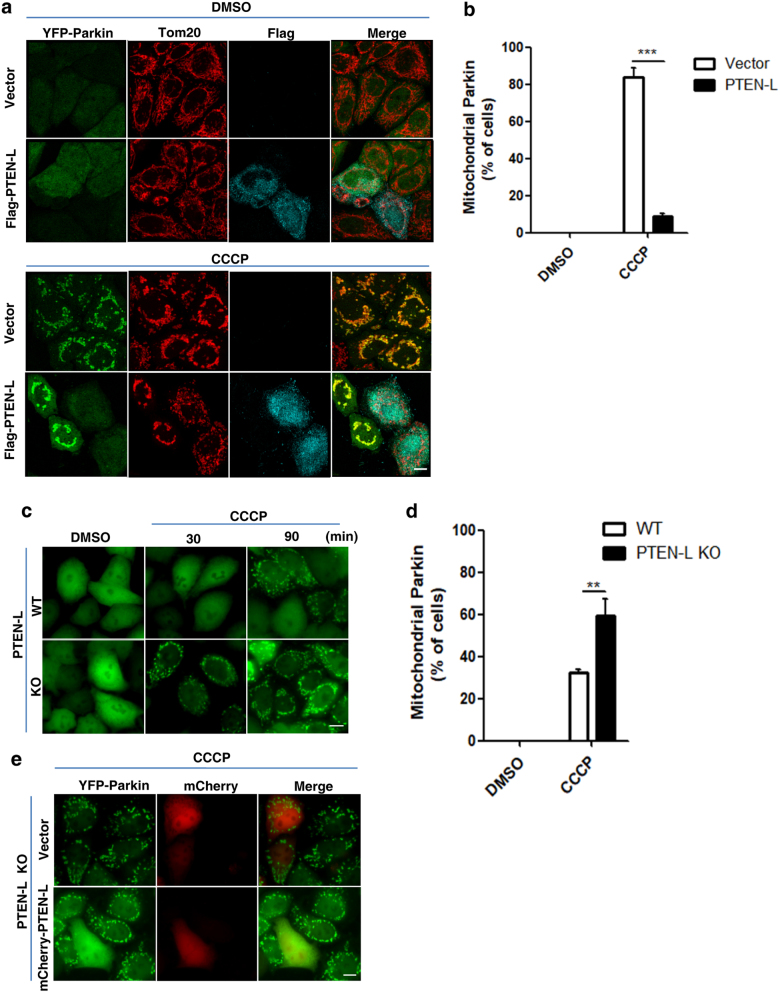
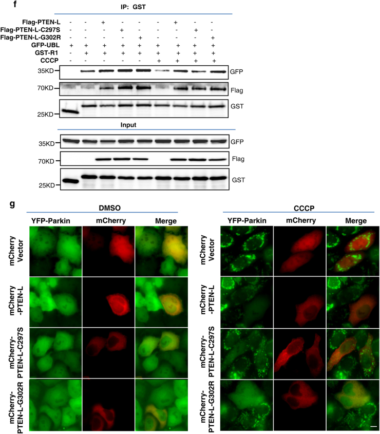
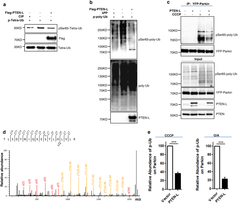
Mitophagy is an important type of selective autophagy for specific elimination of damaged mitochondria. PTEN-induced putative kinase protein 1 (PINK1)-catalyzed phosphorylation of ubiquitin (Ub) plays a critical role in the onset of PINK1-Parkin-mediated mitophagy. Phosphatase and tensin homolog (PTEN)-long (PTEN-L) is a newly identified isoform of PTEN, with addition of 173 amino acids to its N-terminus. Here we report that PTEN-L is a novel negative regulator of mitophagy via its protein phosphatase activity against phosphorylated ubiquitin. We found that PTEN-L localizes at the outer mitochondrial membrane (OMM) and overexpression of PTEN-L inhibits, whereas deletion of PTEN-L promotes, mitophagy induced by various mitochondria-damaging agents. Mechanistically, PTEN-L is capable of effectively preventing Parkin mitochondrial translocation, reducing Parkin phosphorylation, maintaining its closed inactive conformation, and inhibiting its E3 ligase activity. More importantly, PTEN-L reduces the level of phosphorylated ubiquitin (pSer65-Ub) in vivo, and in vitro phosphatase assay confirms that PTEN-L dephosphorylates pSer65-Ub via its protein phosphatase activity, independently of its lipid phosphatase function. Taken together, our findings demonstrate a novel function of PTEN-L as a protein phosphatase for ubiquitin, which counteracts PINK1-mediated ubiquitin phosphorylation leading to blockage of the feedforward mechanisms in mitophagy induction and eventual suppression of mitophagy. Thus, understanding this novel function of PTEN-L provides a key missing piece in the molecular puzzle controlling mitophagy, a critical process in many important human diseases including neurodegenerative disorders such as Parkinson's disease.


Conflict of interest statement

The authors declare no competing interests.

Figures

Fig. 1
Fig. 1
PTEN-L resides at the outer mitochondrial membrane. a Domain structure of PTEN-L protein. b Cell fractionation was performed to isolate mitochondria in HeLa cells. Tim23 and GAPDH were used as mitochondrial and cytosolic markers, respectively. C cytosol, M mitochondria. c Relative ratios of PTEN-L and PTEN in cytosol and mitochondria were calculated with normalization of volume. d Topology assay showing PTEN-L localization at the OMM. Mitochondria were isolated from YFP-Parkin-HeLa cells stably expressing Flag-PTEN-L and treated with different doses of proteinase K and digitonin. e Immunogold EM. YFP-Parkin-HeLa cells stably expressing Flag-PTEN-L were treated without (A, B) or with CCCP (5 µM) (C, D) for 4 h. Cells were subjected to immunoelectron microscopy with anti-Flag antibody. Panels (B) and (D) are the magnified images of boxes in (A) and (C), respectively. Arrows indicate ER structure
Fig. 2
Fig. 2
PTEN-L negatively regulates mitophagy induced by various mitochondria-damaging agents. a YFP-Parkin-HeLa cells with PTEN-L stable expression or control vector were treated with CCCP (5 µM) for 24 h and immunoblotting for mitochondrial proteins was performed as indicated. b Quantification of mitochondrial proteins from a. c Degradation of mtDNA. Representative images of YFP-Parkin-HeLa cells with PTEN-L stable expression and control vector immunostained to label mtDNA (red) after treatment with CCCP (5 µM) for 24 h. d Quantification of mtDNA from > 300 cells per group. Scale bar, 10 µm. e Wild-type (WT) and PTEN-L KO YFP-Parkin-HeLa cells were treated with CCCP (4 µM) for 24 h and immunoblotting was performed as indicated. f Quantification of data from e. Data in b, d, f are presented as mean ± SD from three independent experiments. ***P < 0.001 (two-way ANOVA, b and f), ***P < 0.0001 (Student’s t-test, d)
Fig. 3
Fig. 3
PTEN-L prevents Parkin mitochondrial translocation. a YFP-Parkin-HeLa cells transiently transfected with Flag-PTEN-L or control vector were treated with CCCP (5 µM) for 2 h. YFP-Parkin (green), Tom20 (red), Flag-PTEN-L (cyan). Scale bar, 10 µm. b Percentage of cells with Parkin mitochondrial translocation was quantified by counting at least 300 cells. c Wild-type (WT) and PTEN-L KO YFP-Parkin-HeLa cells were treated with CCCP (4 µM) for 30 and 90 min. YFP-Parkin (green). Scale bar, 10 µm. d Percentage of cells with Parkin mitochondrial translocation from c (CCCP 90 min) was quantified by counting at least 300 cells. e PTEN-L KO YFP-Parkin-HeLa cells were transiently transfected with mCherry-PTEN-L or control vector and treated with CCCP (4 µM) for 90 min. YFP-Parkin (green), mCherry (red). Scale bar, 10 µm. Data in b, d are presented as mean ± SD from three independent experiments. **P < 0.01, ***P < 0.001 (two-way ANOVA test)
Fig. 4
Fig. 4
PTEN-L impairs Parkin E3 ligase activity and reduces pSer65-Parkin. a YFP-Parkin-HeLa cells with PTEN-L stable expression or control vector were treated with CCCP (5 µM) for indicated hours. YFP-Parkin was immunoprecipitated with anti-GFP antibody. Immunoprecipitants (IPs) and whole-cell lysates (WCLs) were analyzed for YFP-Parkin, mitofusin-2 (MFN2), Tom20, PTEN-L, PTEN and tubulin. b YFP-Parkin-HeLa cells with PTEN-L stable expression or control vector were treated with O/A (10 nM and 100 nM) for indicated hours and the WCLs were analyzed by immunoblotting as indicated. c Wild-type (WT) and PTEN-L KO YFP-Parkin-HeLa cells were treated with CCCP (5 µM) for indicated hours and the WCLs were analyzed by immunoblotting as indicated. d Wild-type (WT) and PTEN-L KO YFP-Parkin-HeLa cells were treated with O/A (10 nM and 100 nM) for indicated hours and the WCLs were analyzed by immunoblotting as indicated. e The MS/MS spectra of the Parkin peptide containing phospho-Ser65. YFP-Parkin-HeLa cells were treated with CCCP (10 µM) for 4 h. YFP-Parkin was pulled down with GFP beads and subjected to MS/MS analysis. f pSer65-Parkin was quantified using MS-based relative quantification analysis in YFP-Parkin-HeLa cells with or without PTEN-L stable expression after CCCP (10 µM) and O/A (25 nM and 250 nM) treatment. Data are presented as mean ± SD from 3 independent experiments. **P < 0.01 (Student’s t-test)
Fig. 5
Fig. 5
PTEN-L keeps Parkin in closed conformation by enhancing the interaction of Parkin UBL and RING1 domains in a protein phosphatase activity-dependent manner. a Construction of PTEN-L truncations. PTEN-L contains an ATR region, a phosphatase domain and a C-terminal region with a C2 domain and a C-Tail domain. PTEN-L-C297S is a dual lipid-protein phosphatase-defective mutant, while PTEN-L-G302R is a lipid phosphatase-defective mutant. b, c HEK293T cells transfected with GFP-Parkin and different constructs of Flag-tagged PTEN-L were treated without or with CCCP (5 µM) for 4 h. PTEN-L was immunoprecipitated with anti-Flag beads followed by immunoblotting for GFP and Flag. d Construction of Flag-tagged Parkin truncations, including Parkin-FL (full length) and truncated Parkin constructs: UBL-R0, Exon4, R1-IBR-R2, UBL-R1-IBR-R2 (deletion of R0 domain) and IBR-R2. e HEK293T cells transfected with GFP-PTEN-L and different constructs of Flag-tagged Parkin were treated without or with CCCP (5 µM) for 4 h. PTEN-L was then immunoprecipitated with anti-GFP beads followed by immunoblotting for Flag and GFP. f HEK293T cells were transfected with Flag-PTEN-L or the two Flag-PTEN-L mutants, together with Parkin truncation mutants GFP-UBL and GST-RING1 (R1) or GST empty vector (pEBG). Cells were then treated with or without CCCP (20 µM) for 4 h. RING1 was immunoprecipitated with anti-GST beads followed by immunoblotting for GFP, Flag and GST. g YFP-Parkin-HeLa cells transiently transfected with mCherry-PTEN-L or the two mCherry-PTEN-L mutants were treated with CCCP (5 µM) for 2 h. YFP-Parkin (green), mCherry (red). Scale bar, 10 µm
Fig. 5
Fig. 5
PTEN-L keeps Parkin in closed conformation by enhancing the interaction of Parkin UBL and RING1 domains in a protein phosphatase activity-dependent manner. a Construction of PTEN-L truncations. PTEN-L contains an ATR region, a phosphatase domain and a C-terminal region with a C2 domain and a C-Tail domain. PTEN-L-C297S is a dual lipid-protein phosphatase-defective mutant, while PTEN-L-G302R is a lipid phosphatase-defective mutant. b, c HEK293T cells transfected with GFP-Parkin and different constructs of Flag-tagged PTEN-L were treated without or with CCCP (5 µM) for 4 h. PTEN-L was immunoprecipitated with anti-Flag beads followed by immunoblotting for GFP and Flag. d Construction of Flag-tagged Parkin truncations, including Parkin-FL (full length) and truncated Parkin constructs: UBL-R0, Exon4, R1-IBR-R2, UBL-R1-IBR-R2 (deletion of R0 domain) and IBR-R2. e HEK293T cells transfected with GFP-PTEN-L and different constructs of Flag-tagged Parkin were treated without or with CCCP (5 µM) for 4 h. PTEN-L was then immunoprecipitated with anti-GFP beads followed by immunoblotting for Flag and GFP. f HEK293T cells were transfected with Flag-PTEN-L or the two Flag-PTEN-L mutants, together with Parkin truncation mutants GFP-UBL and GST-RING1 (R1) or GST empty vector (pEBG). Cells were then treated with or without CCCP (20 µM) for 4 h. RING1 was immunoprecipitated with anti-GST beads followed by immunoblotting for GFP, Flag and GST. g YFP-Parkin-HeLa cells transiently transfected with mCherry-PTEN-L or the two mCherry-PTEN-L mutants were treated with CCCP (5 µM) for 2 h. YFP-Parkin (green), mCherry (red). Scale bar, 10 µm
Fig. 6
Fig. 6
PTEN-L dephosphorylates ubiquitin. a YFP-Parkin-HeLa cells with PTEN-L stable expression or control vector were treated with CCCP (5 µM) for 3 h. Whole-cell lysates were analyzed by immunoblotting. b YFP-Parkin-HeLa cells with PTEN-L stable expression or control vector were treated with CCCP (5 µM) for 3 h. Immunofluorescence staining against pSer65-Ub was performed and observed by fluorescent microscopy. pSer65-Ub (red), YFP-Parkin (green), Nucleus (DAPI, blue). Scale bar, 10 µm. c YFP-Parkin-HeLa cells with PTEN-L stable expression or control vector were treated with O/A (25 nM and 250 nM) for indicated hours and immunoblotting was performed. d Wild-type (WT) and PTEN-L KO YFP-Parkin-HeLa cells were treated with O/A (25 nM and 250 nM) for indicated hours and immunoblotting was performed. e In vitro dephosphorylation assay. Purified pSer65-Ub was incubated with purified Flag-PTEN-L, Flag-PTEN-L-C297S or Flag-PTEN-L-G302R in phosphatase reaction buffer and ubiquitin phosphorylation level was evaluated by immunoblotting
Fig. 7
Fig. 7
PTEN-L disrupts the feedforward mechanism in mitophagy by targeting the pSer65-Ub chains. a In vitro dephosphorylation assay using purified pSer65-tetra-Ub. Purified Flag-PTEN-L was incubated with pSer65-tetra-Ub in the phosphatase reaction buffer for 1 h at 30 °C. Calf intestinal phosphatase (CIP) was used as a positive control. b In vitro dephosphorylation assay using purified pSer65-poly-Ub chains, following the same procedure in a. λPP was used as a positive control. c YFP-Parkin-HeLa cells with PTEN-L stable expression or control vector were treated with CCCP (10 µM) for 4 h. YFP-Parkin was pulled down by GFP beads and subjected to immunoblotting. d The MS/MS spectra of the ubiquitin peptide containing phospho-Ser65. YFP-Parkin-HeLa cells were treated with CCCP (10 µM) for 4 h and YFP-Parkin was pulled down with GFP beads. e pSer65-Ub was quantified using MS-based relative quantification analysis in YFP-Parkin-HeLa cells with or without PTEN-L stable expression after CCCP (10 µM) and O/A (25 nM and 250 nM) treatment. Data are presented as mean ± SD from 3 independent experiments. ***P < 0.0001 (Student’s t-test)
Fig. 8
Fig. 8
Illustration of the novel function of PTEN-L as a protein phosphatase in suppression of mitophagy. PTEN-L is able to counteract PINK1-mediated ubiquitin phosphorylation (pSer65-Ub), which then leads to prevention of Parkin recruitment to damaged mitochondria, reduction of pSer65-Parkin level and maintenance of Parkin in its closed inactive conformation to impair Parkin E3 ligase activity, all resulting in the disruption of the feedforward loop to inhibit mitophagy

Comment in

  • PTEN-L puts a brake on mitophagy.
    Wang L, Wang J, Tang Y, Shen HM. Wang L, et al. Autophagy. 2018;14(11):2023-2025. doi: 10.1080/15548627.2018.1502565. Epub 2018 Sep 2. Autophagy. 2018. PMID: 30106322 Free PMC article.

Similar articles

Cited by

References

    1. Lemasters JJ. Selective mitochondrial autophagy, or mitophagy, as a targeted defense against oxidative stress, mitochondrial dysfunction, and aging. Rejuvenation Res. 2005;8:3–5. doi: 10.1089/rej.2005.8.3. - DOI - PubMed
    1. Pickrell AM, Youle RJ. The roles of PINK1, parkin, and mitochondrial fidelity in Parkinson’s disease. Neuron. 2015;85:257–273. doi: 10.1016/j.neuron.2014.12.007. - DOI - PMC - PubMed
    1. Ryan BJ, Hoek S, Fon EA, Wade-Martins R. Mitochondrial dysfunction and mitophagy in Parkinson’s: from familial to sporadic disease. Trends Biochem. Sci. 2015;40:200–210. doi: 10.1016/j.tibs.2015.02.003. - DOI - PubMed
    1. Harper JW, Ordureau A, Heo JM. Building and decoding ubiquitin chains for mitophagy. Nat. Rev. Mol. Cell Biol. 2018;19:93–108. doi: 10.1038/nrm.2017.129. - DOI - PubMed
    1. Pickles S, Vigie P, Youle RJ. Mitophagy and quality control mechanisms in mitochondrial maintenance. Curr. Biol. 2018;28:R170–R185. doi: 10.1016/j.cub.2018.01.004. - DOI - PMC - PubMed

Publication types