

ISCEV extended protocol for the dark-adapted red flash ERG
- PMID: 29934801
- PMCID: PMC6061112
- DOI: 10.1007/s10633-018-9644-z
ISCEV extended protocol for the dark-adapted red flash ERG
Abstract
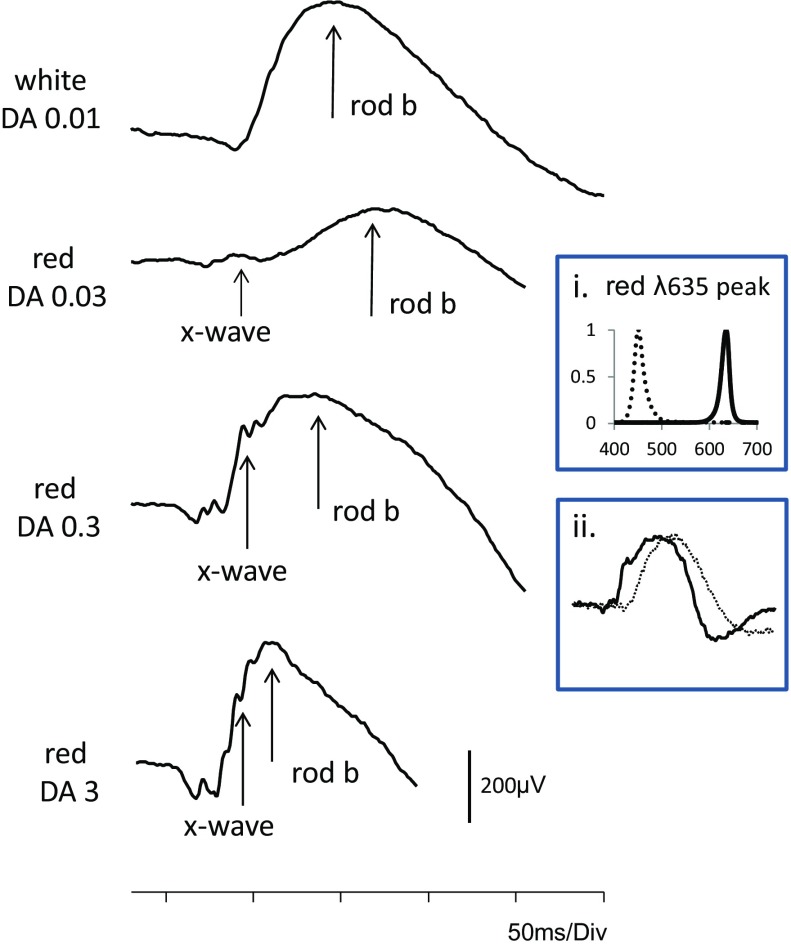
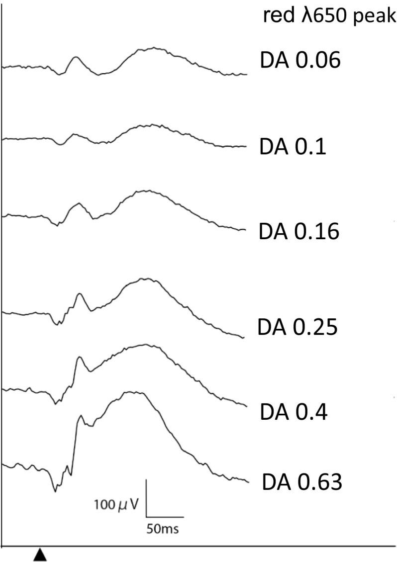
The International Society for Clinical Electrophysiology of Vision (ISCEV) standard for full-field electroretinography (ERG) describes a minimum procedure, but encourages more extensive testing. This ISCEV extended protocol describes an extension to the ERG standard, namely the dark-adapted (DA) red flash ERG. The DA red flash ERG can be incorporated conveniently within the ISCEV standard ERG protocol after a minimum of 20-min DA and recorded after the DA 0.01 ERG to a flash strength of 0.3 phot cd s m-2, eliciting a waveform with two positive peaks in healthy individuals. The first positive component is the cone-mediated x-wave with a peak at 30-50 ms; the second is a rod-mediated b-wave with a peak time of approximately 100 ms. Shorter DA times may be desirable to shorten the recording time or to alter the prominence of the early cone-mediated x-wave relative to the rod-mediated b-wave. The DA red flash ERG is used to aid the diagnosis of achromatopsia (rod monochromacy), cone dystrophy and other forms of cone system dysfunction, including "Bradyopsia" (RGS9/R9AP-retinopathy), when the DA red flash ERG x-wave is preserved in the absence of ISCEV standard LA ERGs. The DA red flash ERG can also help determine the origin of residual DA ERGs in cases of severe rod dysfunction, for example in disorders such as vitamin A deficiency, fundus albipunctatus (RDH5-retinopathy), Oguchi disease (SAG- or GRK1-retinopathy) and some rod-cone dystrophies. To shorter DA periods, the x-wave may be elicited without the following rod b-wave, shown to be helpful in abbreviated protocols for children.
Keywords: Clinical standards; Dark-adapted (DA); Electroretinogram (ERG); Full-field ERG; International Society of Clinical Electrophysiology of Vision (ISCEV); Red flash ERG; Retinal dystrophy.
Conflict of interest statement
Conflict of interest
The authors have no conflicts of interest.
Informed consent
As this article does not contain any studies with human participants performed directly by any of the authors, the concept of informed consent is not applicable.
Statement of human rights
The study involved no research on Human participants and consent is not applicable.
Statement on the welfare of animals
The study involved no research on animals.
Figures
References
-
- Motokawa K, Mita T. Über eine einfachere Untersuchungsmethode und Eigenschaften der Aktionsströme der Netzhaut des Menschen. Tohoku J Exp Med. 1942;42:114–133. doi: 10.1620/tjem.42.114. - DOI
-
- Adrian ED. The rod and cone components in the electrical response of the human eye. J Physiol. 1946;104:84–104. doi: 10.1113/jphysiol.1945.sp004109. - DOI
MeSH terms
LinkOut - more resources
Full Text Sources
Other Literature Sources
