. 2018 Jul;136(1):41-56.
doi: 10.1007/s00401-018-1868-1. Epub 2018 Jun 13.

Alzheimer's disease pathology propagation by exosomes containing toxic amyloid-beta oligomers

Affiliations

Alzheimer's disease pathology propagation by exosomes containing toxic amyloid-beta oligomers

Maitrayee Sardar Sinha et al. Acta Neuropathol. 2018 Jul.

Abstract

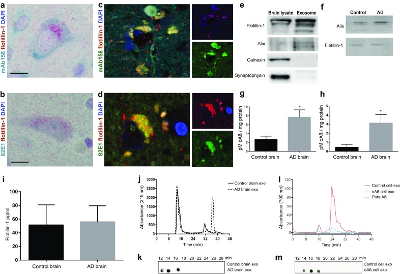
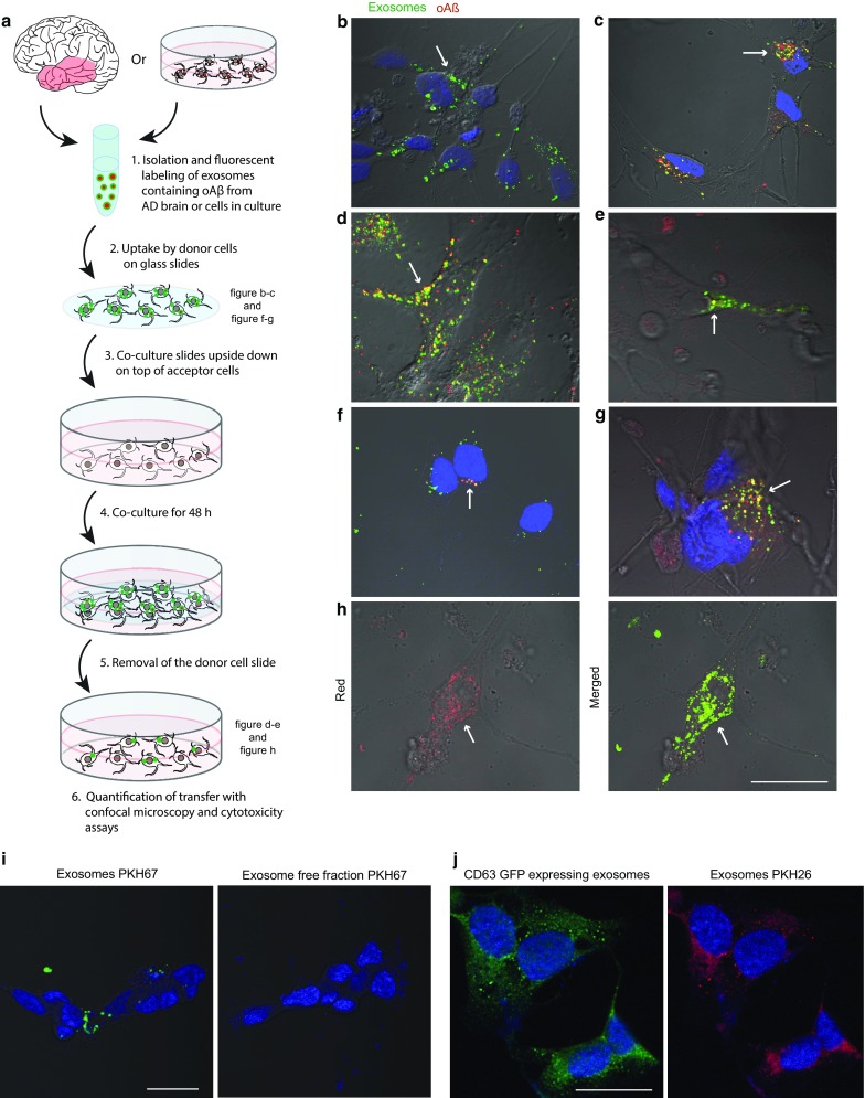
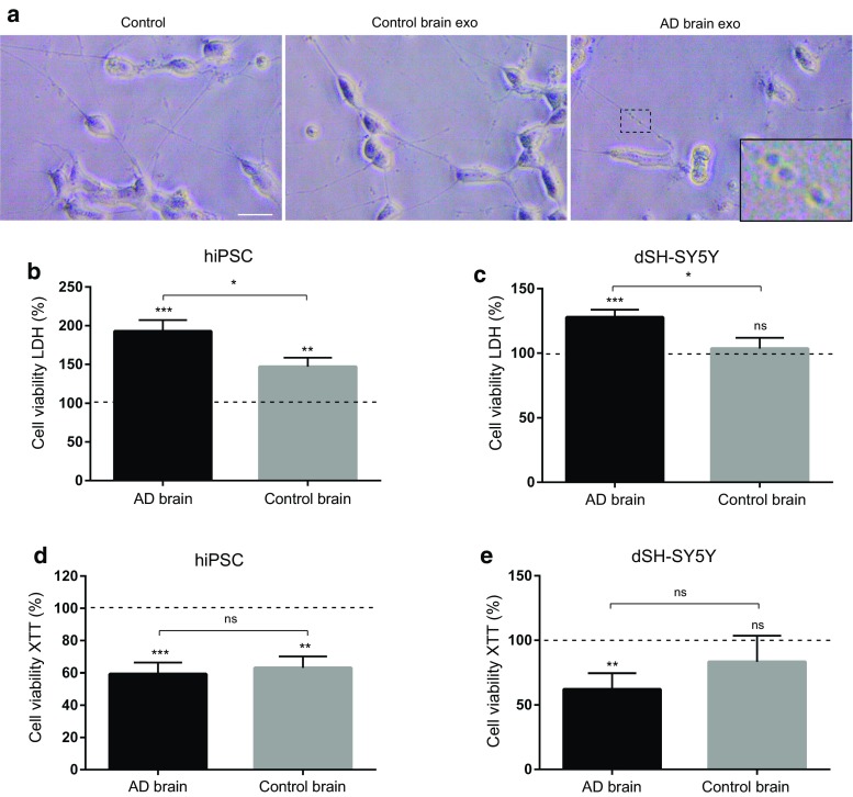
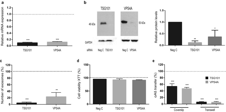
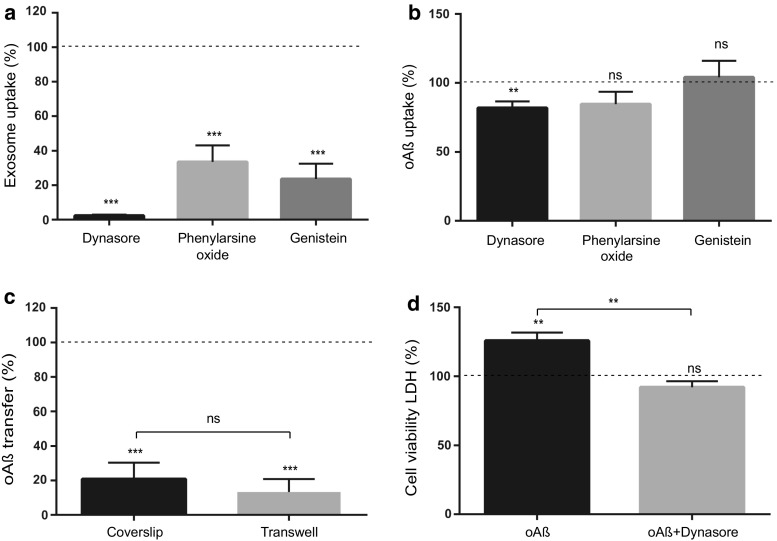
The gradual deterioration of cognitive functions in Alzheimer's disease is paralleled by a hierarchical progression of amyloid-beta and tau brain pathology. Recent findings indicate that toxic oligomers of amyloid-beta may cause propagation of pathology in a prion-like manner, although the underlying mechanisms are incompletely understood. Here we show that small extracellular vesicles, exosomes, from Alzheimer patients' brains contain increased levels of amyloid-beta oligomers and can act as vehicles for the neuron-to-neuron transfer of such toxic species in recipient neurons in culture. Moreover, blocking the formation, secretion or uptake of exosomes was found to reduce both the spread of oligomers and the related toxicity. Taken together, our results imply that exosomes are centrally involved in Alzheimer's disease and that they could serve as targets for development of new diagnostic and therapeutic principles.

Keywords: Alzheimer’s disease; Beta-amyloid; Exosomes; Human; Oligomers; Prion-like; Propagation.


Conflict of interest statement

The authors have declared that no conflict of interest exists.

Figures

Fig. 1
Fig. 1
AD brain exosomes are enriched with oAβ. AD brain sections from temporal neocortex show co-localization of probable oAβ to exosomes. a, b Double-immunostaining with the exosome marker flotillin-1 (labelled red), and two different oligomer-selective (see main text) Aβ antibodies mAb158 and 82E1, respectively (labelled blue-green). Scale bar 10 µm. c, d Co-localization of oAβ to exosomes was performed after immunofluorescence labelling. mAb158 or 82E1, respectively, showed substantial co-localization (yellow) with flotillin-1 inside cells in the AD brain (R2 = 0.78 and 0.86 for mAb158 and 82E1, respectively). Scale bar, 5 µm. e Immunoblot showing flotillin-1, alix, calnexin and synaptophysin in exosome and brain lysate, demonstrating no cellular or synapse vesicle contamination in the exosome preparation. Loading control is shown in Supplementary Fig. S1d. f Immunoblot demonstrating the presence of flotillin-1 and alix, in exosome fractions isolated from control and AD brain. Quantitative ELISA analysis of oAβ in exosomes isolated from AD and control brains, using 82E1 (h) and mAb158 (g) antibodies, respectively. Data are presented as the mean picomolar of oAβ per mg total exosome protein ± SEM (n = 3). *p < 0.05 by two-tailed unpaired Student’s t tests with Welch’s correction. i Quantitative analysis of flotillin-1 by ELISA in control and AD brain exosomes showing equal amount of flotillin-1 between the groups. j Representative SEC profile of lysed exosomes isolated from control and AD brain samples at 215 nm absorbance (general protein detection). k Detection of oAβ in SEC eluate fractions of AD and control brain exosomes by dot blot using mAb158 antibody. l SEC chromatograms of exosomes isolated from conditioned media of control- or oAβ-AF700 treated dSH-SY5Y cells as well as pure oAβ-AF700. Detection at 700 nm absorbance (AF700 detection). m Dot blot detection of oAβ (mAb158 antibody) in SEC eluate fractions of exosomes isolated from conditioned media of control- or oAβ-AF700 treated dSH-SY5Y cells of control and oAβ-AF700 treated dSH-SY5Y cells exosomes. The analysis of SEC fractions confirms the presence of oAβ in exosomes
Fig. 2
Fig. 2
Exosome-mediated uptake and propagation of oAβ in neuronal cells. Exosomes isolated from brain tissue or conditioned media of dSH-SY5Y cells were labelled with the dye PKH67 and added to donor hiPSCs or dSH-SY5Y cells. After 3 h of incubation at 37 °C, donor cells were fixed, stained with mAb158 (for brain exosomes) and analysed by confocal microscopy or donor cells were co-cultured with another set of hiPSCs or dSH-SY5Y (recipient cells). After 48 h of co-culture, donor cells were removed and recipient cells were fixed, stained with mAb158 (for brain exosomes) and analysed by confocal microscopy. a A cartoon illustrating the co-culture model with hiPSC or dSH-SY5Y cells employed to measure the transfer of the brain or cell exosomes containing oAβ. Uptake of b control and c AD brain exosomes (green) containing oAβ (red) in hiPSC donor cells. Transfer of AD brain exosomes (green) containing oAβ (red) to recipient d hiPSCs and e dSH-SY5Y cells. Uptake of exosomes (green) containing oAβ-AF700 (red) in donor f dSH-SY5Y and g hiPSCs. h Transfer of oAβ-AF700 containing exosomes in recipient dSH-SY5Y cells. Super-imposed image of the red (oAβ) and green (exosomes) channels on a DIC image shows co-localization (yellow) of exosomes and oAβ. Arrows indicate exosomes or exosome containing oAβ. i Cellular uptake of isolated brain exosomes and brain exosome free fraction after PKH67 staining showing no PKH67 uptake in the absence of exosomes. j Isolated exosomes from CD63-GFP expressing SH-SY5Y cells, double-labelled with PKH26 (red), were added to dSH-SY5Y cells. The CD63-GFP were intensified using an anti-GFP antibody. Scale bar (bh), j 20 µm, i 10 µm
Fig. 3
Fig. 3
Transfer of AD brain exosomes causes cytotoxicity. Exosomes isolated from control and AD brain tissues were added to donor hiPSCs or dSH-SY5Y cells. After 3 h of incubation at 37 °C, donor cells were washed with PBS and co-cultured with another set of hiPSCs or dSH-SY5Y (recipient cells). After 48 h of co-culture, donor cells were removed. a Morphological changes assessed in recipient dSH-SY5Y showing loss of neurite branching after transfer of AD brain exosomes. Also, neurite beading was seen in dystrophic neurites as shown in magnified insert. The conditioned media was collected for LDH assay (b, c) and recipient cell viability evaluated by XTT (d, e). Values are expressed as percentage of untreated control. Values are mean ± SEM (n = 6 separate experiments). LDH shows that transfer of AD brain exosomes causes significant higher cytotoxicity compared to control brain exosomes in both cell types. NS, not significant; *p < 0.05, **p < 0.01, ***p < 0.001 by two-tailed unpaired Student’s t tests with Welch’s correction
Fig. 4
Fig. 4
Downregulation of exosomal proteins TSG101 and VPS4A inhibits the spread of oAβ. Depletion of TSG101 and VPS4A by siRNA in dSH-SY5Y cells. a Real time PCR analysis of mRNA expressions to show knock down efficiency of transfected cells by TSG101 or VPS4A siRNA. b Representative immunoblot picture of cell lysates after siRNA treatment for 72 h and associated densitometric analysis (n = 3). c Bead flow cytometry analysis of exosomes shows a significant decrease in the number of secreted exosomes after TSG101 or VPS4A siRNA treatment in raSH-SY5Y cells. d No cytotoxicity was detected by XTT assay after 48 h of transfection with siRNA. e Quantification of oAβ transfer in presence of TSG101 or VPS4A siRNA in both coverslip and transwell co-culture model by flow cytometry, shows that both siRNAs significantly inhibit oAβ transfer. Values are expressed as percentage of siRNA negative control and indicated as dotted line. Values are the mean ± SEM (n = 4 separate cultures), *p < 0.05, **p < 0.01, ***p < 0.001 by two-tailed unpaired Student’s t tests with Welch’s correction
Fig. 5
Fig. 5
The uptake of exosomes and the subsequent spreading of oAß is dynamin-dependent. a, b Uptake of PKH67 labelled exosomes or oAβ-AF700 in dSH-SY5Y cells. Cells were pre-incubated with the indicated inhibitors for 30 min, then exposed to exosomes or oAβ-AF700. After 3 h incubation, samples were collected and the proportion of cells with uptake was quantified by flow cytometry and related to untreated control (dotted line). c Flow cytometry analysis of oAβ transfer in presence of dynasore in coverslip and transwell co-culture models. After dynasore treatment there is a significant decrease of the proportion of cells with oAβ transfer in both models (control, dotted line). d Transfer of exosomes isolated from oAβ treated cells causes cytotoxicity in recipient cells compared to untreated control as shown by LDH assay, whereas dynasore treatment significantly reduces the cytotoxic effect versus untreated control (dotted line). Data are represented as the mean ± SEM, NS, not significant; n = 4; **p < 0.01, ***p < 0.001 by two-tailed unpaired Student’s t tests with Welch’s correction

Comment in

References

    1. Agholme L, Lindstrom T, Kagedal K, Marcusson J, Hallbeck M. An in vitro model for neuroscience: differentiation of SH-SY5Y cells into cells with morphological and biochemical characteristics of mature neurons. J Alzheimers Dis. 2010;20:1069–1082. doi: 10.3233/JAD-2010-091363. - DOI - PubMed
    1. Agholme L, Nath S, Domert J, Marcusson J, Kagedal K, Hallbeck M. Proteasome inhibition induces stress kinase dependent transport deficits—implications for Alzheimer’s disease. Mol Cell Neurosci. 2013;58C:29–39. - PubMed
    1. Akrap I, Thavamani A, Nordheim A. Vps4A-mediated tumor suppression upon exosome modulation? Ann Transl Med. 2016;4:180. doi: 10.21037/atm.2016.04.18. - DOI - PMC - PubMed
    1. Asai H, Ikezu S, Tsunoda S, Medalla M, Luebke J, Haydar T, Wolozin B, Butovsky O, Kügler S, Ikezu T. Depletion of microglia and inhibition of exosome synthesis halt tau propagation. Nat Neurosci. 2015 - PMC - PubMed
    1. Bellingham SA, Guo BB, Coleman BM, Hill AF. Exosomes: vehicles for the transfer of toxic proteins associated with neurodegenerative diseases? Front Physiol. 2012;3:124. doi: 10.3389/fphys.2012.00124. - DOI - PMC - PubMed

Publication types

MeSH terms