

Refinement of embolic stroke model in rats: Effect of post-embolization anesthesia duration on arterial blood pressure, cerebral edema and mortality
- PMID: 29935198
- PMCID: PMC6192029
- DOI: 10.1016/j.jneumeth.2018.06.012
Refinement of embolic stroke model in rats: Effect of post-embolization anesthesia duration on arterial blood pressure, cerebral edema and mortality
Abstract
Background: Injection of a clot into the internal carotid artery is an experimental model of ischemic stroke that is considered to closely mimic embolic stroke in humans. In this model, the common carotid artery typically remains temporarily occluded to permit time for stabilization of the clot in the middle cerebral artery. However, the associated lengthening of the anesthesia duration could affect arterial blood pressure and stroke outcome.
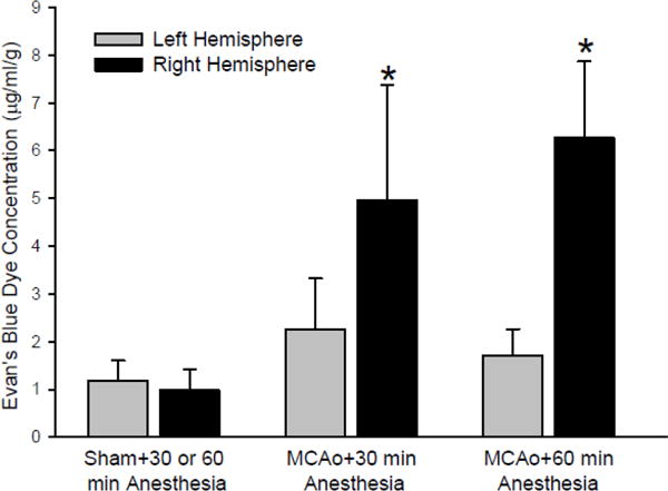
New method: We refined the model by examining how increasing isoflurane anesthesia duration from 30 to 60 min after clot embolization affects mortality, infarct volume, edema, blood-brain barrier permeability, and the 8-h post-ischemic time course of blood pressure, which has not been reported previously in this model.
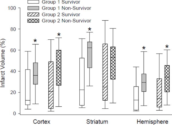
Results: We found that arterial pressure increased after discontinuing anesthesia in both embolized groups and that the increase was greater than in the corresponding non-embolized sham-operated rats. At 24 h, the group with 60-min post-ischemia anesthesia exhibited greater brain water content and a greater ipsilateral-to-contralateral ratio of extravasated Evans blue dye. Mortality was greater in the 60-min group, but infarct volume among survivors was not different from that in the 30-min anesthesia group.
Comparison with existing methods: This study refines the embolic stroke model by demonstrating the importance of minimizing the duration of anesthesia after embolization.
Conclusions: These data indicate that early discontinuation of isoflurane anesthesia after clot embolization permits an earlier hypertensive response that limits edema formation and mortality without significantly affecting infarct volume in survivors, thereby decreasing the required number of animals.
Keywords: Animal model of stroke; Embolic stroke; Isoflurane anesthesia; Middle cerebral artery occlusion; Rat.
Copyright © 2018 Elsevier B.V. All rights reserved.
Conflict of interest statement
The authors declare that they have no conflict of interest.
Figures


References
-
- Astrup J, Symon L, Branston NM, Lassen NA. Cortical evoked potential and extracellular K+ and H+ at critical levels of brain ischemia. Stroke. 1977;8:51–7. - PubMed
-
- Beech JS, Williams SC, Campbell CA, Bath PM, Parsons AA, Hunter AJ, Menon DK. Further characterisation of a thromboembolic model of stroke in the rat. Brain Res. 2001;895:18–24. - PubMed
-
- Busch E, Kruger K, Hossmann KA. Improved model of thromboembolic stroke and rt-PA induced reperfusion in the rat. Brain Res. 1997;778:16–24. - PubMed
-
- Chen ST, Hsu CY, Hogan EL, Maricq H, Balentine JD. A model of focal ischemic stroke in the rat: reproducible extensive cortical infarction 1. Stroke. 1986;17:738–43. - PubMed
Publication types
MeSH terms
Grants and funding
LinkOut - more resources
Full Text Sources
Other Literature Sources
Medical
