. 2018 Jun 23;17(1):148.
doi: 10.1186/s12944-018-0786-5.

Fish oil-derived lipid emulsion induces RIP1-dependent and caspase 8-licensed necroptosis in IEC-6 cells through overproduction of reactive oxygen species

Affiliations

Fish oil-derived lipid emulsion induces RIP1-dependent and caspase 8-licensed necroptosis in IEC-6 cells through overproduction of reactive oxygen species

Jun-Kai Yan et al. Lipids Health Dis. .

Abstract

Background: Excessive cell death of enterocytes has been demonstrated to be partially associated with the intravenously-administrated lipid emulsions (LEs) during parenteral nutrition (PN) support. However, as a new generation of LE, the effect of fish oil-derived lipid emulsion (FOLE) on the death of enterocytes remains elusive.

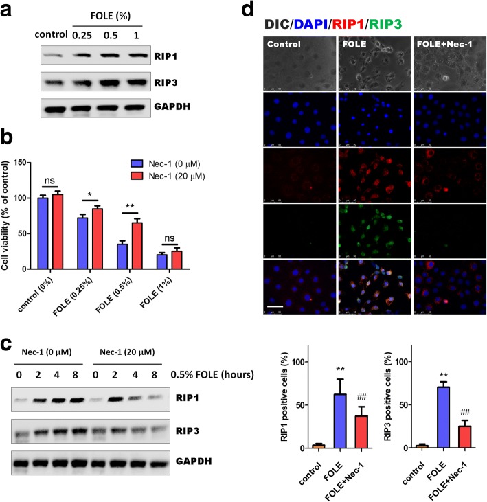
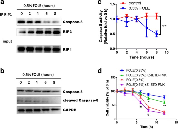
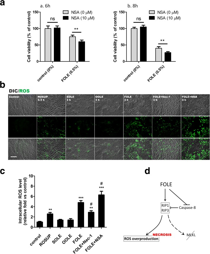
Methods: Intestinal epithelial cells (IEC-6 cell line) were treated with FOLE (0.25-1%) for 24 h. Cell survival was measured by CCK-8 assay, and morphological changes were monitored by time-lapse live cell imaging. The expression of receptor-interacting protein 1/3 (RIP1/3) and caspase 8 was assessed by westernblot, and the formation of necrosome (characterized by the assembly of RIP1/3 complex along with the dissociation of caspase 8) was examined by immunoprecipitation. Additionally, the production of intracellular reactive oxygen species (ROS) was detected by using a ROS detection kit with an oxidation-sensitive probe (DCFH-DA).

Results: FOLE dose-dependently induced non-apoptotic, but programmed necroctic cell death (necroptosis) within 4-8 h after treatment. The assembly of RIP1/3 complex along with the dissociation of caspase 8 from RIP1 was observed in FOLE-treated cells. Moreover, FOLE-induced cell death was significantly alleviated by inhibiting RIP1, and was further aggravated by inhibiting caspase 8. In addition, prior to cell death the accumulation of intracellular ROS was significantly increased in FOLE-treated cells (increased by approximately 5-fold versus control, p < 0.001), which could be attenuated by inhibiting RIP1 (decreased by approximately 35% versus FOLE, p < 0.05).

Conclusions: FOLE induces RIP1-dependent and caspase 8-licensed necroptosis through overproduction of ROS in vitro. Our findings may provide novel insights into the clinical applications of FOLE during PN support.

Keywords: Caspase 8; Fish oil-derived lipid emulsion; IEC-6; Necroptosis; Parenteral nutrition; Reactive oxygen species; Receptor-interacting protein 1.


Conflict of interest statement

Ethics approval and consent to participate

Not applicable.

Competing interests

The authors declare that they have no competing interests.

Publisher’s Note

Springer Nature remains neutral with regard to jurisdictional claims in published maps and institutional affiliations.

Figures

Fig. 1
Fig. 1
FOLE induces necrotic cell death in IEC-6 cells. a Cell viability assessed by CCK-8 assay. IEC-6 cells were treated with 0.25% SOLE, 0.25% OOLE and 0.5% FOLE for 24 h. Note that a significant reduction in the viability was observed in FOLE-treated cells. Data are presented as mean ± SD. **, p < 0.01 compared to control (n = 6–8 wells/group). b Necrotic cell death in FOLE-treated cells. Representative images are shown (Scale bar = 25 μm). c Cell viability assessed by CCK-8 assay. IEC-6 cells were treated with 0.25–1% FOLE for 0–24 h. Notably, FOLE reduced the cell viability of IEC-6 cells in a dose-dependent manner, and rapid reduction was observed at 4–8 h post FOLE treatment. d Time-lapse live cell imaging. Post-confluent IEC-6 cells were treated with 0.5% FOLE, and a 24-h time-lapse live cell imaging was performed as described in material and methods. Representative images are shown. Scale bar = 75 μm. Videos are available as supplementary data (see Additional file 1: IEC-6 cells treated with FOLE.avi). Three experiments were performed that showed similar results
Fig. 2
Fig. 2
FOLE-induced necrotic cell death is dependent on RIP1. a Representative immunoblots of RIP1 and RIP3. IEC-6 cells were treated with FOLE at 0.25–1%, and the indicated proteins were analyzed at 8 h after FOLE treatment. b Cell viability. IEC-6 cells were pre-treated with Nec-1 (20 μM) for 40 min, followed by exposure to 0.25–1% FOLE as described in material and methods. The viability was analyzed 8 h after treatment. Data are presented as mean ± SD. *, p < 0.05, **, p < 0.01 (n = 6–8 wells/group). c Representative immunoblots of RIP1 and RIP3. IEC-6 cells were pre-treated with Nec-1 (20 μM) for 40 min, followed by exposure to 0.5% FOLE. The indicated proteins were analyzed at 2–8 h after treatment. d Representative images of RIP1 and RIP3 staining in IEC-6 cells. Cells were treated as described in (c). Scale bar = 50 μm. Three experiments were performed that showed similar results
Fig. 3
Fig. 3
FOLE-induced cell death requires RIP1/3 complex and is licensed by caspase 8. a Assembly of RIP1/RIP3 complex and dissociation of caspase 8. IEC-6 cells were treated with 0.5% FOLE for 2–8 h. Whole cell extract was immunoprecipitated with anti-RIP1, and the immunoprecipitates were then immunoblotted with anti-RIP3 or caspase 8. b Representative immunoblots of caspase 8. The expression of caspase 8 was analyzed in the cells described above. c Caspase 8 activity. Cells were treated as described above. Data are presented as mean ± SD. **, p < 0.01 compared to control (n = 5–6 wells/group). d Cell viability. IEC-6 cells were pre-treated with Z-IETD-FMK (20 μM) for 40 min, followed by exposure to 0.25–0.5% FOLE for 2–12 h. Data are presented as mean ± SD (n = 6–8 wells/group). *, p < 0.05, **, p < 0.01 compared to FOLE (0.25%); #, p < 0.05 compared to FOLE (0.5%)
Fig. 4
Fig. 4
FOLE-induced necroptosis is mediated via ROS overproduction instead of MLKL. a Cell viability. IEC-6 cells were pre-treated with NSA (10 μM) for 40 min, followed by exposure to 0.5% FOLE for 6 h (a) and 8 h (b). Data are presented as mean ± SD. **, p < 0.01 (n = 6–8 wells/group). b Representative images of ROS production in FOLE-treated cells. Three hours after FOLE-treatment, the levels of intracellular ROS were determined by DCFDA as described in material and methods. Pretreatment with Nec-1 and NSA was performed as described previously. Treatment with ROSUP (30 min) served as positive control. ROS signal is presented as green fluorescence. Scale bar = 100 μm. Three experiments were performed that showed similar results. c Quantification of intracellular ROS levels. Cells were treated as described in (b). Data are presented as mean ± SD **, p < 0.01 ***, p < 0.001 compared to control; #, p < 0.05 compared to FOLE (n = 6–8 wells/group). d A schematic representation showing that ROS overproduction instead of MLKL is at least in part responsible for FOLE-induced necroptosis

Similar articles

Cited by

References

    1. Berger MM. The 2013 Arvid Wretlind lecture: evolving concepts in parenteral nutrition. Clin Nutr. 2014;33(4):563–570. doi: 10.1016/j.clnu.2014.03.005. - DOI - PubMed
    1. Anastasilakis CD, Ioannidis O, Gkiomisi AI, Botsios D. Artificial nutrition and intestinal mucosal barrier functionality. Digestion. 2013;88(3):193–208. doi: 10.1159/000353603. - DOI - PubMed
    1. Xiao YT, Yan WH, Cao Y, Yan JK, Cai W. P38 MAPK pharmacological inhibitor SB203580 alleviates Total parenteral nutrition-induced loss of intestinal barrier function but promotes hepatocyte Lipoapoptosis. Cell Physiol Biochem. 2017;41(2):623–634. doi: 10.1159/000457933. - DOI - PubMed
    1. Feng Y, Barrett M, Hou Y, Yoon HK, Ochi T, Teitelbaum DH. Homeostasis alteration within small intestinal mucosa after acute enteral refeeding in total parenteral nutrition mouse model. Am J Physiol Gastrointest Liver Physiol. 2016;310(4):G273–G284. doi: 10.1152/ajpgi.00335.2015. - DOI - PMC - PubMed
    1. Yamada W, Kaji T, Onishi S, Nakame K, Yamada K, Kawano T, Mukai M, Souda M, Yoshioka T, Tanimoto A, et al. Ghrelin improves intestinal mucosal atrophy during parenteral nutrition: an experimental study. J Pediatr Surg. 2016;51(12):2039–2043. doi: 10.1016/j.jpedsurg.2016.09.035. - DOI - PubMed

MeSH terms