Review
. 2018 Jul:132:235-251.
doi: 10.1016/j.addr.2018.06.011. Epub 2018 Jun 21.

3D bioprinting of functional tissue models for personalized drug screening and in vitro disease modeling

Affiliations
Review

3D bioprinting of functional tissue models for personalized drug screening and in vitro disease modeling

Xuanyi Ma et al. Adv Drug Deliv Rev. 2018 Jul.

Abstract

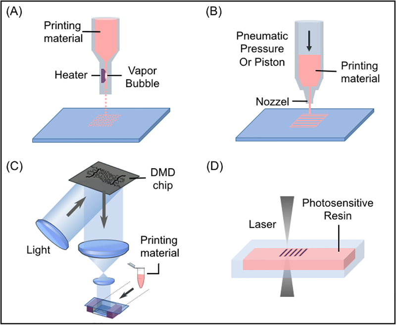
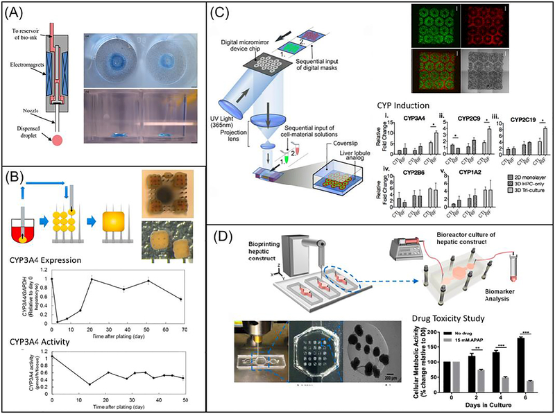
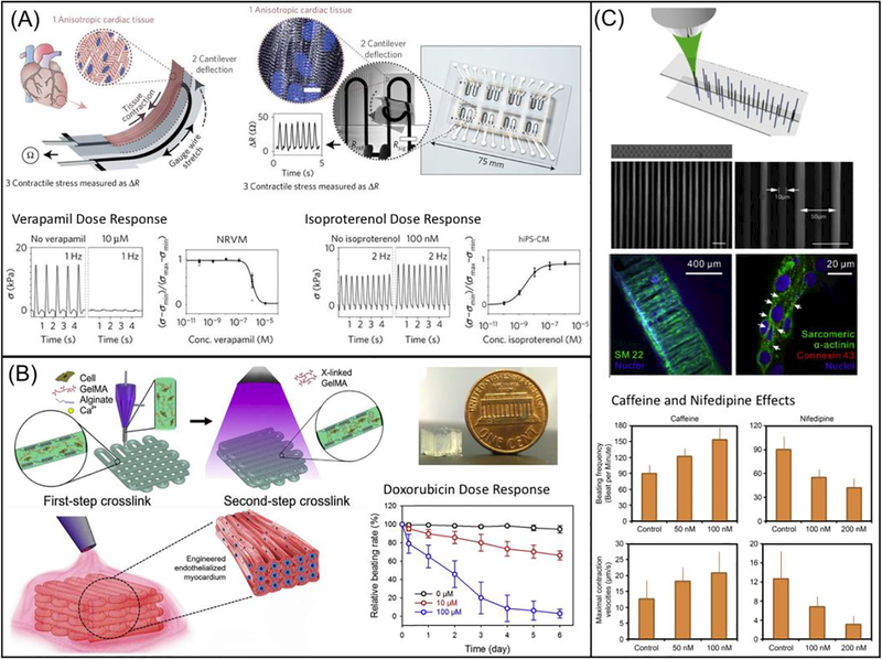
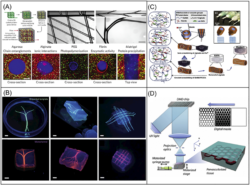
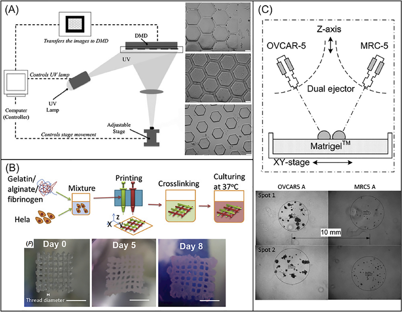
3D bioprinting is emerging as a promising technology for fabricating complex tissue constructs with tailored biological components and mechanical properties. Recent advances have enabled scientists to precisely position materials and cells to build functional tissue models for in vitro drug screening and disease modeling. This review presents state-of-the-art 3D bioprinting techniques and discusses the choice of cell source and biomaterials for building functional tissue models that can be used for personalized drug screening and disease modeling. In particular, we focus on 3D-bioprinted liver models, cardiac tissues, vascularized constructs, and cancer models for their promising applications in medical research, drug discovery, toxicology, and other pre-clinical studies.

Keywords: 3D printing; Biomaterials; Disease model; Drug screening; In vitro culture; Tissue engineering; Tissue model.


Conflict of interest statement

Declaration of interests

All authors declare no competing interests.

Figures

Fig. 1.
Fig. 1.
Schematic diagrams showing the printing approaches: (A) inkjet-based bioprinting systems, (B) extrusion-based bioprinting systems, (C) DLP-based bioprinting and (D) TPP-based bioprinting platforms.
Fig. 2.
Fig. 2.
3D bioprinting of liver tissue models: (A) Schematic diagram showing the inkjet-based printing setup (left) and bright field image showing the printed construct stained in blue (right). (Reprinted from: [196]) (B) Schematic diagram showing the process of building the construct from spheroids, with the top and side views of the construct on the top right. Plots showing expression (middle) and activity (bottom) of CYP3A4 over time (Reprinted from: [198]) (C) Schematic diagram showing the DLP-based bioprinting system, with the fluorescence and bright field images of 3D printed liver construct on the top right. Bar charts showing CYP enzyme induction on lower right. Scale bars are 500 μm. (Reprinted from:[126]) (D) Schematic diagram showing the direct 3D printing within microfluidic chip, with images showing the microfluidic setup and printed structure on lower left. Bar chart showing drug toxicity study on bottom right (Reprinted from:[199])
Fig. 3.
Fig. 3.
3D bioprinting of cardiac tissue models: (A) Schematic diagram showing the design of a multimaterial, patterned, piezoresistive stress sensor that aligns cardiomyocytes in a thick tissue and sense cardiac force output by changes in resistivity during contraction. Scale bar are 10 μm. Plots showing verapamil and isoproterenol dose response are on the bottom. (Reprinted from: [215]) (B) Schematic diagram showing the extrusion-based 3D printing system that generate multimaterial prints of cardiomyocytes and endothelial cells in naturally-based alginate and GelMA scaffold. The image of printed construct is shown on the top right and the doxorubicin dose response is shown on the lower right. (Reprinted from: [216]) (C) Schematic diagram showing TPP-based printing of micron-scale filaments, which was seeded with healthy and Long-QT iPSC-CMs. Fluorescence images in the middle showing the construct in bulk and cell alignment in various conditions. Bar charts on the bottom showing effects of caffeine and nifedipine on beating frequency and maximal contraction (Reprinted from: [217]).
Fig. 4.
Fig. 4.
3D bioprinting of vascularized tissue models. (A) Schematic of a sacrificial bioprinting method using an extrusion-based bioprinter and carbohydrate glass as the sacrificial template, with images showing multiscale structures on top middle and right. Scale bars are 1mm. Bottom cross-sectional fluorescence images showing lumen structures from a variety of cell-laden ECM materials. Scale bars are 200 μm. (Reprinted from: [219]). (B) Fluorescence images showing the bioprinted alginate templates (green) enclosed in GelMA hydrogels and the respective microchannels perfused with a fluorescent microbead suspension (pink) after removal of the alginate templates. Scale bars are 3mm. (Reprinted from: [221]). (C) Schematic of the direct vasculature printing with a multilayered coaxial extrusion-based bioprinter and a blend bioink with two independent crosslinking mechanisms. (Reprinted from: [17]). (D) Schematic of the DLP-based bioprinting system for the rapid printing of prevascularized 3D tissues with direct encapsulation of endothelial and supportive cells in a continuous fashion. (Reprinted from: [127]).
Fig. 5.
Fig. 5.
(A). Schematic diagram of a DLP-based system that uses a programmable DMD to selectively illuminate UV light onto photosensitive monomer solution. Bright-field images on the right showing HeLa cells-seeded PEGDA scaffolds with channels of 25-μm width (top), 45-μm width (middle), and 120-μm width (bottom). Scale bars are 100 μm. (Reprinted from: [240]). (B). Schematic process of an extrusion-based printing of gelatin/alginate/fibrinogen constructs with Hela cells to model cervical tumor. Bright field images on the bottom showing 3D printed Hela cell constructs on day 0, day 5 and day 8. Scale bar are 5 mm. (Reprinted from: [241]). (C). Schematic of an ejection printing platform composed of an automated stage and two nanoliter ejectors to dispense cancer cells (OVCAR-5) and fibroblasts (MRC-5). Bright-field image on the bottom showing 3D printed constructs with OVCAR-5 and MRC-5 cells. (Reprinted from: [149]).

References

    1. Murphy SV, Atala A, 3D bioprinting of tissues and organs., Nat. Biotechnol 32 (2014) 773–785. doi:10.1038/nbt.2958. - DOI - PubMed
    1. Stanton MM, Samitier J, Sánchez S, Bioprinting of 3D hydrogels, Lab Chip. 15 (2015) 3111–3115. doi:10.1039/C5LC90069G. - DOI - PubMed
    1. Irvine S, Venkatraman S, Bioprinting and differentiation of stem cells, Molecules. 21 (2016) 1188. doi:10.3390/molecules21091188. - DOI - PMC - PubMed
    1. Collins SF, Bioprinting is changing regenerative medicine forever, Stem Cells Dev 23 (2014) 79–82. doi:10.1089/scd.2014.0322. - DOI - PubMed
    1. Giannopoulos AA, Mitsouras D, Yoo S-J, Liu PP, Chatzizisis YS, Rybicki FJ, Applications of 3D printing in cardiovascular diseases, Nat. Rev. Cardiol 13 (2016) 701–718. doi:10.1038/nrcardio.2016.170. - DOI - PubMed

Publication types