

Fetal tracheal occlusion in mice: a novel transuterine method
- PMID: 29937007
- PMCID: PMC6387659
- DOI: 10.1016/j.jss.2018.04.028
Fetal tracheal occlusion in mice: a novel transuterine method
Abstract
Background: Fetal tracheal occlusion (TO) is an emerging surgical therapy in congenital diaphragmatic hernia that improves the fetal lung growth. Different animal models of congenital diaphragmatic hernia and TO present advantages and disadvantages regarding ethical issues, cost, surgical difficulty, size, survival rates, and available genetic tools. We developed a minimally invasive murine transuterine TO model, which will be useful in defining how TO impacts lung molecular biology, cellular processes, and overall lung physiology.
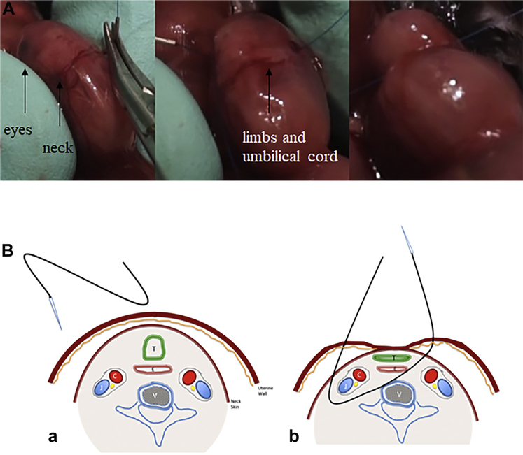
Materials and methods: Time-mated C57BL/6 mice underwent laparotomy at embryonic day 16.5 (E16.5) with transuterine TO performed on two fetuses in each uterine horn. At E18.5, dams were sacrificed and fetuses harvested. The lungs of the TO fetuses were compared with the nonmanipulated counterparts by morphometric and histologic analysis.
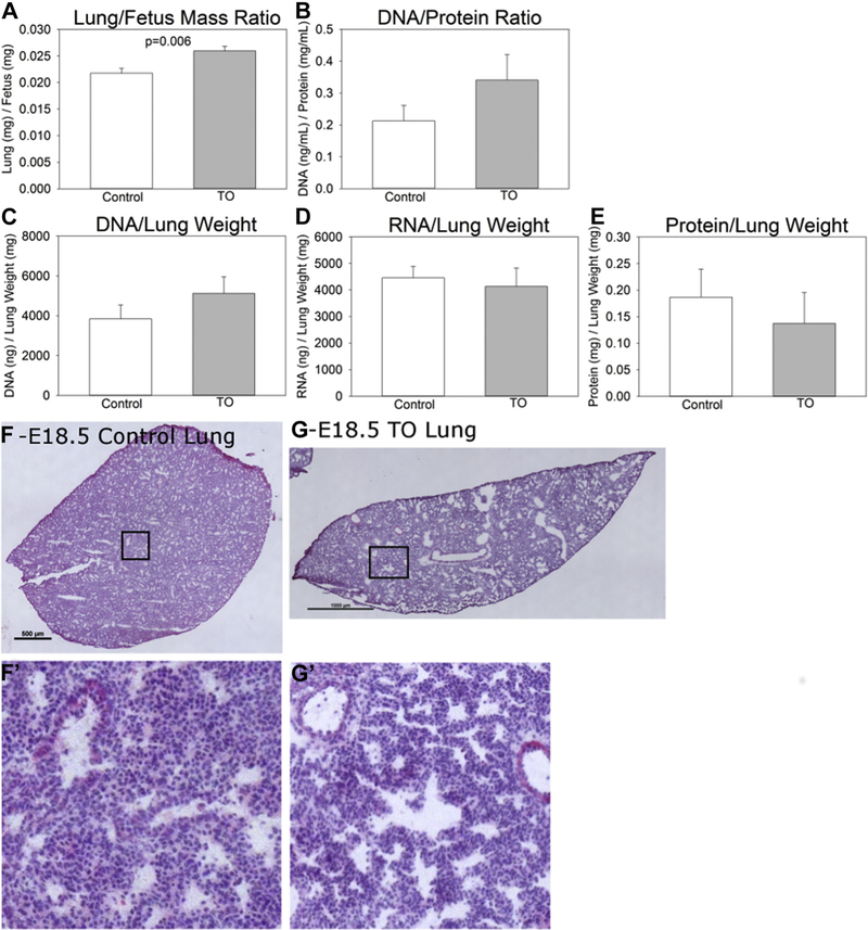
Results: Successful TO was confirmed in 16 of 20 TO fetuses. Twelve of them survived to E18.5 (75%). Fetal weights were comparable, but lung weights were significantly greater in TO (28.41 ± 5.87 versus 23.38 ± 3.09, P = 0.043). Lung to body weight ratio was also greater (0.26 ± 0.003 versus 0.22 ± 0.002, P = 0.006). E18.5 TO lungs demonstrated dilated central and distal airspaces with increased cellularity. DNA/protein and DNA/lung weight ratios were elevated while protein/lung weight ratio was lower in TO compared to control.
Conclusions: Mice fetal transuterine TO is feasible with comparable outcomes to other current animal models. The increase in the lung weight, lung to body weight ratio and the DNA/protein ratio indicate organized lung growth rather than edema or cell hypertrophy.
Keywords: CHAOS; Congenital diaphragmatic hernia; Fetal lung development; Fetal tracheal occlusion; Lung growth; Mice.
Copyright © 2018 Elsevier Inc. All rights reserved.
Conflict of interest statement
Financial disclosure: There is no financial disclosure to be declared.
Figures
References
-
- Beck V, Davey MG, Mayer S, et al. A longer tracheal occlusion period results in increased lung growth in the nitrofen rat model. Prenat Diagn. 2012;32:39–44. - PubMed
-
- Muensterer OJ, Nicola T, Farmer S, Harmon CM, Ambalavanan N. Temporary fetal tracheal occlusion using a gel plug in a rabbit model of congenital diaphragmatic hernia. J Pediatr Surg. 2012;47:1063–1066. - PubMed
-
- Kitano Y, Davies P, von Allmen D, Adzick NS, Flake AW. Fetal tracheal occlusion in the rat model of nitrofen-induced congenital diaphragmatic hernia. J Appl Physiol (1985). 1999;87:769–775. - PubMed
-
- Sananès N, Ruano R, Weingertner A-S, et al. Experimental fetal endoscopic tracheal occlusion in rhesus and cynomolgus monkeys: nonhuman primate models. J Matern Fetal Neonatal Med 2015;28:1822–1827. - PubMed
-
- Maltais F, Seaborn T, Guay S, Piedboeuf B. In vivo tracheal occlusion in fetal mice induces rapid lung development without affecting surfactant protein C expression. Am J Physiol Lung Cell Mol Physiol. 2003;284:L622–L632. - PubMed
MeSH terms
Grants and funding
LinkOut - more resources
Full Text Sources
Other Literature Sources
