. 2018 Jul 11;24(1):97-108.e4.
doi: 10.1016/j.chom.2018.05.009. Epub 2018 Jun 21.

Lipid Peroxidation Drives Gasdermin D-Mediated Pyroptosis in Lethal Polymicrobial Sepsis

Affiliations

Lipid Peroxidation Drives Gasdermin D-Mediated Pyroptosis in Lethal Polymicrobial Sepsis

Rui Kang et al. Cell Host Microbe. .

Abstract

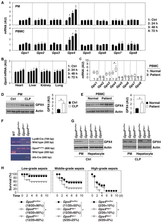
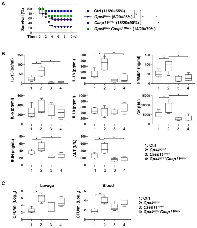
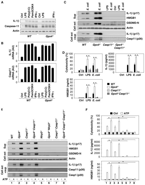
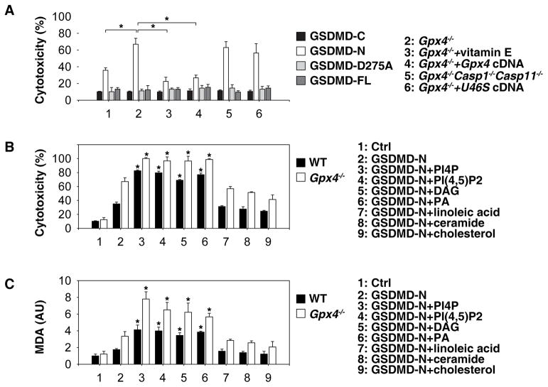
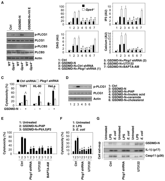
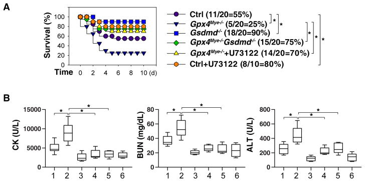
Sepsis is a life-threatening condition caused by pathogen infection and associated with pyroptosis. Pyroptosis occurs upon activation of proinflammatory caspases and their subsequent cleavage of gasdermin D (GSDMD), resulting in GSDMD N-terminal fragments that form membrane pores to induce cell lysis. Here, we show that antioxidant defense enzyme glutathione peroxidase 4 (GPX4) and its ability to decrease lipid peroxidation, negatively regulate macrophage pyroptosis, and septic lethality in mice. Conditional Gpx4 knockout in myeloid lineage cells increases lipid peroxidation-dependent caspase-11 activation and GSDMD cleavage. The resultant N-terminal GSDMD fragments then trigger macrophage pyroptotic cell death in a phospholipase C gamma 1 (PLCG1)-dependent fashion. Administration of the antioxidant vitamin E that reduces lipid peroxidation, chemical inhibition of PLCG1, or genetic Caspase-11 deletion or Gsdmd inactivation prevents polymicrobial sepsis in Gpx4-/- mice. Collectively, this study suggests that lipid peroxidation drives GSDMD-mediated pyroptosis and hence constitutes a potential therapeutic target for lethal infection.

Keywords: GPX4; GSDMD; caspase-11; ferroptosis; immunometabolism; inflammasome; lipid peroxidation; pyroptosis; sepsis.


Conflict of interest statement

Declaration of Interests

The authors declare no conflicts of interest or financial interests.

Figures

Fig. 1
Fig. 1. Gpx4 deficiency in myeloid cells results in increased susceptibility to polymicrobial infection
(A) Q-PCR analysis of Gpx1-8 mRNA in PMs or PBMCs from septic mice at 24–72 hours (n=3 mice/group; data are expressed as means ± SD, *, P<0.05 versus control group, t test). (A) Q-PCR analysis of Gpx1-8 mRNA in the indicated tissues from septic mice at 24–72 hours (n=3 mice/group; data are expressed as means ± SD, *, P<0.05 versus control group, t test). (C) Box plots comparing Gpx1-8 mRNA levels in PBMC samples from septic patients (n=16) and healthy controls (n=16). The mRNAs are presented as median value (black line), interquartile range (box), and minimum and maximum of all data (black line). *, P<0.05 versus control group, t test. (D) Western blot analysis of GPX4 protein expression in mouse PMs from control group or septic mice at 72 hours (n=3 mice/group; data expressed as means ± SD, *, P<0.05, t test). (E) Western blot analysis of GPX4 protein expression in human PBMCs or septic patients at hospital admission (n=3 patient/group; data expressed as means ± SD, *, P<0.05, t test). (F) Genotype identification of transgenic mice based on reverse transcription polymerase chain reaction. (G) Western blot analysis of GPX4 expression in PMs and hepatocytes from Gpx4 flox/flox, Gpx4Hep−/−, or Gpx4Mye−/− mice with or without CLP for 72 hours. (H) Mice with the indicated genotypes were subjected to CLP with syringe needles with gauges ranging from 27 (A, “low-grade sepsis”), 22 (B, “middle-grade sepsis”), to 17 (C, “high-grade sepsis”). Animal survival was assayed (n=20 mice/group; *, P<0.05, Kaplan-Meier survival analysis).
Fig. 2
Fig. 2. Effects of vitamin E and cell death inhibitors on sepsis in mice
(A) Analysis of animal survival in mice with or without repeated intraperitoneal administration of vitamin E (500 mg/kg) at three, 24, 48, and 72 hours after CLP (22-gauge needle)-induced sepsis (n=5–10 mice/group; *, P<0.05, Kaplan-Meier survival analysis). (B) In parallel, serum levels of MDA, 4-HNE, CK, BUN, and ALT were assayed at 72 hours after CLP (n=5 mice/group; *, P<0.05, ANOVA LSD test). Data are presented as median value (black line), interquartile range (box), and minimum and maximum of all data (black line). (C) Analysis of animal survival in mice with or without repeated intraperitoneal administration of Z-VAD-FMK (“ZVF”, 5 mg/kg), wedelolactone (“Wed”, 20 mg/kg), ferrostatin-1 (“Fer”, 10 mg/kg), or necrostatin-1 (“Nec”, 5 mg/kg) at three, 24, 48, and 72 hours after CLP (22-gauge needle)-induced sepsis (n=10 mice/group; *, P<0.05, Kaplan-Meier survival analysis). (D) In parallel, serum levels of IL-1β, IL-18, HMGB1, IL-6, IL-10, IL-17, and IL-12 were assayed at 72 hours after CLP (n=5 mice/group; *, P<0.05, ANOVA LSD test). Data are presented as median value (black line), interquartile range (box), and minimum and maximum of all data (black line).
Fig. 3
Fig. 3. Caspase-11-dependent pyroptosis mediates septic death in Gpx4Mye−/− mice
(A) Mice with the indicated genotypes were subjected to CLP with 22-gauge syringe needles (middle-grade sepsis) and animal survival was monitored (n=20 mice/group; *, P<0.05, Kaplan-Meier survival analysis). Gpx4Mye−/− mice data was the same control as Fig. 1H (middle-grade sepsis). (B) Analysis of serum levels of IL-1β, IL-18, HMGB1, IL-6, IL-10, CK, BUN, and ALT in middle grade septic mice at 72 hours (n=5 mice/group; *, P<0.05, ANOVA LSD test). Data are presented as median value (black line), interquartile range (box), and minimum and maximum of all data (black line). (C) Analysis of bacterial loading in middle grade septic mice at 72 hours (n=5 mice/group; *, P<0.05, ANOVA LSD test). Data are presented as median value (black line), interquartile range (box), and minimum and maximum of all data (black line).
Fig. 4
Fig. 4. GPX4 blocks GSDMD cleavage during inflammasome activation
(A) Western blot analysis of IL-1β and caspase-11 protein expression in BMDMs following treatment with LPS (500 ng/ml), poly (I:C) (5 μg/ml), Pam3CSK4 (1 μg/ml), or IFN-γ (10/ng/ml) for six hours. (B) Q-PCR analysis of IL-1β and caspase-11 mRNA expression in BMDMs following treatment with LPS (500 ng/ml), poly (I:C) (5 μg/ml), Pam3CSK4 (1 μg/ml), or IFN-γ (10/ng/ml) for six hours. (C) Western blot analysis of indicated proteins in BMDMs recovered from mice with the indicated genotypes following LPS electroporation or E. coli (MOI=25) infection for 16 hours. Sup= supernatants. (D) In parallel, cytotoxicity (LDH release) and levels of IL-1β and HMGB1 in the supernatant were assayed (n=3 wells/group; data expressed as means ± SD, *, P<0.05, ANOVA LSD test). (E) Western blot analysis of LPS (500 ng/ml)-primed indicated BMDMs following treatment with ATP (5 mM) for one hour. Sup=supernatants. (F) In parallel, cytotoxicity and levels of IL-1β and HMGB1 in the supernatant were assayed (n=3 wells/group; data expressed as means ± SD, *, P<0.05, ANOVA LSD test).
Fig. 5
Fig. 5. Oxidation of phospholipids promote GSDMD-N-mediated pyroptosis
(A) WT and Gpx4−/− BMDMs were pretreated with vitamin E (100 μM) for three hours before transfection with GSDMD-C, GSDMD-N, GSDMD-D275A, or GSDMD-FL expression constructs. Cell viability was assayed within 24 hours of transfection (n=3 wells/group; means ± SD, *, P<0.05, t test). (B) WT and Gpx4−/− BMDMs were pretreated with indicated lipid components (20 μM) for three hours, and then transfected with GSDMD-N. Cell viability was assayed within 24 hours of transfection (n=3 wells/group; means ± SD, *, P<0.05 versus GSDMD-N group, t test). (C) WT and Gpx4−/− BMDMs were pretreated with indicated lipid components (20 μM) for three hours and then transfected with GSDMD-N. Intracellular MDA level was assayed within 24 hours of transfection (n=3 wells/group; means ± SD, *, P<0.05 versus GSDMD-N group, t test).
Fig. 6
Fig. 6. PLC activation contributes to GSDMD-mediated pyroptosis
(A) WT and Gpx4−/− BMDMs were pretreated with or without vitamin E (100 μM) for three hours, and then transfected with GSDMD-N. Protein expression was assayed using western blot. (B) Analysis of GSDMD-N-mediated cytotoxicity, DAG, IP3, and calcium production at 24 hours in WT and Gpx4−/− BMDMs in the absence or presence of Plcg1-shRNA, U73122 (10 μM), or BAPTA-AM (10 μM) (n=3 wells/group; data expressed as means ± SD, *, P<0.05 versus GSDMD-N group, t test). (C) Analysis of GSDMD-N-mediated cytotoxicity at 24 hours in THP1, HL-60, and HeLa cell lines (n=3 wells/group; data expressed as means ± SD, *, P<0.05, t test). (D) WT BMDMs were pretreated with indicated lipid components (20 μM) for three hours, and then transfected with GSDMD-N for 24 hours. Protein expression was assayed using western blot. (E) Analysis of GSDMD-N/PI4P- or GSDMD-N/PI(4,5)P2-mediated cytotoxicity at 24 hours in BMDMs in the absence or presence of Plcg1-shRNA, U73122 (10 μM), or BAPTA-AM (10 μM) (n=3 wells/group; data expressed as means ± SD, *, P<0.05 versus control group, t test). (F) Analysis of LPS electroporation- or E. coli (MOI=25) infection-mediated cytotoxicity at 16 hours in BMDMs in the absence or presence of Plcg1-shRNA or U73122 (10 μM) (n=3 wells/group; data expressed as means ± SD, *, P<0.05 versus control group, t test). (G) Analysis of LPS electroporation- or E. coli (MOI=25) infection-mediated protein expression at 16 hours in BMDMs in the absence or presence of Plcg1-shRNA or U73122 (10 μM).
Fig. 7
Fig. 7. PLC activation contributes to GSDMD-mediated septic death
(A) Analysis of animal survival in mice with or without repeated intraperitoneal administration of U73122 (30 mg/kg) at three, 24, 48, and 72 hours after CLP (22-gauge needle)-induced sepsis (n=10–20 mice/group; *, P<0.05, Kaplan-Meier survival analysis). Gpx4Mye−/− mice data was the same control as Fig. 1H (middle-grade sepsis). (B) In parallel, quantitation of serum CK, BUN, and ALT in middle grade septic mice at 72 hours (n=5–8 mice/group; *, P<0.05, ANOVA LSD test). Data are presented as median value (black line), interquartile range (box), and minimum and maximum of all data (black line).

Comment in

References

    1. Aachoui Y, Leaf IA, Hagar JA, Fontana MF, Campos CG, Zak DE, Tan MH, Cotter PA, Vance RE, Aderem A, et al. Caspase-11 protects against bacteria that escape the vacuole. Science. 2013;339:975–978. - PMC - PubMed
    1. Aglietti RA, Estevez A, Gupta A, Ramirez MG, Liu PS, Kayagaki N, Ciferri C, Dixit VM, Dueber EC. GsdmD p30 elicited by caspase-11 during pyroptosis forms pores in membranes. Proc Natl Acad Sci U S A. 2016;113:7858–7863. - PMC - PubMed
    1. Andujar I, Rios JL, Giner RM, Miguel Cerda J, del Recio MC. Beneficial effect of shikonin on experimental colitis induced by dextran sulfate sodium in BALB/c mice. Evid Based Complement Alternat Med. 2012;2012:271606. - PMC - PubMed
    1. Boyum A. Isolation of mononuclear cells and granulocytes from human blood. Isolation of monuclear cells by one centrifugation, and of granulocytes by combining centrifugation and sedimentation at 1 g. Scand J Clin Lab Invest Suppl. 1968;97:77–89. - PubMed
    1. Brigelius-Flohe R, Maiorino M. Glutathione peroxidases. Biochimica et biophysica acta. 2013;1830:3289–3303. - PubMed

Publication types

MeSH terms