. 2018 Sep 4;28(3):449-462.e5.
doi: 10.1016/j.cmet.2018.05.026. Epub 2018 Jun 21.

The Drosophila Immune Deficiency Pathway Modulates Enteroendocrine Function and Host Metabolism

Affiliations

The Drosophila Immune Deficiency Pathway Modulates Enteroendocrine Function and Host Metabolism

Layla Kamareddine et al. Cell Metab. .

Abstract

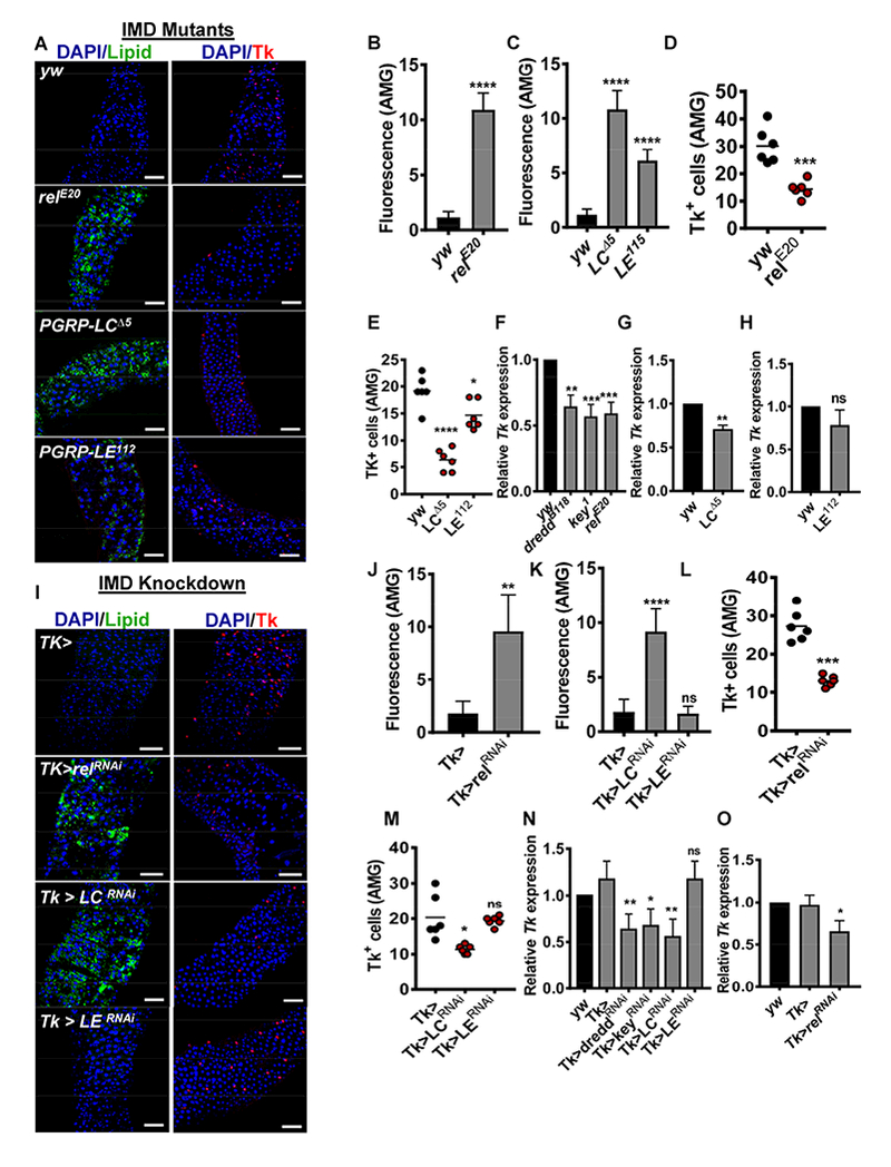
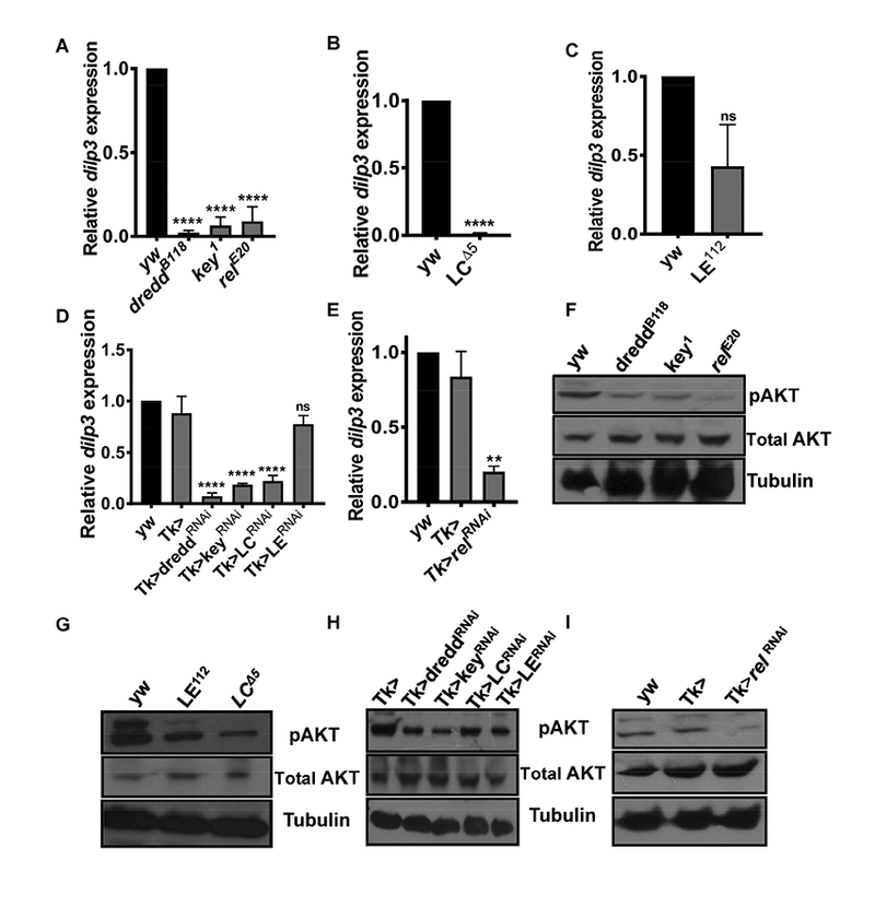
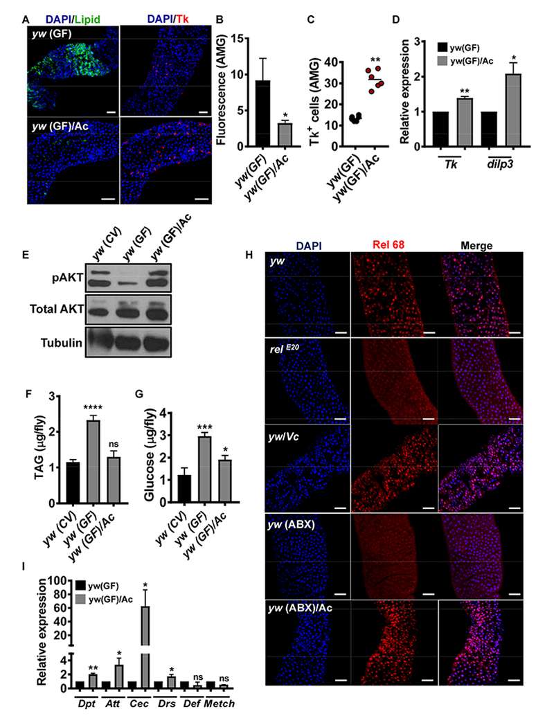
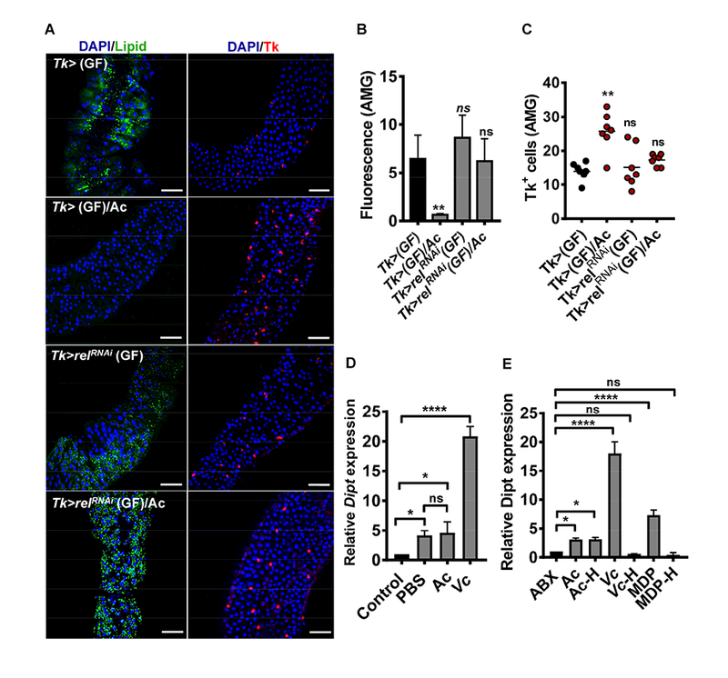
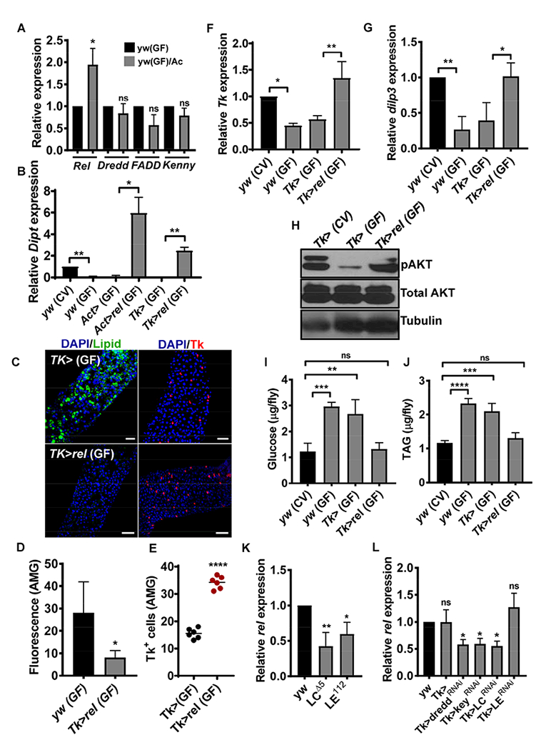
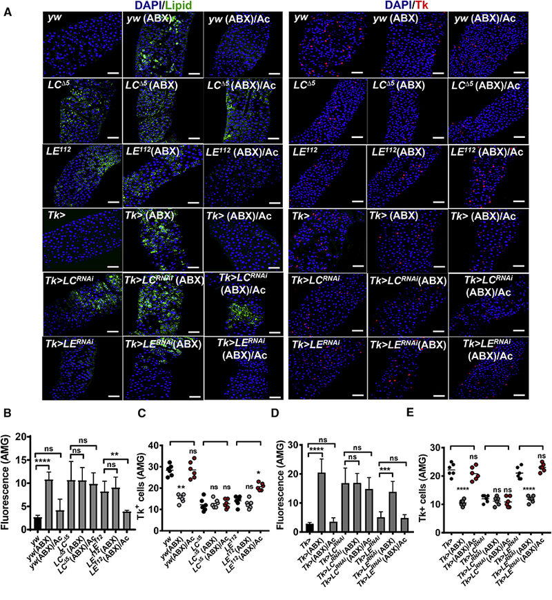
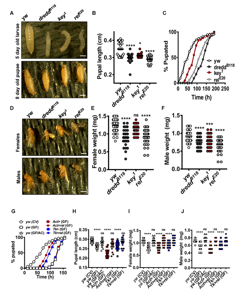
Enteroendocrine cells (EEs) are interspersed between enterocytes and stem cells in the Drosophila intestinal epithelium. Like enterocytes, EEs express components of the immune deficiency (IMD) innate immune pathway, which activates transcription of genes encoding antimicrobial peptides. The discovery of large lipid droplets in intestines of IMD pathway mutants prompted us to investigate the role of the IMD pathway in the host metabolic response to its intestinal microbiota. Here we provide evidence that the short-chain fatty acid acetate is a microbial metabolic signal that activates signaling through the enteroendocrine IMD pathway in a PGRP-LC-dependent manner. This, in turn, increases transcription of the gene encoding the endocrine peptide Tachykinin (Tk), which is essential for timely larval development and optimal lipid metabolism and insulin signaling. Our findings suggest innate immune pathways not only provide the first line of defense against infection but also afford the intestinal microbiota control over host development and metabolism.

Keywords: Drosophila insulin-like peptide 3; PGRP-LC; enteroendocrine cell; enteroendocrine peptide; immune deficiency pathway; innate immunity; lipid droplet; metabolism; short-chain fatty acid; tachykinin.


Conflict of interest statement

Declaration of Interests: The authors declare no competing interests.

Figures

Figure 1:
Figure 1:. Enteroendocrine IMD pathway signaling regulates intestinal lipid droplet accumulation and Tk expression.
(A) Representative fluorescence images showing DAPI and Bodipy staining (DAPI/Lipid) or DAPI staining and Tachykinin immunofluorescence (DAPI/Tk) in the anterior midgut of control (yw) and IMD pathway mutant (relE20, PGRP-LCΔ5, and PGRP-LE112) flies. Ten intestines were examined for each genotype. Scale bar, 50 pm. Quantification of fluorescing cells in the anterior midgut of the indicated control or mutant flies after (B-C) Incubation with a fluorescent Bodipy dye (D-E) incubation with an anti-tachykinin antibody and a fluorescent secondary antibody (Tk+ cells). (F-H) Quantification of Tk transcription by qRT-PCR in the intestines of control (yw) or mutant flies (dreddB118, key1, relE20 relE20, PGRP-LCΔ5, and PGRP-LE112) flies. (I) Representative fluorescence images showing DAPI and Bodipy staining (DAPI/Lipid) or DAPI staining and Tachykinin immunofluorescence (DAPI/Tk) in the anterior midgut of driver-only control (Tk>) or rel (Tk>relRNAi), PGRP-LC (Tk>LCRNAi) and PGRP-LE (Tk>LERNAi) knockdown flies. Scale bars, 50 pm. Ten intestines were examined for each genotype. Quantification of fluorescing cells in the anterior midgut of the indicated control or knockdown flies after (J-K) Incubation with a fluorescent Bodipy dye (L-M) incubation with an anti-tachykinin antibody and a fluorescent secondary antibody (Tk+ cells). (N-O) Quantification of Tk transcription by qRT-PCR in the intestines of control or RNAi knockdown flies as indicated. An unpaired t test (B,D,J), a one-way ANOVA with Dunnett’s multiple comparison test (C,E,F,K,N,O), a Welch’s t test (G,H,L), and a Kruskal-Wallis with Dunn’s multiple comparisons test (M) was used to evaluate statistical significance.*p<0.05,**p<0.01, ***p<0.001,****p<0.0001. ns not significant. See also Fig S1–2 and Tables S1–S5.
Figure 2:
Figure 2:. IMD pathway signaling in EEs controls Tk transcription, dilp3 transcription, and insulin signaling.
(A-C) Quantification of dilp3 transcription by qRT-PCR in the intestines of control or IMD pathway mutant flies with the indicated genotypes. (D-E) Quantification of dilp3 transcription by qRT-PCR in the intestines of control or RNAi knockdown flies as indicated. A Welch’s t-test (B,C) or a one-way ANOVA with Dunnett’s multiple comparison test (A,D,E) was used to evaluate statistical significance. *p<0.05, **p<0.01, ***p<0.001, ****p<0.0001. ns not significant. (F-I) Western blot analysis of total and phosphorylated AKT (pAKT) levels in flies with the indicated genotypes. Samples were prepared from ten whole flies. Tubulin was used as a loading control. See also Fig S3–4 and Table S5.
Figure 3:
Figure 3:. Dietary acetate activates IMD pathway signaling and restores metabolic homeostasis to axenic flies.
(A) Representative fluorescence images showing DAPI staining and Bodipy staining (DAPI/Lipid) and Tachykinin immunofluorescence (DAPI/Tk) in the anterior midgut of axenic (GF) control flies (yw) fed fly food alone or supplemented with 50mM acetate (Ac). Ten intestines were examined for each genotype. Scale bar, 50 pm. Quantification of fluorescing cells in the anterior midgut of the indicated flies (B) Incubated with a fluorescent Bodipy dye (C) incubated with an antitachykinin antibody and a fluorescent secondary antibody (Tk+ cells). (D) Quantification of Tk and dilp3 transcription by qRT-PCR in the intestines of axenic flies treated as in (A). Measurements were normalized to transcript levels of unsupplemented flies and represent the mean of three independent biological replicates. Error bars represent the standard deviation. Ten intestines were used for each replicate. (E) Western blot analysis of total and phosphorylated AKT (pAKT) levels in conventionally raised (CV) and axenic (GF) flies treated as indicated in (A). Ten flies were used for preparation of each sample. Tubulin was used as a loading control. (F-G) Quantification of systemic triglycerides and glucose levels in conventionally (CV) raised and axenic (GF) flies treated as indicated in (A). Five flies were used in each of three independent biological replicates. Measurement represents the mean, and error bars represent the standard deviation. yw (CV) was used as the reference to assess statistical significance. (H) Representative immunofluorescence images of the intestines of flies obtained using an antibody that recognizes the Rel 68 cleavage product and stained with DAPI. Control (yw) or relE20 mutants were examined. Flies were raised conventionally and examined directly (CV) or after antibiotic treatment (ABX). Additional treatments included dietary acetate supplementation (Ac) and oral infection with a quorum sensing-competent V. cholerae strain (Vc). Ten intestines were examined for each condition. Scale bar, 50 pm. (I) Quantification of Diptericin (Dpt), Attacin (Att), Cecropin (Cec), Drosocin (Drs), Defensin (Def), and Metchnikovin (Metch) transcription by qRT-PCR in the intestines of flies treated as noted in (A). Measurements were normalized to the transcript levels of flies that were not administered acetate. Ten intestines were used for each replicate. Measurements represent the mean of three independent biological replicates. Error bars represent the standard deviation. A Welch’s t test (B,C,D,I) or a one-way ANOVA followed by Dunnett’s multiple comparisons test (F,G) was used to assess statistical significance.*p<0.05, **p<0.01 ***p<0.001, ****p<0.0001. ns not significant. See also Figure S5 and Table S5.
Figure 4:
Figure 4:. IMD pathway knockdown in EE prevents the metabolic and transcriptional response of axenic flies to acetate.
(A) Representative fluorescence images showing DAPI staining and Bodipy staining (DAPI/Lipid) and Tachykinin immunofluorescence (DAPI/Tk) in the anterior midgut of axenic (GF) driver-only flies (Tk>) or Tk-expressing EE-specific rel knockdown (Tk>re/RNAi) flies fed fly food alone or supplemented with 50mM acetate (Ac). Ten intestines were examined for each genotype. Scale bar, 50 pm. Quantification of fluorescing cells in the anterior midgut of the indicated flies (B) Incubated with a fluorescent Bodipy dye (C) incubated with an anti-tachykinin antibody and a fluorescent secondary antibody (Tk+ cells). (D) Quantification of Diptericin (Dipt) transcription by qRT-PCR in control, PBS or acetate supplemented, and V. cholera infected flies. Acetate, PBS, and V. cholerae were introduced into the hemolymph by septic injury. Measurements were normalized to transcript levels of control flies. Measurements represent the mean of three independent biological replicates. Error bars represent the standard deviation. Ten flies were used for each replicate. (E) Quantification of Diptericin (Dipt) transcription by qRT-PCR in flies administered Antibiotic (ABX), Acetate (Ac), acid hydrolyzed Acetate (Ac-H), Vibrio cholerae (Vc), acid hydrolyzed Vc (Vc-H), muramyl dipeptide (MDP), or acid hydrolyzed MDP (MDP-H). Measurements were normalized to transcript levels of ABX treated flies. Measurements represent the mean of three independent biological replicates. Error bars represent the standard deviation. Ten flies were used for each replicate. A one-way ANOVA followed by a Dunnett’s test (B,C,D,E) was used to calculate significance.*p<0.05, **p<0.01,****p<0.0001. ns not significant. See also Table S5.
Figure 5:
Figure 5:. Constitutive rel transcription in Tk-expressing EEs is sufficient to restore metabolic homeostasis to axenic flies.
(A) Quantification of transcription of the indicated IMD pathway components by qRT-PCR in the intestines of axenic flies (GF) with or without dietary supplementation with 50 mM acetate (Ac). (B) Quantification of Diptericin (Dipt) transcription by qRT-PCR in the intestines of conventional (CV) or axenic (GF) control flies (yw), driver only flies (Act> and Tk>), or flies expressing full length rel either ubiquitously (Act>rel) or in Tk-expressing EE (Tk>rel). Measurements were normalized to transcript levels of conventionally-raised control flies. Measurements represent the mean of three independent biological replicates. Error bars represent the standard deviation. Ten intestines were used for each replicate. (C) Representative fluorescence images of DAPI staining and Bodipy staining (DAPI/Lipid) and Tachykinin (Tk) immunofluorescence (DAPI/Tk) in the anterior midgut of axenic driver-only control (Tk>) flies or flies with rel constitutively transcribed in Tk-expressing EEs (Tk>Rel). Scale bar, 50 pm. Ten intestines were examined for each genotype. Quantification of fluorescing cells in the anterior midgut of driver only flies (Tk>) or flies expressing full length rel in Tk-expressing EE (Tk>rel) (D) Incubated with a fluorescent Bodipy dye (E) Incubated with an anti-tachykinin antibody and a fluorescent secondary antibody (Tk+ cells). (F-G) Transcript levels of Tk and dilp3 measured by qRT-PCR in the intestines of conventionally-raised (CV) or axenic flies (GF) with the genotypes described in (B). Measurements were normalized to those of conventionally raised yw flies and represent the mean of three independent biological replicates. Ten intestines were used for each replicate. Error bars represent the standard deviation. (H) Western blot analysis of total and phosphorylated AKT (pAKT) levels in conventionally raised (CV) or axenic (GF) flies with the genotypes as noted in (B). Samples were prepared from ten flies for each condition. Tubulin was used as a loading control. (I-J) Quantification of systemic triglycerides and glucose levels in conventionally raised (CV) or axenic (GF) flies with the genotypes described in (B). Five flies were used for each of three biological replicates. Measurements represent the mean, and error bars represent the standard deviation. Quantification of rel transcription by qRT-PCR in the intestines of (K) control (yw) and IMD pathway mutants PGRP-LCΔ5 (LCΔ5), PGRP-LE112 (LE112) (L) EE-specific driver-only control (Tk>), or dredd (Tk>dreddRNAi), key (Tk>keyRNAi), PGRP-LC (Tk>PGRP-LCRNAi), PGRP-LE and (Tk>PGRP-LERNAi) knockdown flies. For pooled data representing three biological replicates, the mean and SD are shown. Ten intestines were used for each biological replicate. A Welch’s t test (A,B,D,F,G), an unpaired t test (E), or a one-way ANOVA followed by Dunnett’s multiple comparisons test (I,J,K,L) was used to assess statistical significance.*p<0.05, **p<0.01 ***p<0.001, ****p<0.0001. ns not significant. See also Table S5.
Figure 6:
Figure 6:. Acetate functions upstream of PGRP-LC to activate the IMD pathway.
(A) Representative fluorescence images showing DAPI staining and Bodipy staining (DAPI/Lipid) and Tachykinin immunofluorescence (DAPI/Tk) in the anterior midgut of conventionally-raised flies. Where noted, flies were treated with antibiotics (ABX) and acetate (Ac). The following genotypes were examined: control (yw), PGRP-LCΔ5 (LCΔ5), and PGRP-LE112 (LE112) mutant flies as well as Tk> driver only flies, Tk>PGRP-LCRNAi (Tk>LCRNAi), and Tk> PGRP-LERNAi (Tk>LERNAi). Ten intestines were examined for each genotype. Scale bar, 50 pm. Quantification of fluorescing cells in the anterior midgut of the indicated flies (B-C) Incubated with a fluorescent Bodipy dye (D-E) incubated with an anti-tachykinin antibody and a fluorescent secondary antibody (Tk+ cells). A one-way ANOVA followed by Dunnett’s multiple comparisons test (B,D) or a Kruskal-Wallis with Dunn’s multiple comparisons test (C,E) was used to assess statistical significance. **p<0.01 ***p<0.001, ****p<0.0001. ns not significant. See also Figure S6.
Figure 7:
Figure 7:. IMD pathway activation in EEs is essential for normal development.
(A) Representative images of 5 and 8 day old larvae and pupae, respectively. (B) Quantification of pupal lengths of control (yw) and IMD pathway mutant (dreddB118, key1, and relE20). Horizontal bars represent the mean measurement. (C) Time to larval pupation in hours (h) for yw control and noted IMD pathway mutant flies. (D) Representative images of five-day old control (yw) and IMD pathway mutant adult female and male flies. (E-F) Quantification of the body weight of five-day old control (yw) and IMD pathway mutant adult female and male flies. Horizontal bars represent the mean measurement. (G) Time to larval pupation in hours (h) of axenic control flies (yw), driver only flies (Act> and Tk>), flies expressing full length rel either ubiquitously (Act>rel) or in Tk-expressing EE (Tk>rel). Flies were either fed fly food alone or supplemented with acetate (Ac). (H) Quantification of pupal lengths of flies with genotype and treatment as indicated in (G). Horizontal bars represent the mean measurement. (I-J) Quantification of the body weight of five-day old adult female and male flies with genotype and treatment as indicated in (G). Horizontal bars represent the mean measurement. A Kruskal-Wallis with Dunn’s multiple comparisons test (B,E,F,H,I,J) was used to calculate statistical significance.**p<0.01 ***p<0.001, ****p<0.0001. ns not significant. See also Figure S7.

Similar articles

Cited by

References

    1. Aggrawal K, and Silverman N (2007). Peptidoglycan recognition in Drosophila. Biochem Soc Trans 35, 1496–1500. - PubMed
    1. Amcheslavsky A, Song W, Li Q, Nie Y, Bragatto I, Ferrandon D, Perrimon N, and Ip YT (2014). Enteroendocrine cells support intestinal stem-cell-mediated homeostasis in Drosophila. Cell Rep 9, 32–39. - PMC - PubMed
    1. Berkey CD, Blow N, and Watnick PI (2009). Genetic analysis of Drosophila melanogaster susceptibility to intestinal Vibrio cholerae infection. Cell Microbiol 11, 461–474. - PMC - PubMed
    1. Bogunovic M, Dave SH, Tilstra JS, Chang DT, Harpaz N, Xiong H, Mayer LF, and Plevy SE (2007). Enteroendocrine cells express functional Toll-like receptors. Am J Physiol Gastrointest Liver Physiol 292, G1770–1783. - PMC - PubMed
    1. Bohni R, Riesgo-Escovar J, Oldham S, Brogiolo W, Stocker H, Andruss BF, Beckingham K, and Hafen E (1999). Autonomous control of cell and organ size by CHICO, a Drosophila homolog of vertebrate IRS1–4. Cell 97, 865–875. - PubMed

Publication types

MeSH terms