. 2018 May 1;119(2):182-192.
doi: 10.1038/s41416-018-0119-5.

Interaction of WBP2 with ERα increases doxorubicin resistance of breast cancer cells by modulating MDR1 transcription

Affiliations

Interaction of WBP2 with ERα increases doxorubicin resistance of breast cancer cells by modulating MDR1 transcription

Shuai Chen et al. Br J Cancer. .

Abstract

Background: Surgery combined with new adjuvant chemotherapy is the primary treatment for early stage invasive and advanced stage breast cancer. Growing evidence indicates that patients with ERα-positive breast cancer show poor response to chemotherapeutics. However, ERα-mediated drug-resistant mechanisms remain unclear.

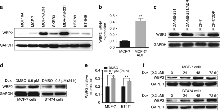
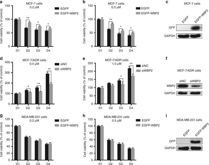
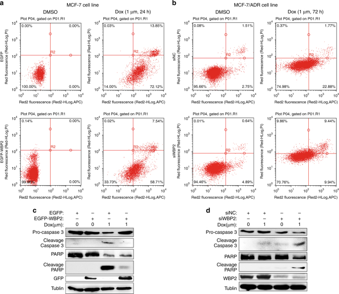
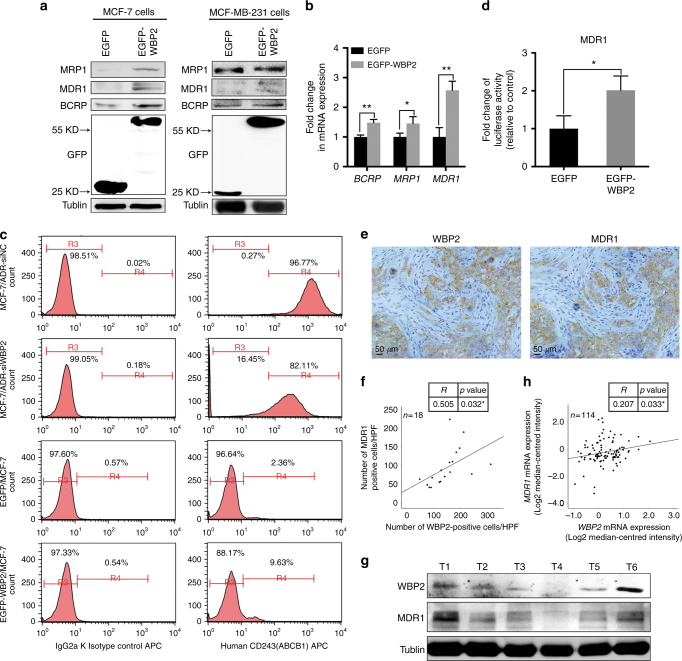
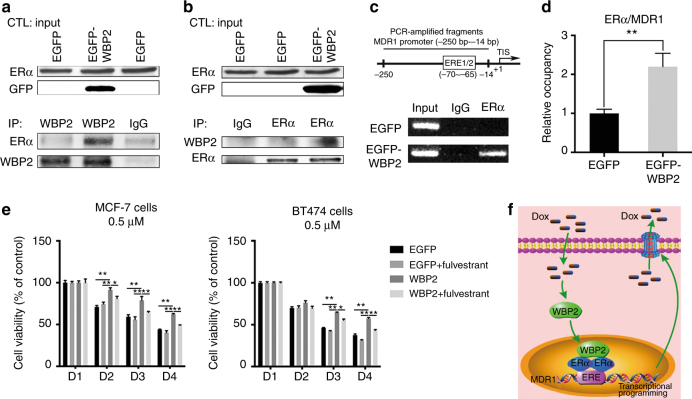
Methods: Levels of WW domain-binding protein 2 (WBP2) and drug-resistant gene were determined by western blotting and RT-PCR, respectively. Cell viability was measured by preforming MTT assay. CD243 expression and apoptosis rate were evaluated by flow cytometry. Interactions of WBP2/ERα and ERα/MDR1 were detected by co-immunoprecipitation and chromatin immunoprecipitation (ChIP) assay, respectively.

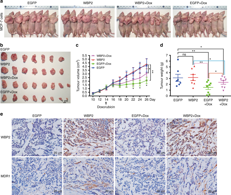
Results: There was an intrinsic link between WBP2 and ERα in drug-resistant cancer cells. Upregulation of WBP2 in MCF7 cells increased the chemoresistance to doxorubicin, while RNAi-mediated knockdown of WBP2 in MCF7/ADR cells sensitised the cancer cells to doxorubicin. Further investigation in in vitro and in vivo models demonstrated that WBP2 expression was directly correlated with MDR1, and WBP2 could directly modulate MDR1 transcription through binding to ERα, resulting in increased chemotherapy drug resistance.

Conclusions: Our finding provides a new mechanism for the chemotherapy response of ERα-positive breast tumours, and WBP2 might be a key molecule for developing new therapeutic strategies to treat chemoresistance in breast cancer patients.


Conflict of interest statement

The authors declare no competing interests.

Figures

Fig. 1
Fig. 1
Correlation between WBP2 and doxorubicin resistance in ER-positive breast cancer cells. a Protein levels of WBP2 in normal breast epithelial cells (MCF10A) and breast cancer cells (MCF-7, MCF-7/ADR, SKBR3, MDA-MB-231, HS578t and BT-549). b Relative expression of WBP2 mRNA in MCF-7 and MCF-7/ADR cells. c Protein levels of WBP2 in ER-negative breast cancer cells (MDA-MB-231 and MDA-MB-231/ADR) and ER-positive breast cancer cells (MCF-7 and MCF-7/DDP). Differential protein expression (d) and mRNA expression (e) of WBP2 in MCF-7 and BT474 cells treated with DMSO or 0.5 μM doxorubicin for 24 h. f Differential protein expression of WBP2 in MCF-7 and BT474 cells treated with 0.2 μM doxorubicin for 0, 24, 48 and 72 h. Dox indicates doxorubicin. **P < 0.01
Fig. 2
Fig. 2
Effects of WBP2 on cell viability in doxorubicin-treated MCF-7 and MCF-7/ADR cell viability. Measurement of cell viability using MTT assay in control MCF-7 cells and WBP2-overexpressing MCF-7 cells treated with 0.2 μM (a) and 0.5 μM (b) doxorubicin for 0, 24, 48 and 72 h. c WBP2 overexpression efficiency was verified by performing western blotting in MCF-7 cells. Measurement of cell viability using MTT assay in MCF-7/ADR cells and RNAi-mediated WBP2 knockdown in MCF-7/ADR cells treated with 0.5 μM (d) and 1.0 μM (e) doxorubicin for 0, 24, 48 and 72 h. f Validation of WBP2 knockdown efficiency in MCF-7/ADR cells. Test of cell viability through MTT assay in control MDA-MB-231 cells and WBP2-overexpressing MDA-MB-231 cells treated with 0.2 μM (g) and 0.5 μM (h) doxorubicin for 0, 24, 48 and 72 h. i Validation of WBP2 overexpression efficiency in MDA-MB-231 cells. EGFP, control MCF-7 cells; EGFP-WBP2, WBP2-overexpressing MCF-7 cells; siNC, control MCF-7/ADR cells; siWBP2, RNAi-mediated WBP2 knockdown in MCF-7/ADR cells. *P < 0.05, **P < 0.01
Fig. 3
Fig. 3
Role of WBP2 in doxorubicin-induced cell apoptosis in MCF-7 and MCF-7/ADR cells. a Cell apoptosis analysis utilising flow cytometry in control MCF-7 cells and WBP2-overexpressing MCF-7 cells treated with 1.0 μM doxorubicin for 24 h. b Detection of cell apoptosis utilising flow cytometry in MCF-7/ADR cells and RNAi-mediated WBP2 knockdown in MCF-7/ADR cells treated with 1.0 μM doxorubicin for 72 h. c Cell lysates from the control MCF-7, control MCF-7-WBP2, drug-treated MCF-7 and drug-treated MCF-7-WBP2 cells were separated by 8–12% SDS-PAGE, blotted and probed with antibodies against caspase-3, PARP, cleaved PARP, GFP and WBP2. Tubulin served as the internal control. d The expression of above four antibodies (caspase-3, PARP, cleaved PARP and WBP2) were also detected in the control MCF-7/ADR, control MCF-7/ADR-siWBP2, drug-treated MCF-7/ADR and drug-treated MCF-7/ADR-siWBP2 cells by western blotting. Tubulin served as the internal control. EGFP, control MCF-7 cells; EGFP-WBP2, WBP2 stable expression MCF-7 cells; siNC, control MCF-7/ADR cells; siWBP2, RNAi-mediated WBP2 knockdown in MCF-7/ADR cells
Fig. 4
Fig. 4
Drug resistance of WBP2 to doxorubicin in nude mice bearing breast cancer tumours. a All mice were killed by cervical dislocation and imaged. b The tumours of mice from the four groups were harvested and imaged. c The tumour growth curve of xenograft mice after initial cell injection and drug treatment. d The differences of tumour weight among the four groups. e IHC staining of tumours with WBP2 and MDR1 antibodies in the four groups. Scale bar, 1 cm. ns no significant. *P < 0.05, **P < 0.01
Fig. 5
Fig. 5
Effects of WBP2 on the expression of MDR1 in ER-positive MCF-7 cells. a Cell lysates from control MCF-7 and WBP2-overexpressing MCF-7 cells were separated by 10% SDS-PAGE, blotted and probed with antibodies against MRP1, MDR1, BCRP and GFP. Tubulin served as the internal control. b Transcription levels of MRP1, MDR1 and BCRP were tested by using real-time quantitative PCR. GAPDH served as the internal control. c Detection of MDR1 expression in MCF-7/ADR-siNC, MCF-7/ADR-siWBP2, control MCF-7 and WBP2-overexpressing MCF-7 cells by indirect fluorescence staining and flow cytometry analysis. d Luciferase activity of MDR1-reporter gene in control MCF-7 and WBP2 stable expression MCF-7 cells. e IHC staining with WBP2 and MDR1 antibodies in clinical breast cancer specimens. f Correlation analysis between the number of WBP2-positive cells and MDR1-expressed cells. Averages of five HPFs per sample was used to evaluate the Pearson correlation (n = 18). R indicates Pearson’s coefficient. g The protein levels of WBP2 and MDR1 in clinical breast cancer specimens were detected by western blotting. h Correlation analysis between WBP2 and MDR1 expression in chemotherapy treatment patients (n = 114). R indicates Pearson’s coefficient. Tubulin served as the internal control. EGFP, control MCF-7 cells; EGFP-WBP2, WBP2-overexpressing MCF-7 cells. Scale bar, 50 μm. *P < 0.05, **P < 0.01
Fig. 6
Fig. 6
WBP2 mediates the upregulation of MDR1 in MCF-7 cells. The interaction between WBP2 and ERα were detected using antibodies against WBP2 (a) and ERα (b) by performing co-immunoprecipitation assay in control MCF-7 and WBP2-overexpressing MCF-7 cells. c ChIP assay was performed to determine the interaction between the half-ERE motif of MDR1 promoter region and ERα using anti-ERα antibody in control MCF-7 and WBP2 stable expression MCF-7 cells. d The association of ERα with the MDR1 promoter was confirmed by qChIP assay. e After pre-treated with fulvestrant for 48 h, cell viability was measured using MTT assay in control MCF-7/BT474 cells and WBP2-overexpressing MCF-7/BT474 cells under the treatment with 0.5 μM doxorubicin for 0, 24, 48 and 72 h. f The proposed model of WBP2-mediated drug resistance in ER-positive breast cancer cells. EGFP, control MCF-7 cells; EGFP-WBP2, WBP2-overexpressing MCF-7 cells. *P < 0.05, **P < 0.01

References

    1. DeSantis CE, et al. Cancer treatment and survivorship statistics, 2014. CA Cancer J. Clin. 2014;64:252–271. doi: 10.3322/caac.21235. - DOI - PubMed
    1. Regan MM, Gelber RD. Predicting response to systemic treatments: learning from the past to plan for the future. Breast. 2005;14:582–593. doi: 10.1016/j.breast.2005.08.021. - DOI - PubMed
    1. Pruthi S, et al. A multidisciplinary approach to the management of breast cancer, part 2: therapeutic considerations. Mayo Clin. Proc. 2007;82:1131–1140. doi: 10.4065/82.9.1131. - DOI - PubMed
    1. Sledge GW, et al. Phase III trial of doxorubicin, paclitaxel, and the combination of doxorubicin and paclitaxel as front-line chemotherapy for metastatic breast cancer: an intergroup trial (E1193) J. Clin. Oncol. 2003;21:588–592. doi: 10.1200/JCO.2003.08.013. - DOI - PubMed
    1. Ning YZ, et al. ATP-binding cassette transporter enhances tolerance to DDT in tetrahymena. Sci. China Life Sci. 2015;58:297–304. doi: 10.1007/s11427-014-4743-x. - DOI - PubMed

Publication types