. 2018 Jun;67(5):1177-1193.
doi: 10.1111/ppa.12834. Epub 2018 Feb 14.

Characterization of the pathogenicity of strains of Pseudomonas syringae towards cherry and plum

Affiliations

Characterization of the pathogenicity of strains of Pseudomonas syringae towards cherry and plum

M T Hulin et al. Plant Pathol. 2018 Jun.

Abstract

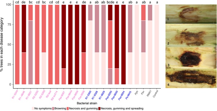
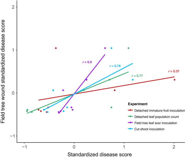
Bacterial canker is a major disease of Prunus avium (cherry), Prunus domestica (plum) and other stone fruits. It is caused by pathovars within the Pseudomonas syringae species complex including P. syringae pv. morsprunorum (Psm) race 1 (R1), Psm race 2 (R2) and P. syringae pv. syringae (Pss). Psm R1 and Psm R2 were originally designated as the same pathovar; however, phylogenetic analysis revealed them to be distantly related, falling into phylogroups 3 and 1, respectively. This study characterized the pathogenicity of 18 newly genome-sequenced P. syringae strains on cherry and plum, in the field and laboratory. The field experiment confirmed that the cherry cultivar Merton Glory exhibited a broad resistance to all clades. Psm R1 contained strains with differential specificity on cherry and plum. The ability of tractable laboratory-based assays to reproduce assessments on whole trees was examined. Good correlations were achieved with assays using cut shoots or leaves, although only the cut shoot assay was able to reliably discriminate cultivar differences seen in the field. Measuring bacterial multiplication in detached leaves differentiated pathogens from nonpathogens and was therefore suitable for routine testing. In cherry leaves, symptom appearance discriminated Psm races from nonpathogens, which triggered a hypersensitive reaction. Pathogenic strains of Pss rapidly induced disease lesions in all tissues and exhibited a more necrotrophic lifestyle than hemibiotrophic Psm. This in-depth study of pathogenic interactions, identification of host resistance and optimization of laboratory assays provides a framework for future genetic dissection of host-pathogen interactions in the canker disease.

Keywords: Pseudomonas; non‐host; pathogenicity; resistance.


Figures

Figure 1
Figure 1
Bayesian phylogenetic tree of Pseudomonas syringae. The phylogeny was constructed using a concatenated alignment of seven genes (acnB, fruK, gapA, gltA, gyrB, pgi and rpoD). A subset of strains from the bacterial canker‐causing clades P. syringae pv. syringae, P. syringae pv. morsprunorum race 1 (R1) and P. syringae pv. morsprunorum race 2 (R2) was selected for analysis. Strains isolated from cherry are in pink, whilst those from plum are in blue. Phylogroups are labelled P1 to P3. The pathogenicity of strains in bold was tested in this study. Scale bar shows substitutions per site. Bootstrap support values are >99% unless otherwise indicated.
Figure 2
Figure 2
Percentage of inoculations in each disease score category after wound inoculation of cherry cv. Van with strains of Pseudomonas syringae: P. syringae pv. syringae (Pss), P. syringae pv. morsprunorum (Psm) race 1 (R1) and Psm race 2 (R2), selected out‐group nonpathogen strains (P. syringae pv. phaseolicola 1448A, Pph; P. syringae pv. avellanae 631, Psv; P. syringae RMA1) and a 10 mm MgCl2 control. Data presented are the percentage of replicates (= 5) for each strain in each disease category. This complete experiment was performed once. Disease symptoms were scored on a ordinal scale as illustrated: 1, no symptoms; 2, limited browning; 3, brown/black symptoms and gumming; 4, brown/black symptoms, gumming and spread from site of inoculation. Strains isolated from cherry are labelled in pink, whilst those from plum are in blue. Statistical Tukey‐HSD (= 0.05; confidence level: 0.95) groupings of bacterial strains determined by a proportional odds model (POM) analysis are presented above the bar. Full statistical analysis can be found in Table S5.
Figure 3
Figure 3
Field inoculation of cherry cultivars with selected strains of Pseudmonas syringae. Data presented are the disease score and length of disease symptoms observed 7 months after inoculation. (a) Leaf scar inoculation; (b) wound inoculation. The plots are ordered by host of isolation (cherry, plum, other plant species and 10 mm MgCl2 control) and P. syringae clade (P. syringae pv. morsprunorum (Psm) race 1 (R1), Psm race 2 (R2), P. syringae pv. syringae (Pss), P. syringae pv. phaseolicola 1448A (Pph), P. syringae RMA1). (1) Percentage of replicates for each strain (= 10) in each disease score category (colour‐coded as in Figure 2). (2) Boxplot of length of symptoms associated with each strain on the four cultivars. Boxplots are colour‐coded for each strain based on clade R1 (blue), R2 (green), Pss (red), nonpathogens (orange) and 10 mm MgCl2 control (black). All data points (= 10) are presented. This complete experiment was performed once. Strains isolated from cherry are labelled in pink, whilst those from plum are in blue. For disease score a proportional odds model (POM) analysis indicated that there was a significant difference between inoculation method (< 0.01, d.f. = 1), between P. syringae strains (< 0.01, d.f. = 8) and between cultivars (< 0.01, d.f. = 3). As there was no interaction between strain and cultivar in the POM analysis, the post hoc groupings were the same across the plots. For symptom length, REML analysis indicated there were significant differences between strains and cultivars for both the leaf scar and wound experiments (< 0.01, d.f. = 8 and < 0.01, d.f. = 3 respectively). Tukey‐HSD (= 0.05, confidence level: 0.95) groups are presented above each strain for each cultivar. Full statistical analysis can be found in Tables S6 and S7 (POM disease score analysis) and Tables S8 and S9 (REML symptom length analysis).
Figure 4
Figure 4
Field inoculations of different plum cultivars with selected strains of Pseudomonas syringae. Data are presented exactly as in Figure 3; strains are P. syringae pv. morsprunorum (Psm) race 1 (R1), Psm race 2 (R2), P. syringae pv. syringae (Pss), P. syringae pv. phaseolicola 1448A (Pph) and P. syringae RMA1. Strains isolated from cherry are labelled in pink, whilst those from plum are in blue. For disease score, proportional odds model (POM) analysis indicated there were significant differences between inoculation method (< 0.01, d.f. = 1), strains (< 0.01, d.f. = 8) and cultivars (< 0.01, d.f. = 1). For symptom length, REML analysis indicated there were significant differences between strains in both inoculation experiments (< 0.01, d.f. = 8) but not between host cultivars (= 0.20, d.f. = 1 for leaf scar, = 0.35, d.f. = 1 for wound). Tukey‐HSD (= 0.05, confidence level: 0.95) groups are presented above each strain for each cultivar. Full statistical analysis can be found in Tables S10 and S11 (POM disease score analysis) and Tables S12 and S13 (REML symptom length analysis).
Figure 5
Figure 5
Lesion development on cut shoots of cherry and plum cultivars following inoculation with Pseudomonas syringae. The plots are ordered by host of isolation (cherry, plum, other plant species and the 10 mm MgCl2 control). (a) Boxplot of percentage area of black/brown discolouration in the top 30 mm associated with different P. syringae strains on four cherry cultivars (a) and two plum cultivars (b). All data points for each treatment (= 10) are presented. This experiment was performed once. The bar chart is colour‐coded based on clade: P. syringae pv. morsprunorum (Psm) race 1 (R1), blue; Psm race 2 (R2), green; P. syringae pv. syringae (Pss), red; nonpathogens (P. syringae pv. phaseolicola 1448A, Pph; P. syringae pv. avellanae 631, Psv; P. syringae RMA1), orange; control, black. (c) Representative images of the symptoms on shoots of cherry cv. Napoleon inoculated with Pss 9097 (1–4) or the 10 mm MgCl2 control (5). Strains isolated from cherry are labelled in pink, whilst those from plum are in blue. An ANOVA revealed there were significant differences between bacterial strains (< 0.001, d.f. = 8), no significant difference between the susceptibility of the two Prunus species (= 0.57, d.f. = 1) and there was a significant interaction between Prunus species and P. syringae strain (< 0.01, d.f. = 8) as well as interactions between strain and individual cultivars (< 0.01, d.f. = 36). Tukey‐HSD (= 0.05; confidence level: 0.95) significance groups for the different strains for each separate cultivar are presented above each boxplot. Full statistical analysis can be found in Table S14.
Figure 6
Figure 6
Boxplot to show diameter of brown/black lesions caused by different Pseudomonas syringae strains on immature cherry fruits 10 days post‐inoculation. (a) All strains used in this study. Strains isolated from cherry are labelled in pink, whilst those from plum are in blue. The boxplot is colour‐coded, Pseudomonas syringae pv. morsprunorum (Psm) race 1 (R1), blue; Psm race 2 (R2), green; P. syringae pv. syringae (Pss), red; nonpathogens, orange; control, black. All data points for each treatment (= 5) are shown. This complete experiment was performed once. Representative images are presented: 1, Psm R1; 2, Psm R2; 3, Pss; 4, nonpathogens; 5, 10 mm MgCl2 control. An ANOVA revealed significant differences between strains (< 0.01, d.f. = 21). Tukey‐HSD (= 0.05, confidence level: 0.95) significance groups are presented above each bar. (b) Boxplot of diameter of brown/black symptoms caused by cherry pathogens on four cherry cultivars using immature cherry fruits. Data presented are all values (= 20) per treatment of two independent experiments. An ANOVA revealed significant differences between strains (< 0.01, d.f. = 3), cultivars (< 0.01, d.f. = 3) and a significant interaction (< 0.01, d.f. = 9). Tukey‐HSD (= 0.05, confidence level: 0.95) significance groups for the different strains for each separate cultivar are presented above each boxplot. Full statistical analysis can be found in Tables S15 and S16.
Figure 7
Figure 7
Population counts of different strains over time on cherry cv. Van (a) and plum cv. Victoria (b) leaves. Strains isolated from cherry are labelled in pink, whilst those from plum are in blue. Line colours for each strain are presented in the key and are colour‐coded by clade: Pseudomonas syringae pv. morsprunorum (Psm) race 1 (R1), blue; Psm race 2 (R2), green; P. syringae pv. syringae (Pss), red; nonpathogens, orange. Population counts are log CFU mL −1. Data presented are the mean values (= 9), with error bars showing standard error above and below the mean. This complete experiment was performed once. An ANOVA revealed significant differences between strains (< 0.01, d.f. = 8). Tukey‐HSD (= 0.05, confidence level: 0.95) significance groups for the different strains (based on day‐10 populations) are presented. Full statistical analysis can be found in Table S17.
Figure 8
Figure 8
Pathogenicity of different strains, assessed by population counts on cherry and plum leaves and on different cherry cultivars. (a) Boxplots of day‐10 population counts on cherry cv. Van and plum cv. Victoria. Strains isolated from cherry are labelled in pink, whilst those from plum are in blue. Boxplots are colour‐coded by clade: Pseudomonas syringae pv. morsprunorum (Psm) race 1 (R1), blue; Psm race 2 (R2), green; P. syringae pv. syringae (Pss), red; nonpathogens, orange. Population counts are log CFU mL −1. Data presented are all the values from two independent experiments (= 18), although Pss 9293 and Ps 9643 were assessed only once. REML analysis for both cherry and plum revealed significant differences between strains (< 0.01, d.f. = 8). Tukey‐HSD (= 0.05, confidence level: 0.95) significance groups for the different strains are presented. (b) Representative images of symptom development at inoculation sites on cherry cv. Van leaves 10 dpi. Black pen dots mark the edge of inoculations. The pathogens Psm R1‐5244, Psm R2‐leaf and Pss 9097 caused brown/black disease lesions with some yellowing around the lesion edge; nonpathogens Psm R1‐5300 and RMA1 failed to produce any symptoms. (c) Boxplot of day‐10 population counts of three pathogenic P. syringae strains on different cherry cultivars. Data presented are all the values for each treatment of two independent experiments (= 18). Tukey‐HSD (= 0.05, confidence level: 0.95) significance groups for the different strains on each separate cultivar are presented. An ANOVA revealed significant differences between strains (< 0.01, d.f. = 2), cultivars (< 0.01, d.f. = 3) and a significant interaction (< 0.01, d.f. = 6). Tukey‐HSD groups comparing the different cultivars are also presented. Full statistical analysis can be found in Tables S18 (cherry day‐10 populations), S19 (plum day‐10 populations) and S20 (cherry cultivars).
Figure 9
Figure 9
Symptom score analysis on cherry and plum. (a) Symptom development over time after inoculation of various Pseudmonas syringae strains in leaves of cherry cv. Van at different concentrations. Strains isolated from cherry are labelled in pink, whilst those from plum are in blue. Symptoms were scored from 0 to 5: 0, no symptoms; 1, limited browning; 2, browning <50% of inoculated site; 3, browning >50% of inoculated site; 4, complete browning; 5, spread from site of inoculation. Data presented are the means (= 4). This experiment was performed once. Symptom development over time (0–48 h) was analysed using AUDPC. An ANOVA revealed significant differences between strains (< 0.01, d.f. = 4), concentrations (< 0.01, d.f. = 3) and a significant interaction (< 0.01, d.f. = 12). Tukey‐HSD (= 0.05, confidence level: 0.95) significance groups are presented. (b) Typical symptom development 24 and 168 h post‐inoculation (hpi): 1, Psm R1‐5244; 2, Pss‐9097; 3, RMA1. (c) Symptom development over time for multiple strains on cherry and plum leaves. Symptoms were scored as in (a), but there is no score 5 as no spreading was observed in these experiments. Data presented are the mean values from two independent experiments (= 8). ANOVAs of AUDPC values (0–48 hpi) for cherry and plum revealed significant differences between strains (< 0.01, d.f. = 8). Tukey‐HSD (= 0.05, confidence level: 0.95) significance groups are presented in the table next to the plot. Full statistical analysis can be found in Tables S22 (variable concentrations), S23 (AUDPC cherry leaves 0–48 h) and S24 (AUDPC plum leaves 0–48 h). AUDPC data can be found in Tables S25, S26 and S27 (variable concentration, cherry and plum experiments, respectively).
Figure 10
Figure 10
Correlation between results from various pathogenicity tests with the length of symptoms recorded after wound inoculation of field trees. The wound inoculation on field trees was assumed to be most representative of the natural disease. The datasets included field leaf scar symptom length (Fig. 3a), cut shoot percentage area of brown/black symptoms (Fig. 5), immature cherry fruit symptom diameter (Fig. 6a) and bacterial population counts in CFU mL −1 (Fig. S6). Each dataset included the results for seven bacterial strains on cherry cv. Van. Data were standardized (mean⁄SD) to allow correlation analysis to be performed. A linear model line was plotted for each experiment and Pearson's correlation coefficients are presented. Data used and the correlation results are presented in Tables S28 and S29.

References

    1. American Pomological Society , 1966. Merton cherries from England. Journal of Fruit Varieties and Horticultural Digest 20, 46.
    1. Andrews S, 2010. FastQC: A quality control tool for high throughput sequence data. [http://www.bioinformatics.babraham.ac.uk/projects/fastqc]. Accessed 12 December 2017.
    1. Arnold DL, Lovell HC, Jackson RW, Mansfield JW, 2011. Pathogen profile Pseudomonas syringae pv. phaseolicola: from ‘has bean’ to supermodel. Molecular Plant Pathology 12, 617–27. - PMC - PubMed
    1. Aziz RK, Bartels D, Best AA et al, 2008. The RAST Server: rapid annotations using subsystems technology. BMC Genomics 9, 75. - PMC - PubMed
    1. Bankevich A, Nurk S, Antipov D et al, 2012. SPades: a new genome assembly algorithm and its applications to single‐cell sequencing. Journal of Computational Biology 19, 455–77. - PMC - PubMed

LinkOut - more resources