Review
. 2016 Dec;11(4):145-156.
doi: 10.1016/j.joto.2016.10.001. Epub 2016 Oct 27.

Ototoxic effects and mechanisms of loop diuretics

Affiliations
Review

Ototoxic effects and mechanisms of loop diuretics

Dalian Ding et al. J Otol. 2016 Dec.

Abstract

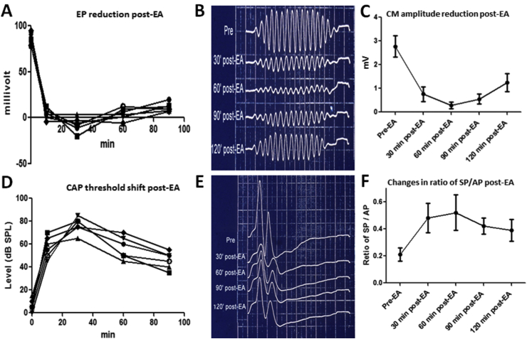
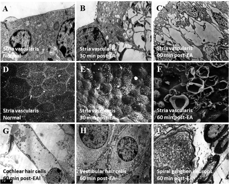
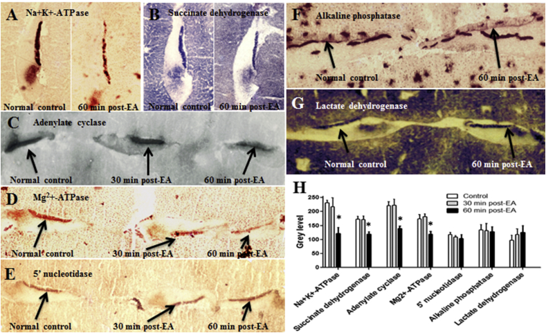
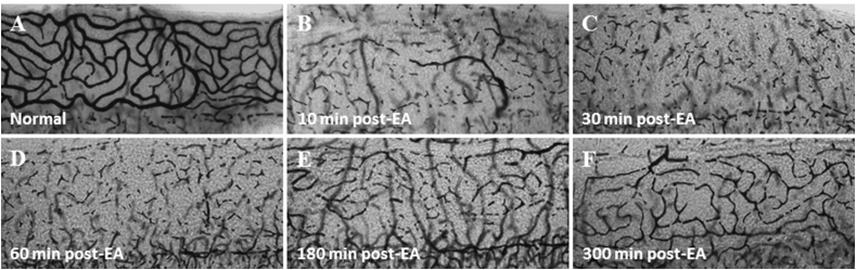
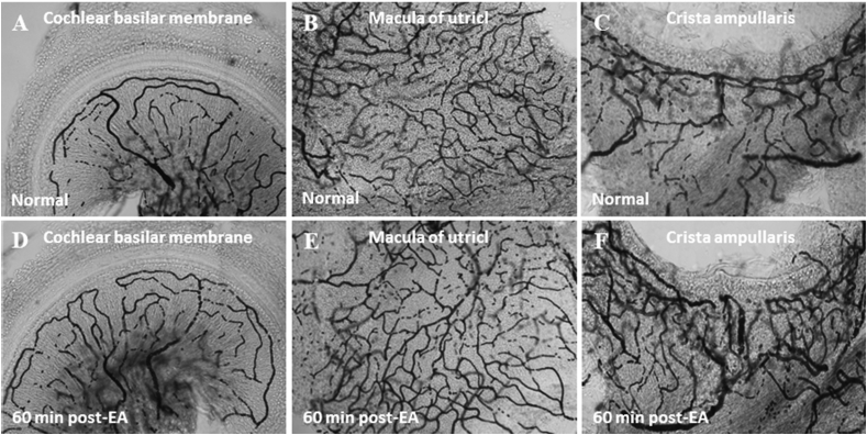
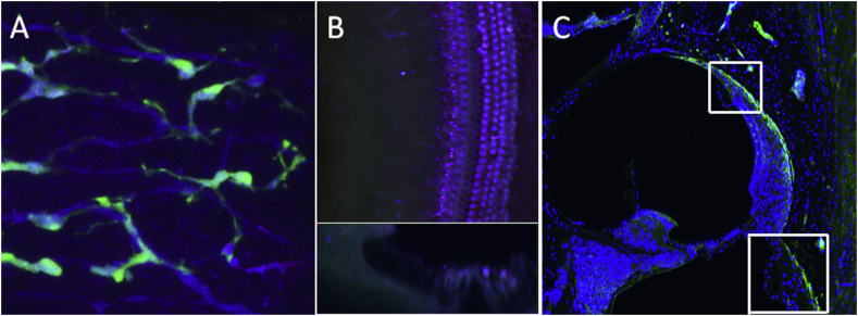
Over the past two decades considerable progress has been made in understanding the ototoxic effects and mechanisms underlying loop diuretics. As typical representative of loop diuretics ethacrynic acid or furosemide only induces temporary hearing loss, but rarely permanent deafness unless applied in severe acute or chronic renal failure or with other ototoxic drugs. Loop diuretic induce unique pathological changes in the cochlea such as formation of edematous spaces in the epithelium of the stria vascularis, which leads to rapid decrease of the endolymphatic potential and eventual loss of the cochlear microphonic potential, summating potential, and compound action potential. Loop diuretics interfere with strial adenylate cyclase and Na+/K+-ATPase and inhibit the Na-K-2Cl cotransporter in the stria vascularis, however recent reports indicate that one of the earliest effects in vivo is to abolish blood flow in the vessels supplying the lateral wall. Since ethacrynic acid does not damage the stria vascularis in vitro, the changes in Na+/K+-ATPase and Na-K-2Cl seen in vivo may be secondary effects results from strial ischemia and anoxia. Recent observations showing that renin is present in pericytes surrounding stria arterioles suggest that diuretics may induce local vasoconstriction by renin secretion and angiotensin formation. The tight junctions in the blood-cochlea barrier prevent toxic molecules and pathogens from entering cochlea, but when diuretics induce a transient ischemia, the barrier is temporarily disrupted allowing the entry of toxic chemicals or pathogens.

Keywords: Diuretics; Ischemia; Pericytes; Renin; Stria vascularis.


Figures

Fig. 1
Fig. 1
Cochlear dysfunction post-EA. (A) Endocochlear potential (EP) measured from six guinea pigs as a function of time after EA injection. (B) Waveform changes of the cochlear microphonic potential (CM) stimulated by 2 kHz tone presented at 80 dB SPL post-EA. (C) Amplitude of CM was greatly reduced post-EA. (D) Threshold shift of click-evoked compound action potentials (CAP) from six guinea pigs as a function of time after EA injection. (E) Waveform changes of summating potentials (SP) and CAP complexes at 90 dB SPL post-EA. (F) Changes in ratio of SP/CAP amplitude post-EA.
Fig. 2
Fig. 2
Ethacrynic acid-induced ultrastructural lesions in the inner ear. (A) Epithelium of stria vascularis in normal control animal. (B) At 30 min post-EA, strial edema present in region of intermediate cells. (C) At 60 min post-EA, cytoplasm of epithelium in stria vascularis disrupted by edema; note splitting and vacuolization. (D) Edge of marginal cells in stria vascularis is smooth and flat in normal control animal. (E) At 30 min post-EA, the surface of stria vascularis was uneven due to cellular swelling. (F) At 60 min post-EA, the marginal cells on the surface of stria vascularis were destroyed. (G, H, I). The cochlear hair cells (G), vestibular hair cells (H), and spiral ganglion neurons (I) exhibited were normal 60 min after EA injection.
Fig. 3
Fig. 3
EA-induced inactivation of enzymes within epithelium of stria vascularis. (A) Frozen section of spiral ligament embedded in the liver tissue showing intense Na+, K+-ATPase labeling in stria vascularis in normal control animal. In contrast, Na+, K+-ATPase expression in stria vascularis was greatly reduced 60 min after EA injection. (B) Frozen section of spiral ligament shows intense succinate dehydrogenase labeling in stria vascularis in normal animal. However, the staining of succinate dehydrogenase was decreased 60 min post-EA. (C) Frozen section of spiral ligament shows intense adenylate cyclase activity in stria vascularis in normal controls and 30 min post-EA treated animals. Labeling of adenylate cyclase was decreased 60 min after EA injection. (D) Frozen section of spiral ligament shows intense Mg++-ATPase in stria vascularis in normal and 30 min post-EA treated animals. However, the staining of Mg++-ATPase was reduced 60 min post-EA. (E) Frozen section of spiral ligament shows intense 5'nucleotidase in the stria vascularis in normal control and 30 min, and 60 min after EA injection. (F) Frozen section of spiral ligament shows moderate expression of alkaline phosphatase in the stria vascularis in normal control and 60 min after EA injection. (G) Frozen section of spiral ligament shows intense lactate dehydrogenase in the stria vascularis in normal control and 60 min after EA injection. (H) Mean gray level of enzyme expression in the region of stria vascularis. In comparison to normal controls. Activities of Na+, K+-ATPase, succinate dehydrogenase, adenylate cyclase, and Mg++-activated ATPase were significantly inhibited 60 min after EA injection. Activity of 5' nucleotidase, alkaline phosphatase and lactate dehydrogenase were not affected by EA injection. *Significantly different from normal control by ANOVA statistical analysis and posthoc test (Tukey) using the GraphPad Prism (P < 0.05).
Fig. 4
Fig. 4
Surface preparations of rat stria vascularis after EA injection. (A) normal control; (B) 10 min post-EA; (C) 30 min post-EA; (D) 60 min post-EA; (E) 180 min post-EA; (F) 300 min post-EA. At 10, 30 and 60 min, the blood supply to stria vascularis was greatly abolished whereas at 180–300 min post-EA the blood supply was in the early stage of recovery.
Fig. 5
Fig. 5
Surface preparations of rat inner ear with or without EA treatment. (A) Blood supply to cochlear basilar membrane in normal control. (B) Blood supply to macula of utricle in normal control. (C) Blood supply to crista ampullaris in normal control. (D) At 60 min post-EA, blood supply to cochlear basilar membrane was unaffected. (E) At 60 min post-EA, blood supply to macula of utricle was unaffected. (F) At 60 min post-EA, blood supply to crista ampullaris was unaffected. The appearance of vessels at 60 min post EA was not notably different from the appearance at any earlier or later time point.
Fig. 6
Fig. 6
Gentamicin concentration (μg/ml; mean ± S.D. for the groups) in perilymph at the peak value of 2.5 h and the half-life of 12 h after (1) a single gentamicin injection, (2) 20 daily gentamicin injections or (3) one treatment of ethacrynic acid gentamicin. The concentration of gentamicin in perilymph after one co-administration of ethacrynic acid/gentamicin was significantly increased (p < 0.05) to a level was equivalent to the gentamicin accumulation in perilymph produced by 20 daily gentamicin treatments.
Fig. 7
Fig. 7
Dose-response of EA to epithelium of stria vascularis in vitro. The surface structure of marginal cells of stria vascularis were stained with phalloidin in green, and the nuclei of cells in the epithelium were labeled with ToPro-3 in red. (A) Stria vascularis was cultured in standard serum-free medium without EA for 24 h as normal control. (B) Stria vascularis cultured in 10 μM EA for 24 h. (C) Stria vascularis cultured in 100 μM EA for 24 h. (D) Stria vascularis cultured in 1000 μM EA for 24 h. EA had little or no effect on stria vascularis morphology.
Fig. 8
Fig. 8
Localization of renin cells (green) in the cochlea. (A) Pericytes surrounding capillaries in stria vascularis express green fluorescence in renin-gfp mouse. (B) Capillaries in cochlear basilar membrane do not express renin-gfp. (Top figure is a surface view of cochlear basilar membrane. Bottom figure is Z axis image plane showing the section of cochlear basilar membrane). These results suggest that pericytes around capillaries in the cochlear lateral wall contains the renin-angiotensin aldosterone system whereas pericytes in the vessels in the modiolus and cochlear basilar membrane do not contain renin. (C) Renin immunolabeling in rat was only present in the vessels of the cochlear lateral wall, but in the vessels in the modiolus or cochlear basilar membrane.

References

    1. Akiyoshi M. Effect of loop-diuretics on hair cells of the cochlea in Guinea pigs. Histological and histochemical study. Scand. Audiol. Suppl. 1981;(14 Suppl):185–199. - PubMed
    1. Arnold W., Nadol J.B., Jr., Weidauer H. Ultrastructural histopathology in a case of human ototoxicity due to loop diuretics. Acta Oto-Laryngol. 1981;91:399–414. - PubMed
    1. Berg A.C., Chernavvsky-Sequeira C., Lindsey J., Gomez R.A., Sequeira-Lopez M.L. Pericytes synthesize renin. World J. Nephrol. 2013;2:11–16. - PMC - PubMed
    1. Bergers G., Song S. The role of pericytes in blood-vessel formation and maintenance. Neuro Oncol. 2005;7:452–464. - PMC - PubMed
    1. Bleich M., Greger R. Mechanism of action of diuretics. Kidney Int. Suppl. 1997;59:S11–S15. - PubMed