

Apoptosis in inner ear sensory hair cells
- PMID: 29937851
- PMCID: PMC6002637
- DOI: 10.1016/j.joto.2017.08.001
Apoptosis in inner ear sensory hair cells
Abstract
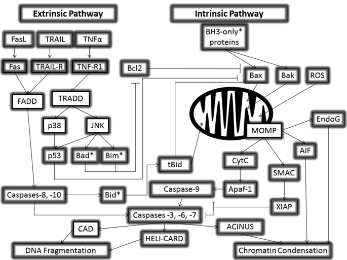
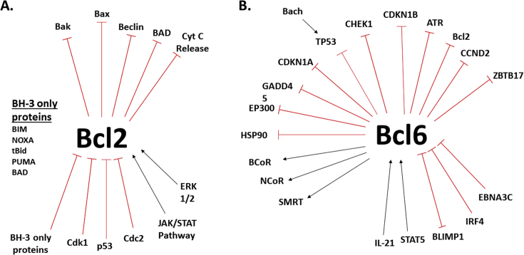
Apoptosis, or controlled cell death, is a normal part of cellular lifespan. Cell death of cochlear hair cells causes deafness; an apoptotic process that is not well understood. Worldwide, 1.3 billion humans suffer some form of hearing loss, while 360 million suffer debilitating hearing loss as a direct result of the absence of these cochlear hair cells (Worldwide Hearing, 2014). Much is known about apoptosis in other systems and in other cell types thanks to studies done since the mid-20th century. Here we review current literature on apoptosis in general, and causes of deafness and cochlear hair cells loss as a result of apoptosis. The family of B-cell lymphoma (Bcl) proteins are among the most studied and characterized. We will review current literature on the Bcl2 and Bcl6 protein interactions in relation to apoptosis and their possible roles in vulnerability and survival of cochlear hair cells.
Keywords: Apoptosis; Bcl2; Bcl6; Hair cell.
Figures
Similar articles
-
Anti-apoptotic factor z-Val-Ala-Asp-fluoromethylketone promotes the survival of cochlear hair cells in a mouse model for human deafness.Neuroscience. 2010 Jul 14;168(3):851-7. doi: 10.1016/j.neuroscience.2010.04.011. Epub 2010 Apr 13. Neuroscience. 2010. PMID: 20394804
-
Recent advances in cochlear hair cell regeneration-A promising opportunity for the treatment of age-related hearing loss.Ageing Res Rev. 2017 Jul;36:149-155. doi: 10.1016/j.arr.2017.04.002. Epub 2017 Apr 13. Ageing Res Rev. 2017. PMID: 28414155 Review.
-
Cochlear pathology, sensory cell death and regeneration.Br Med Bull. 2002;63:25-38. doi: 10.1093/bmb/63.1.25. Br Med Bull. 2002. PMID: 12324382 Review.
-
The zinc finger transcription factor Gfi1, implicated in lymphomagenesis, is required for inner ear hair cell differentiation and survival.Development. 2003 Jan;130(1):221-32. doi: 10.1242/dev.00190. Development. 2003. PMID: 12441305
-
Peroxynitrite induces apoptosis of mouse cochlear hair cells via a Caspase-independent pathway in vitro.Apoptosis. 2017 Nov;22(11):1419-1430. doi: 10.1007/s10495-017-1417-8. Apoptosis. 2017. PMID: 28900799
Cited by
-
Fursultiamine Prevents Drug-Induced Ototoxicity by Reducing Accumulation of Reactive Oxygen Species in Mouse Cochlea.Antioxidants (Basel). 2021 Sep 26;10(10):1526. doi: 10.3390/antiox10101526. Antioxidants (Basel). 2021. PMID: 34679662 Free PMC article.
-
Primed to die: an investigation of the genetic mechanisms underlying noise-induced hearing loss and cochlear damage in homozygous Foxo3-knockout mice.Cell Death Dis. 2021 Jul 7;12(7):682. doi: 10.1038/s41419-021-03972-6. Cell Death Dis. 2021. PMID: 34234110 Free PMC article.
-
Oral Antioxidant Vitamins and Magnesium Limit Noise-Induced Hearing Loss by Promoting Sensory Hair Cell Survival: Role of Antioxidant Enzymes and Apoptosis Genes.Antioxidants (Basel). 2020 Nov 25;9(12):1177. doi: 10.3390/antiox9121177. Antioxidants (Basel). 2020. PMID: 33255728 Free PMC article.
-
Congenital Diseases of DNA Replication: Clinical Phenotypes and Molecular Mechanisms.Int J Mol Sci. 2021 Jan 18;22(2):911. doi: 10.3390/ijms22020911. Int J Mol Sci. 2021. PMID: 33477564 Free PMC article. Review.
-
Prevention and Rehabilitation of Old Age Deafness.Indian J Otolaryngol Head Neck Surg. 2020 Dec;72(4):524-531. doi: 10.1007/s12070-020-01856-3. Epub 2020 Apr 16. Indian J Otolaryngol Head Neck Surg. 2020. PMID: 33088786 Free PMC article.
References
-
- Agrawal Y., Platz E.A., Niparko J.K. Risk factors for hearing loss in US adults: data from the National Health and Nutrition Examination Survey, 1999 to 2002. Otol. Neurotol. 2009;30:139–145. - PubMed
-
- Alam S.A., Oshima T., Suzuki M., Kawase T., Takasaka T., Ikeda K. The expression of apoptosis-related proteins in the aged cochlea of Mongolian gerbils. Laryngoscope. 2001;111:528–534. - PubMed
-
- Anatomybody-charts . 2016. Anatomy Body Charts.http://anatomybody-charts.us/inner-ear-anatomy/inner-ear-anatomy-2/
Publication types
LinkOut - more resources
Full Text Sources
Other Literature Sources
