Platelet distribution width as a novel indicator of disease activity in systemic lupus erythematosus
- PMID: 29937910
- PMCID: PMC5996572
- DOI: 10.4103/jrms.JRMS_1038_16
Platelet distribution width as a novel indicator of disease activity in systemic lupus erythematosus
Abstract
Background: Significance of platelet distribution width (PDW) and mean platelet volume (MPV) in assessing disease activity of systemic lupus erythematosus (SLE) remains unclear. This study was aimed to evaluate PDW and MPV as potential disease activity markers in adult SLE patients.
Materials and methods: A total of 204 study participants, including 91 SLE patients and 113 age- and gender-matched healthy controls, were selected in this cross-sectional study. They were classified into three groups: control group (n = 113), active SLE group (n = 54), and inactive SLE group (n = 37). Demographic, clinical, and laboratory data were analyzed.
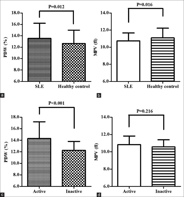
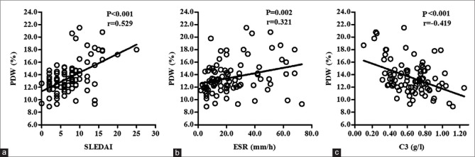
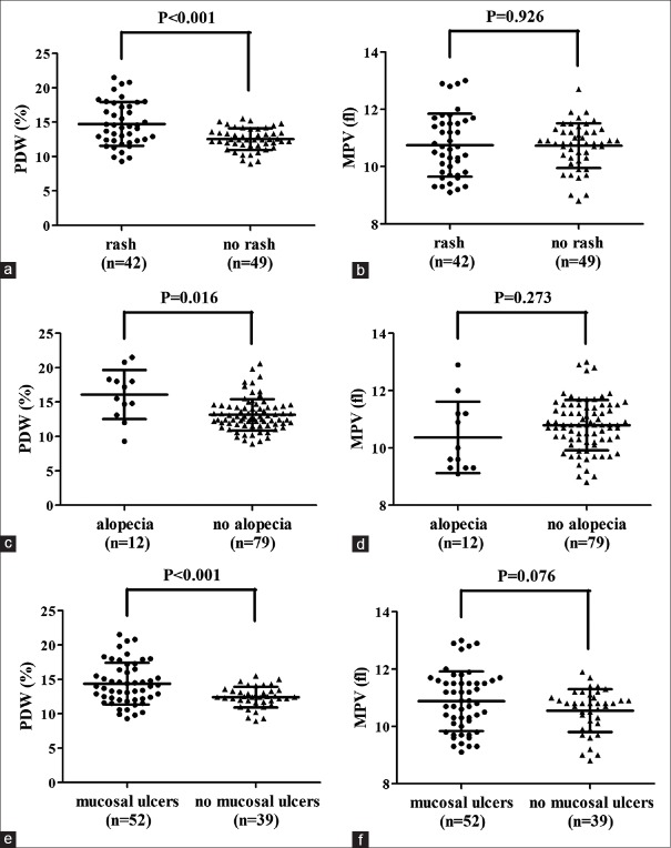
Results: In patient group, PDW was statistically higher than that in control group (13.54 ± 2.67 vs. 12.65 ± 2.34, P = 0.012), and in active group, PDW was significantly increased compared to inactive group (14.31 ± 2.90 vs. 12.25 ± 1.55, P < 0.001). However, MPV was significantly lower in SLE group than in control group (10.74 ± 0.94 vs. 11.09 ± 1.14, P = 0.016). PDW was positively correlated with SLE disease activity index (P < 0.001, r = 0.529) and erythrocyte sedimentation rate (P = 0.002, r = 0.321) and negatively correlated with C3 (P < 0.001, r = -0.419). However, there was no significant association between MPV and these study variables. A PDW level of 11.85% was determined as a predictive cutoff value of SLE diagnosis (sensitivity 76.9%, specificity 42.5%) and 13.65% as cutoff of active stage (sensitivity 52.6%, specificity 85.3%).
Conclusion: This study first associates a higher PDW level with an increased SLE activity, suggesting PDW as a novel indicator to monitor the activity of SLE.
Keywords: Biomarkers; blood platelets; systemic lupus erythematosus.
Conflict of interest statement
There are no conflicts of interest.
Figures

Similar articles
-
Platelet Distribution Width Level in Patients With Systemic Lupus Erythematosus-Associated Pulmonary Arterial Hypertension and Its Diagnostic Value.Arch Rheumatol. 2020 Feb 7;35(3):394-400. doi: 10.46497/ArchRheumatol.2020.7791. eCollection 2020 Sep. Arch Rheumatol. 2020. PMID: 33458663 Free PMC article.
-
Mean platelet volume to lymphocyte ratio and platelet distribution width to lymphocyte ratio in Iraqi patients diagnosed with systemic lupus erythematosus.Reumatologia. 2022;60(3):173-182. doi: 10.5114/reum.2022.117837. Epub 2022 Jul 13. Reumatologia. 2022. PMID: 35875718 Free PMC article.
-
Mean platelet volume as an indicator of disease activity in juvenile SLE.Clin Rheumatol. 2014 May;33(5):637-41. doi: 10.1007/s10067-014-2540-3. Epub 2014 Feb 25. Clin Rheumatol. 2014. PMID: 24567240
-
Association between Mean Platelet Volume and Systemic Lupus Erythematosus: A Meta-Analysis.Iran J Public Health. 2024 May;53(5):978-987. doi: 10.18502/ijph.v53i5.15578. Iran J Public Health. 2024. PMID: 38912146 Free PMC article. Review.
-
Lack of association between mean platelet volume and disease activity in systemic lupus erythematosus patients: a systematic review and meta-analysis.Rheumatol Int. 2018 Sep;38(9):1635-1641. doi: 10.1007/s00296-018-4065-6. Epub 2018 May 29. Rheumatol Int. 2018. PMID: 29845430
Cited by
-
Platelet volume indices correlate to severity of heart failure and have prognostic value for both cardiac and thrombotic events in patients with congenital heart disease.Heart Vessels. 2022 Dec;37(12):2107-2118. doi: 10.1007/s00380-022-02112-0. Epub 2022 Jun 27. Heart Vessels. 2022. PMID: 35761122
-
Clinical characteristics and prognosis in systemic lupus erythematosus-associated pulmonary arterial hypertension based on consensus clustering and risk prediction model.Arthritis Res Ther. 2023 Aug 23;25(1):155. doi: 10.1186/s13075-023-03139-y. Arthritis Res Ther. 2023. PMID: 37612772 Free PMC article.
-
Platelets in Skin Autoimmune Diseases.Front Immunol. 2019 Jul 4;10:1453. doi: 10.3389/fimmu.2019.01453. eCollection 2019. Front Immunol. 2019. PMID: 31333641 Free PMC article. Review.
-
Triglycerides as Biomarker for Predicting Systemic Lupus Erythematosus Related Kidney Injury of Negative Proteinuria.Biomolecules. 2022 Jul 5;12(7):945. doi: 10.3390/biom12070945. Biomolecules. 2022. PMID: 35883502 Free PMC article.
-
The Lung Is Not a Primary Site of Platelet Biogenesis.Physiol Res. 2025 Apr 30;74(2):263-273. doi: 10.33549/physiolres.935477. Physiol Res. 2025. PMID: 40432441 Free PMC article.
References
LinkOut - more resources
Full Text Sources
Other Literature Sources
Miscellaneous