

Anatomo-Physiologic Basis for Auricular Stimulation
- PMID: 29937968
- PMCID: PMC6011382
- DOI: 10.1089/acu.2017.1254
Anatomo-Physiologic Basis for Auricular Stimulation
Abstract
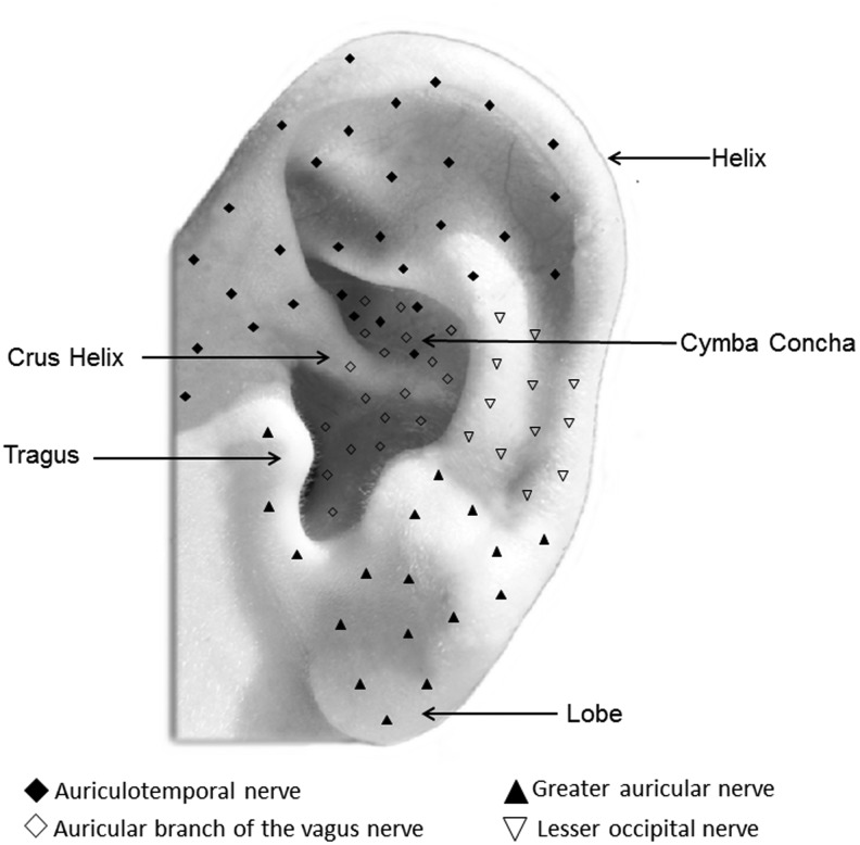
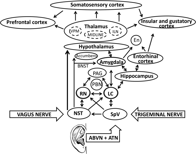
Introduction: Stimulation of cranial nerves modulates central nervous system (CNS) activity via the extensive connections of their brainstem nuclei to higher-order structures. Clinical experience with vagus-nerve stimulation (VNS) demonstrates that it produces robust therapeutic effects, however, posing concerns related to its invasiveness and side-effects. Discussion: Trigeminal nerve stimulation (TNS) has been recently proposed as a valid alternative to VNS. The ear presents afferent vagus and trigeminal-nerve distribution; its innervation is the theoretical basis of different reflex therapies, including auriculotherapy. An increasing number of studies have shown that several therapeutic effects induced by invasive VNS and TNS, can be reproduced by noninvasive auricular-nerve stimulation. However, the sites and neurobiologic mechanisms by which VNS and TNS produce their therapeutic effects are not clear yet. Conclusions: Accumulating evidence suggests that VNS and TNS share multiple levels and mechanisms of action in the CNS.
Keywords: auricular stimulation; brainstem; central nervous system; forebrain; neuromodulation; trigeminal nerve stimulation; vagus nerve stimulation.
Conflict of interest statement
No competing financial interests exist.
Figures
References
-
- Sale MV, Mattingley JB, Zalesky A, Cocchi L. Imaging human brain networks to improve the clinical efficacy of non-invasive brain stimulation. Neurosci Biobehav Rev. 2015;57:187–198 - PubMed
-
- Taghva AS, Malone DA, Rezai AR. Deep brain stimulation for treatment-resistant depression. World Neurosurg. 2012;80(3–4):S27.e17–S27.e24 - PubMed
-
- Hoy KE, Fitzgerald PB. Brain stimulation in psychiatry and its effects on cognition. Nat Rev Neurol. 2010;6(5):267–275 - PubMed
-
- Albert GC, Cook CM, Prato FS, Thomas AW. Deep brain stimulation, vagal nerve stimulation and transcranial stimulation: An overview of stimulation parameters and neurotransmitter release. Neurosci Biobehav Rev. 2009;33(7):1042–1060 - PubMed
Publication types
LinkOut - more resources
Full Text Sources
Other Literature Sources
Medical