. 2018 Apr;3(1):16-28.
doi: 10.1002/jin2.35. Epub 2018 Apr 16.

The anticoagulant properties of cadmium telluride quantum dots

Affiliations

The anticoagulant properties of cadmium telluride quantum dots

Ciarán M Maguire et al. J Interdiscip Nanomed. 2018 Apr.

Abstract

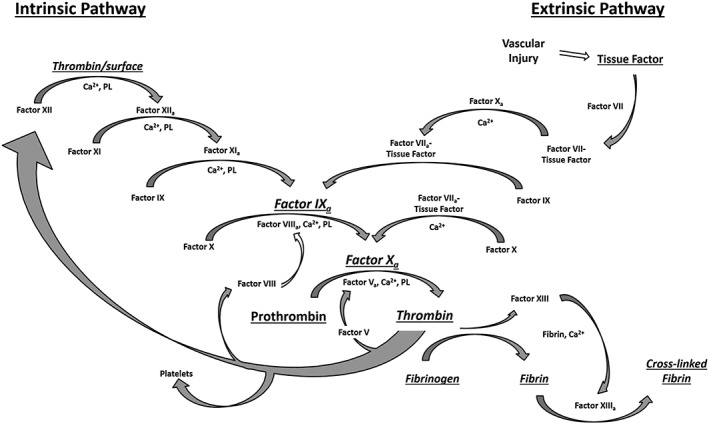
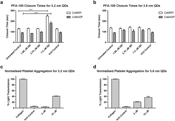
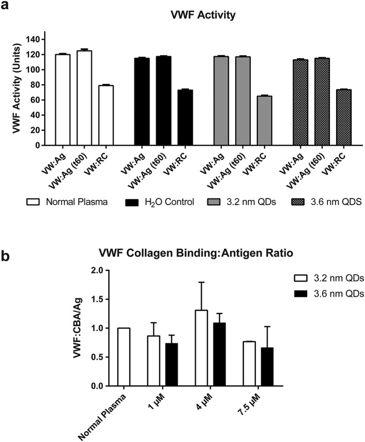
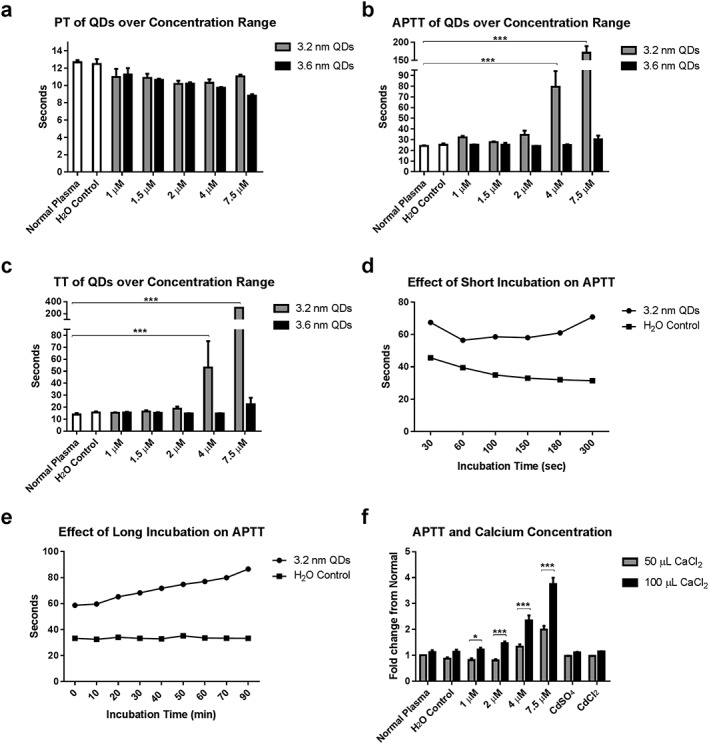
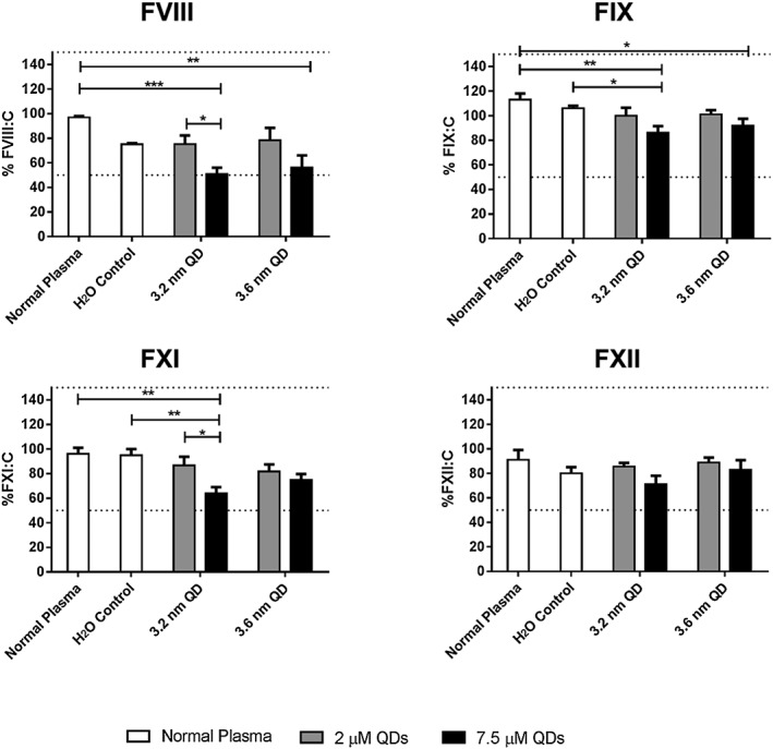
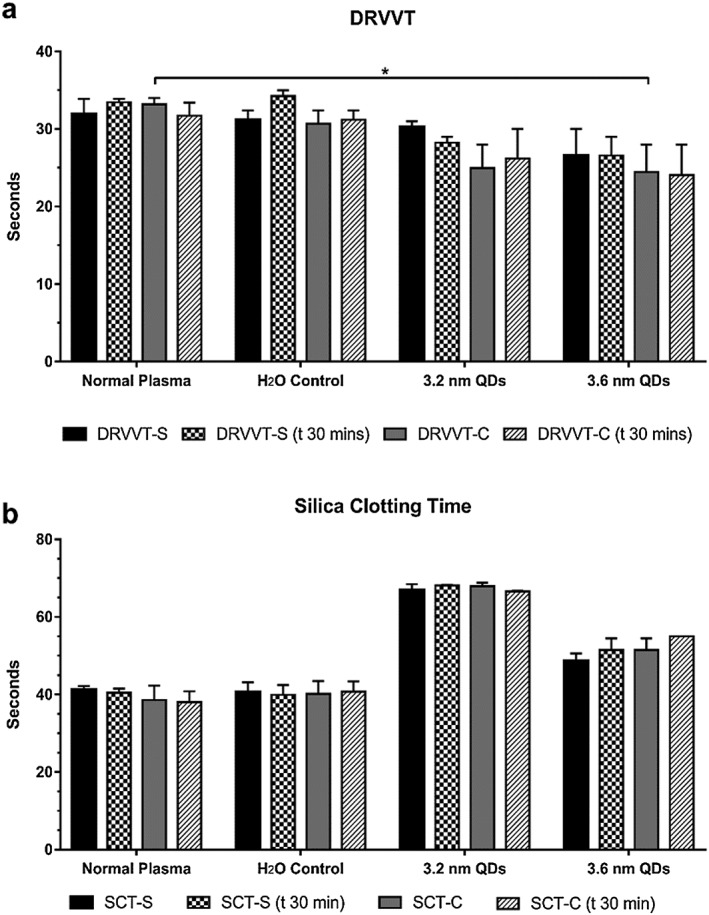
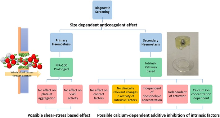
The size-dependent optical properties of quantum dots (QDs) are frequently exploited for use in medical imaging and labelling applications. Similarly, presented here, they also elicit profound size-dependent anticoagulant properties. Cadmium telluride quantum dot (QDs) (3.2 nm) were shown to have a dramatic anticoagulant effect centred on around the intrinsic coagulation pathway, compared to their 3.6 nm counterparts. Several clinically relevant diagnostic tests were carried out over a concentration range of the QDs and demonstrated that the 3.2 nm QDs elicited their response on the intrinsic pathway as a whole, yet the activity of the individual intrinsic coagulation factors was not affected. The mechanism appears also to be strongly influenced by the concentration of calcium ions and not cadmium ions leached from the QDs. Static and shear-based primary haemostasis assays were also carried out, demonstrating a profound anticoagulant effect which was independent of platelets and phospholipids. The data presented here suggest that the physical-chemical properties of the QDs may have a role in the modulation of haemostasis and the coagulation cascade, in a yet not fully understood mechanism. This study has implications for the use of similar QDs as diagnostic or therapeutic tools in vivo, and for the occupational health and safety of those working with such materials.

Keywords: Blood coagulation factors; Von Willebrand factor; platelet function tests; quantum dots.


Figures

Figure 1
Figure 1
The coagulation cascade is the pathway is responsible for maintaining the balance between a pro‐thrombotic and anti‐thrombotic state. Contact with a negatively charged surface or following vascular injury results in the release of tissue factor, platelet activation and activation of Factor X, followed by the conversion of fibrinogen to fibrin and the formation of a cross‐linked fibrin clot. Feedback loops cause amplification of factor activation. Ca2+: calcium ions; PL: phospholipids.
Figure 2
Figure 2
Platelet Function Analyser‐100 (A and B) and platelet aggregation (C and D) results illustrating the size‐dependent effects of quantum dots on primary haemostasis. Aperture closure time is significantly extended outside the normal clinical range at a 7.5‐μM concentration of the 3.2‐nm QDs (A) for both collagen/epinephrine and collagen/ADP (*** P = 0.001, one‐way ANOVA with Dunnett's, n = 3). No prolongation in closure time is observed at equal concentrations of the 3.6 nm variant (B). Minimal platelet aggregation is observed for both sizes of QDs up to a concentration of 10 μM.
Figure 3
Figure 3
Effect of 3.2 and 3.6‐nm QDs on VWF activity. A: Von Willebrand factor activity measured following interaction with QDs at 7.5 μM, at time zero and after 1‐h incubation. The results show VWF antigen levels (VWF:Ag) and VWF ristocetin cofactor levels (VWF:RC). Data represent mean ± SEM (N = 3). No changes in VWF antigen or ristocetin cofactor levels are observed across all treatment groups. B: Normalised von Willebrand factor collagen binding/antigen ratios for normal plasma treated with 3.2 and 3.6 nm QDs at varying concentrations. No statistically significant changes were observed. Data represents mean ± SEM (N = 5).
Figure 4
Figure 4
Size and concentration‐dependent effect of QDs on prothrombin time (PT), thrombin time (TT) and activated partial thromboplastin time (APTT) assays (A‐C). No change from normal was observed for the PT. Results of the APTT and TT demonstrate a profound size‐dependent intrinsic pathway centred anticoagulant effect (*** P < 0.001, two‐way ANOVA with Dunnett's, n = 4). The effect of incubation time on the APTT was also examined (D‐E). Short incubation time (D) results in APTT being sensitive to levels and activity of contact factors, with long incubation times (E) remove this sensitivity. Increasing calcium concentration also results in a further prolongation in the APTT for 3.2 nm QDs (F) (* P < 0.05, *** P < 0.001, two‐way ANOVA with Bonferroni's multiple comparison test, n = 4). Data represents mean ± SEM for all graphs.
Figure 5
Figure 5
Intrinsic factor screen for activity of Factor VIII, IX, XI and XII. The IFS was carried out at 2 and 7.5 μM QD concentration. All factor activities remain within the normal clinical range of 50–150%, or 0.5–1.5 IU/mL (dashed lines), with the greatest reduction in activity being observed at 7.5 μM of the 3.2 nm QDs. Data represent mean ± SEM. * P < 0.05, ** P < 0.01, *** P < 0.001 ANOVA with Tukey's post‐test (N = 3).
Figure 6
Figure 6
Interaction of QDs with phospholipids. A: Dilute Russell's viper venom time (DRVVT) for plasma exposed to QDs and measured at time zero and following 30 min incubation. Results are shown for high (DRVVT‐C) and low (DRVVT‐S) phospholipid concentrations). B: Silica clotting times (SCT) for plasma treated with QDs at 7.5 μM. The assay was carried out at time zero and following 30‐min incubation with high (SCT‐C) and low (SCT‐S) phospholipid concentration. Data represent mean ± SEM (N = 3), * P < 0.05, two‐way ANOVA with Dunnett's.
Figure 7
Figure 7
3.2‐nm cadmium telluride quantum dots (CdTe QDs) interact with a number of components involved in haemostasis. In a shear‐based platelet function analyser system, 3.2 nm QDs cause the prolongation in platelet aggregation time. Under static conditions, it was determined that the QDs do not inhibit platelet aggregation or VWF activity. The effect of the QDs was centred on the intrinsic pathway of the coagulation cascade. The anticoagulant properties observed are calcium ion concentration dependent, and activator‐type and phospholipid concentration independent. No effect was observed that may be due to interaction with the contact factors, and the activities of the intrinsic factors were within normal clinical ranges. The observed anticoagulant activities of the QDs may be due to an additive effect of somewhat reduced Factor VIII activity and inhibited coagulation complex formation. Colour coding: Green – confirmed effect; Red – no effect; Yellow – possible effect.

References

    1. Baumgartner, H. R. , and Haudenschild, C. 1972. Adhesion of platelets to subendothelium. Ann N Y Acad Sci 201(1):22–36. - PubMed
    1. Bhattacharya, D. K. 2002. Functional status of platelets and hereditary platelet disorders. Int J Hum Genet 2(3):197–204.
    1. Carson, S. D. , and Konigsberg, W. H. 1980. Cadmium increases tissue factor (coagulation factor III) activity by facilitating its reassociation with lipids. Science 208(4441):307–309. - PubMed
    1. Choong, G. , Liu, Y. , and Templeton, D. M. 2014. Interplay of calcium and cadmium in mediating cadmium toxicity. Chem Biol Interact 211:54–65. - PubMed
    1. Cochrane, C. , Revak, S. , and Wuepper, K. 1973. Activation of Hageman factor in solid and fluid phases. A critical role of kallikrein. J Exp Med 138(6):1564–1583. - PMC - PubMed

LinkOut - more resources