. 2018 Jun 18;5(3):ENEURO.0345-17.2018.
doi: 10.1523/ENEURO.0345-17.2018. eCollection 2018 May-Jun.

Locomotor- and Reward-Enhancing Effects of Cocaine Are Differentially Regulated by Chemogenetic Stimulation of Gi-Signaling in Dopaminergic Neurons

Affiliations

Locomotor- and Reward-Enhancing Effects of Cocaine Are Differentially Regulated by Chemogenetic Stimulation of Gi-Signaling in Dopaminergic Neurons

Annika H Runegaard et al. eNeuro. .

Abstract

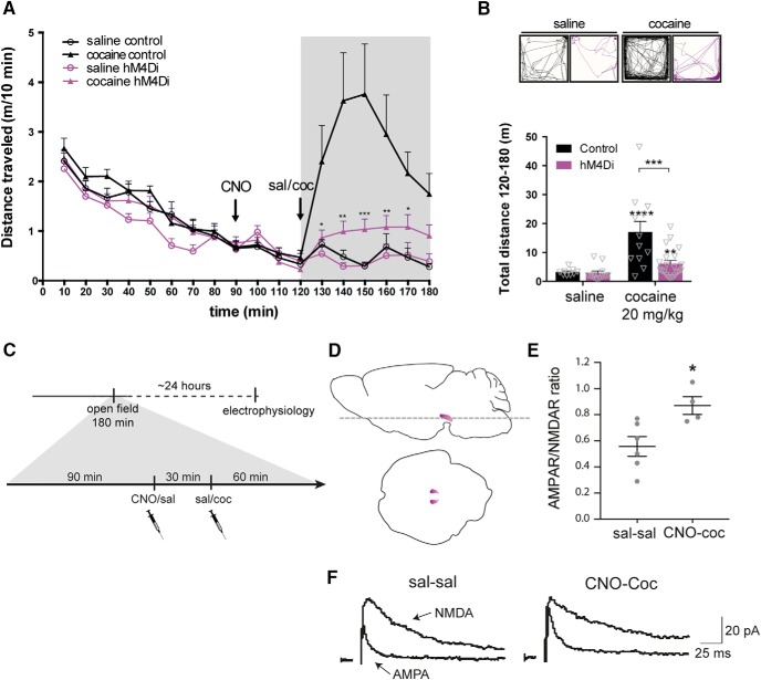
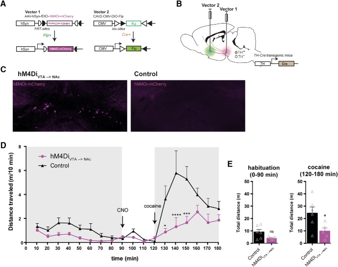
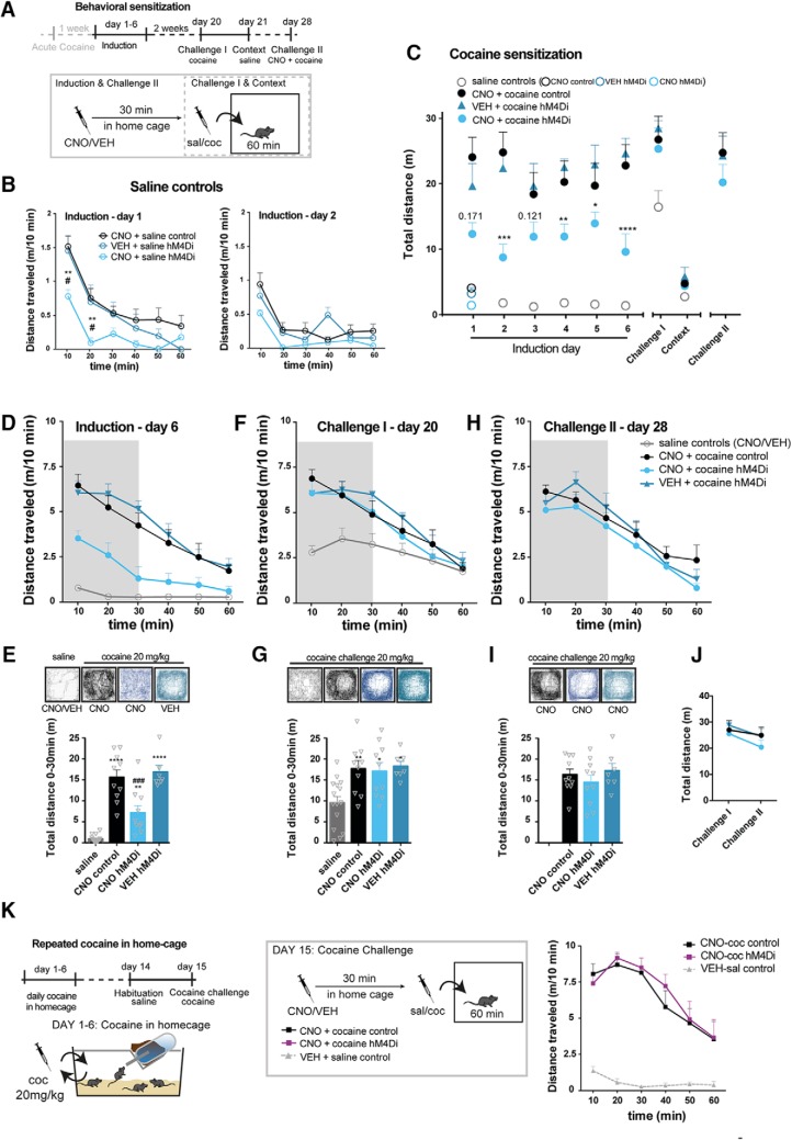
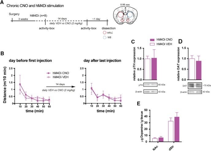
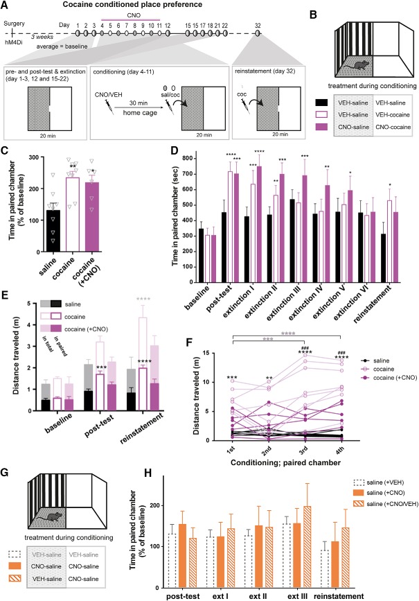
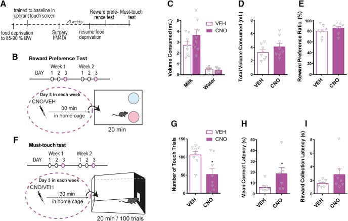
Dopamine plays a key role in the cellular and behavioral responses to drugs of abuse, but the implication of metabotropic regulatory input to dopaminergic neurons on acute drug effects and subsequent drug-related behavior remains unclear. Here, we used chemogenetics [Designer Receptors Exclusively Activated by Designer Drugs (DREADDs)] to modulate dopamine signaling and activity before cocaine administration in mice. We show that chemogenetic inhibition of dopaminergic ventral tegmental area (VTA) neurons differentially affects locomotor and reward-related behavioral responses to cocaine. Stimulation of Gi-coupled DREADD (hM4Di) expressed in dopaminergic VTA neurons persistently reduced the locomotor response to repeated cocaine injections. An attenuated locomotor response was seen even when a dual-viral vector approach was used to restrict hM4Di expression to dopaminergic VTA neurons projecting to the nucleus accumbens. Surprisingly, despite the attenuated locomotor response, hM4Di-mediated inhibition of dopaminergic VTA neurons did not prevent cocaine sensitization, and the inhibitory effect of hM4Di-mediated inhibition was eliminated after withdrawal. In the conditioned place-preference paradigm, hM4Di-mediated inhibition did not affect cocaine-induced place preference; however, the extinction period was extended. Also, hM4Di-mediated inhibition had no effect on preference for a sugar-based reward over water but impaired motivation to work for the same reward in a touchscreen-based motivational assay. In addition, to support that VTA dopaminergic neurons operate as regulators of reward motivation toward both sugar and cocaine, our data suggest that repeated cocaine exposure leads to adaptations in the VTA that surmount the ability of Gi-signaling to suppress and regulate VTA dopaminergic neuronal activity.

Keywords: G protein–coupled receptors; addiction; chemogenetics; cocaine; dopamine; mouse behavior.


Figures

Figure 1.
Figure 1.
Targeting and modulating VTA dopaminergic signaling with DREADDs. A, Injection of Cre-dependent DREADD virus (AAV-hSyn-DIO-hM4Di-mCherry or AAV-hSyn-DIO-rM3Ds-mCherry) in the midbrain of TH-Cre mice results in expression of the DREADD transgene in TH-positive VTA neurons and is distributed along their projections to target regions including ventral striatum. B, C, Cre-mediated control of expression demonstrated by representative images of mCherry fluorescence in midbrain slices from TH-Cre (upper panels) and WT (lower panels) mice injected with AAV carrying Cre-dependent hM4Di-mCherry (B) or rM3Ds-mCherry (C). D, Schematics of coronal sections and area indicated by the light gray square of striatum (left) and midbrain (right), for which immunofluorescence of TH (turquoise) and hM4Di-mCherry (magenta) is shown individually below, together with a merged zoom of the dashed rectangular areas. Scale bar = 50 μm (representative sections, n = 9). Numbers over schematic sections indicate distance from bregma. E, F, Representative immunohistochemical images of mCherry in midbrain (upper panels) and striatum (lower panels) of TH-Cre mice injected with Cre-dependent hM4Di-mCherry (E) or mCherry (F) in the midbrain (as shown in A) demonstrate that transgene translocation and expression pattern depend on the transgene. G, Inhibition of action potentials in VTA DA neurons is accomplished only in slices expressing hM4Di, not mCherry only, following CNO application. Upper panel: Coronal section of midbrain slice with dashed rectangle and green arrow indicating area of patch-recording which is shown in a 10×-magnification picture of VTA neurons expressing mCherry-tagged hM4Di (magenta), of which one is patched and shown in 40× magnification with the position of the patch and CNO puff pipette. Lower panel: Upper trace, representative current clamp recording from a hM4Di-expressing neuron demonstrating transient inhibition of evoked action potentials following focal application of CNO (30 μM); lower trace, CNO (30 μm) application had no effect on an mCherry-expressing VTA neuron.
Figure 2.
Figure 2.
Bidirectional effects on locomotor habituation by hM4Di and rM3Ds stimulation in VTA. A, AAV carrying hM4Di or rM3Ds was injected into the VTA of TH-Cre or WT mice. B, Timeline of the behavioral setup applied to assess habituation of novelty-induced locomotor activity. CNO or vehicle (VEH) was injected i.p. in the home cage 30 min before placement into the center of an open field where locomotor activity was recorded and tracked for 90 min. C, Time course of the 90-min habituation of novelty-induced locomotor activity in an open field 30 min after VEH (left) or CNO (right) injections in control (black), hM4Di-(magenta), or rM3Ds-(blue) expressing mice. To ease visual inspection, VEH- and CNO-treated mice are shown in different graphs. To aid comparison, the control mice treated with VEH have been added to the graph showing CNO-treated mice as a dashed gray line. While no significant difference was found between WT, hM4Di, and rM3Ds mice treated with VEH, CNO demonstrates bidirectional control of novelty-induced exploratory activity during habituation in hM4Di and rM3Ds mice while leaving WT mice unaffected (FTIME(17,1173) = 47.95, p < 0.0001, FGROUP(2,69) = 12.18, p < 0.0001, FTREATMENT(1,69) = 0.50, p = 0.48, and FGROUP × TREATMENT(2,69) = 7.36, p = 0.0013, mixed-model approach with group and treatment as between-subjects factors and time as repeated measure, followed by pairwise comparisons using the planned comparison procedure; ****, p < 0.0001, ***, p < 0.001, **, p < 0.01, *, p < 0.05 relative to CNO control, n: 12 hM4Di VEH, 17 rM3Ds VEH, 6 control VEH, 15 hM4Di CNO, 12 rM3Ds CNO, 13 control CNO). D, Representative tracks and graphs of total distance traveled during the habituation. Analysis of the total distance traveled in VEH- and CNO-treated mice shows significant reduction and increase in novelty-induced exploration following CNO in hM4Di and rM3Ds mice, respectively, compared to WT controls (FTREATMENT(1,69) = 0.02, p = 0.89, FGROUP(2,69) = 14.93, p < 0.0001, FTREATMENTxGROUP(2,69) = 5.37, p = 0.0068, two-way ANOVA, followed by planned comparisons between-groups; *, p < 0.05, **, p < 0.01 relative to CNO control, and within-group; #, p < 0.05 and p = 0.06 relative to VEH, n: 12 hM4Di VEH, 17 rM3Ds VEH, 6 control VEH, 15 hM4Di CNO, 12 rM3Ds CNO, 13 control CNO). Data are shown as mean + SEM.
Figure 3.
Figure 3.
hM4Di-mediated inhibition inhibits the acute locomotor response to cocaine but does not prevent potentiation of VTA DA neurons following cocaine. A, Acute cocaine-induced locomotor activity following habituation assessed in an open field in WT and TH-Cre mice injected with Cre-dependent hM4Di in the VTA. Distance traveled is shown as meters traveled per 10 min (m/10 min) and reveals significantly diminished cocaine-induced locomotor response following CNO treatment in TH-Cre mice expressing hM4Di compared to WT mice that did not express hM4Di (control; 120–180 min; FTIME(6,300) = 11.57, p < 0.0001, FGROUP(1,50) = 7.68, p = 0.0078 and FTREATMENT(1,50) = 36.17, p < 0.0001, mixed-model approach followed by planned comparisons; ***, p < 0.001, **, p < 0.01, *, p < 0.05 compared to cocaine control, n: 9 saline control, 12 cocaine control, 12 saline hM4Di, 21 cocaine hM4Di). B, Representative tracks and graphs with total distance traveled after cocaine injection (120–180 min). Control mice showed clear cocaine-induced hyperlocomotor activity, which is impeded in hM4Di mice (FGROUP(1,50) = 9.71, p = 0.003, FTREATMENT(1,50) = 25, p < 0.0001, mixed-model approach followed by planned comparisons; ****, p < 0.0001 cocaine control versus saline control, **, p = 0.0097 cocaine hM4Di compared to saline hM4Di, ***, p = 0.0005 cocaine hM4Di compared to cocaine controls, n: 9 saline control, 12 cocaine control, 12 saline hM4Di, 21 cocaine hM4Di). C, Timeline of experimental setup to assess the influence of hM4Di-mediated inhibition on the effect of cocaine on AMPAR/NMDAR in VTA DA neurons. As in A, mice expressing hM4Di were placed in an open field and, after 90-min habituation, injected with CNO, 30 min before cocaine (20 mg/kg). Control mice were given saline injection. 60 min after cocaine injection, mice were placed back in home cage until the following day, when mice were sacrificed, and horizontal midbrain sections (as illustrated in D) were prepared for electrophysiology. E, Dot plot of individual AMPAR/NMDAR ratios 1 d after acute cocaine injection in hM4Di mice treated with CNO compared to hM4Di mice treated with saline. Stimulation of hM4Di did not prevent cocaine-induced potentiation of VTA DA neurons (t8 = 2.89, p = 0.02, unpaired t test, n: 6 sal-sal, 4 CNO-cocaine). F, Representative traces of AMPA and NMDA currents. Data are mean ± SEM.
Figure 4.
Figure 4.
Stimulation of hM4Di selectively in VTA to NAc projecting DA neurons recapitulates the inhibitory effect on cocaine locomotion. A, B, The two viral vectors used in the dual-viral approach to specify expression of hM4Di to DA neurons projecting from VTA to NAc; the Flp-dependent DREADD virus, AAV-hSyn-fDIO-hM4Di-mCherry (Vector 1) was injected into the midbrain of TH-Cre mice and the Cre-dependent CAV2-DIO-Flp (Vector 2) was injected in the ventral striatum to specify hM4Di expression to TH-positive neurons projecting from VTA to the ventral striatum. C, Representative immunofluorescent images of hM4Di-mCherry (magenta) in coronal midbrain sections of TH-Cre (hM4DiVTA→NAc) and WT control. D, Time course of open field session in which the acute response to cocaine (20 mg/kg) was assessed after 90-min habituation and 30 min with CNO (2 mg/kg), revealing a significant attenuation of the immediate locomotor response to cocaine (FGROUP(1,9) = 6.754, p = 0.0288, two-way repeated-measures ANOVA, n: 6 WT control, 5 hM4DiVTA→NAc. Bonferroni’s post hoc test; ****, p < 0.0001, *** p < 0.001, and *, p < 0.05 relative to WT control). E, Total distance traveled during habituation (0–90 min; left) and after cocaine administration (120–180 min; right) demonstrates that while no significant difference between groups was revealed during habituation (t9 = 2.072, p = 0.0681, unpaired t test), activation of hM4Di specifically in the VTA neurons projecting to NAc is sufficient to dampen the cocaine locomotor response compared to WT controls (t9 = 2.439, p = 0.037, unpaired t test). Data are mean + SEM.
Figure 5.
Figure 5.
Repeated cocaine exposure diminishes the inhibitory effect of hM4Di on locomotor activity. A, Timeline of behavioral sensitization paradigm conducted 1 wk after the acute cocaine experiment in the open field test. Locomotor activity was tracked for 60 min in activity boxes immediately after cocaine/saline administration on each day of induction (days 1–6) challenge- and context test days. During induction control (WT) or hM4Di (TH-Cre) mice were treated for six consecutive days with cocaine or saline 30 min after CNO or VEH pretreatment defining the following groups; saline controls, CNO + cocaine control (WT), CNO + cocaine hM4Di and VEH + cocaine hM4Di. Saline control mice were WT mice pretreated with CNO, hM4Di mice pretreated with CNO, or hM4Di mice pretreated with VEH. On the Challenge I and context test days, there were no pretreatments, and mice were placed in the activity boxes immediately after cocaine or saline injections. On the Challenge II test day, all mice were pretreated with CNO 30 min before the cocaine challenge. B, Locomotor activity of saline-treated mice on the first days of induction, day 1 (left) and 2 (right), shown as meters traveled per 10 min (m/10 min). Only in the initial phase of habituation on induction day 1, CNO-treated hM4Di mice demonstrated reduced locomotion during habituation (induction day 1; F(2,12) = 4.688, p = 0.031, two-way repeated-measures ANOVA, Bonferroni’s post hoc test; **, p < 0.01 compared to CNO control, #, p < 0.05 compared to VEH hM4Di. Induction day 2; F(2,12) = 3.249, p = 0.075, two-way repeated-measures ANOVA, n: 7 CNO control, 4 VEH hM4Di, 4 CNO hM4Di). From induction day 2, there was no difference, and saline control groups were pooled for further analysis. C, Total locomotor activity during induction, challenges, and context control days of cocaine and saline control groups (the latter shown as one group as from induction day 2). Locomotor activity after cocaine administration was reduced when hM4Di mice were pretreated with CNO during induction (FGROUP(3,38) = 35.84, p < 0.0001, ****, p < 0.0001, ***, p < 0.001, **, p < 0.01 *, p < 0.05 compared to VEH + cocaine hM4Di, Bonferroni’s post hoc tests after significant two-way repeated-measures ANOVA, n: 15 saline controls (pooled 7 controls and 4 hM4Di pretreated with CNO and 4 hM4Di pretreated with VEH) and the following cocaine groups: 10 CNO control, 10 CNO hM4Di, 7 VEH hM4Di). D, Locomotor activity on the last day of induction, day 6, shown as m/10 min. E, Representative tracks and graphs of total distance traveled during the initial 30 min of the test shown in D. On the last day of induction, CNO-treated hM4Di mice moved significantly less than CNO-treated controls and VEH treated hM4Di mice (F(3,39) = 37.22, p < 0.0001, one-way ANOVA. Bonferroni’s post hoc test; **** p < 0.0001, **, p < 0.01 relative to saline control, ###, p < 0.001 relative to cocaine CNO control). Likewise, locomotor activity and representative tracks are shown for challenge I (in F and G), where the mice received a challenge-dose of cocaine 20 mg/kg immediately before testing and for challenge II (in H and I), where all mice were pretreated with CNO (2 mg/kg) 30 min before the cocaine challenge. G, On Challenge I, all mice treated with cocaine during induction demonstrated significant increased locomotor activity to the challenge dose compared to mice that during induction were treated with saline (F(3,38) = 6.385, p = 0.0013, one-way ANOVA, Bonferroni’s post hoc test; **, p < 0.01, *, p < 0.05 relative to saline control). I, hM4Di stimulation had no effect on locomotor activity in sensitized mice on a challenge dose of cocaine, as there was no difference in the cocaine-induced response between groups (FGROUP(2,24) = 0.7356, p = 0.4897, one-way ANOVA, or in the response between challenge I and challenge II as shown in J. J, Total distance traveled during the 60 min of each challenge (FTIME(1,24) = 4.145, p = 0.053, two-way repeated-measures ANOVA. K, Left: timeline of repeated cocaine administration in home cage and subsequent assessment of CNO effects on cocaine-induced locomotion in a cocaine challenge. Middle: groups and experimental setup of the cocaine challenge. Right: cocaine induced locomotor activity of CNO pretreated hM4Di and control mice that have received repeated cocaine injections in home cage (6 daily injections, the last given a week before the test), shown as m/10 min. The cocaine-induced hyperlocomotor response in an open field was unaltered by CNO in hM4Di-expressing mice when they have been treated repeatedly with cocaine in an unrelated environment (home cage), with no difference between hM4Di and control mice treated with cocaine (FGROUP(1,8) = 0.1655, p = 0.6948, two-way repeated-measures ANOVA). Data are shown as mean + SEM.
Figure 6.
Figure 6.
Chronic CNO administration does not alter basal behavior and DA homeostasis. A, Timeline of the experimental setup to assess behavioral and cellular effects of repeated CNO administration in TH-Cre mice expressing hM4Di within the DA system. Locomotor activity was assessed in activity boxes before and after 14 days of daily CNO or VEH injections. Subsequently, brains were dissected and striatal areas subject to HPLC and WB assays to assess DA homeostatic parameters. B, Locomotion in activity boxes the day before first injection, and the day after the last injection of 14-days repeated injections of VEH or CNO in mice expressing hM4Di in VTA DA neurons. Repeated stimulation of hM4Di did not induce adaptations affecting locomotion in the activity box the day after last injection (FGROUP(1,6) = 0.1646, P = 0.699 by two-way repeated-measures ANOVA, n = 4). C–E, The brains were dissected, and molecular homeostasis of DA was assayed. There was no change in protein levels of pTH (C) and DAT (D) in dStr as assessed by WB (pTH: t(6) = 0.1014, p = 0.9225; DAT: t(6) = 0.3857, p = 0.7130, unpaired t test, n = 4), nor in DA levels of NAc and dStr (E) as assessed by HPLC (NAc: t(6) = 0.63, p = 0.547; dStr: t(6) = 0.807, p = 0.45, unpaired t test, n = 4). Data are shown as mean + SEM. Note, in this experiment, non-DREADD CNO controls were not included.
Figure 7.
Figure 7.
hM4Di-stimulation segregates hyperlocomotion from reward in a CPP paradigm. A, Timeline and experimental protocol for cocaine-induced CPP in mice with hM4Di expression in VTA DA neurons. Below the timeline is an upside-down illustration of the biased two-compartment apparatus (shown in B) and treatment during the various components of the CPP. During pretest, posttest, extinction, and reinstatement sessions, partitions with a small opening separated the light and dark compartments, which allowed mice to freely move between the two compartments during the 20-min sessions. During conditioning, mice were pretreated with CNO or VEH 30 min before injection of saline or cocaine, after which they were placed in and confined to one of the compartments for 30 min. Cocaine was always given in the least preferred (light) compartment, but treatment and sequence were alternated between groups. A 3-day pretest period provided a stable baseline for comparison. Eight days of conditioning was followed by a posttest and active extinction training session until the time spent in the paired compartment was not significantly different from baseline levels. Ten days after the last extinction day, the mice received a primed cocaine dose of 5 mg/kg to assess reinstatement. B, Illustration of the CPP apparatus with a light (white walls and smooth floor) and a dark (black and gray striped walls and gray Lego-grip floor) compartment. Treatment during conditioning (in each compartment) for each of the three groups is shown below. C, Time spent in cocaine-paired (light) compartment in percentage of baseline (average of pretests for each group) shows significant conditioning of both cocaine groups when compared to saline-treated mice (F(2,19) = 6.549, p = 0.0069, one-way ANOVA, n: 8 saline, 6 cocaine (+ CNO), 8 cocaine. Bonferroni’s post hoc test; **, p < 0.01, *, p < 0.05 relative to saline). D, Time spent in the cocaine-paired compartment during baseline, posttest, extinction, and reinstatement demonstrates that while hM4Di-stimulation (i.e. CNO treatment) before cocaine during conditioning does not prevent cocaine-induced place preference, it prolongs the extinction phase, as these mice took more sessions before they lost their cocaine-induced place preference (FINTERACTION(16,152) = 1.833, p = 0.0315, p < 0.0001, ***, p < 0.001, **, p < 0.01 compared to baseline, Bonferroni’s post hoc test after significant two-way repeated-measures ANOVA). E, Distance traveled in total (shadowed) and cocaine-paired (solid) compartment during the 20-min sessions of baseline, posttest, and reinstatement. hM4Di-stimulation before cocaine during conditioning prevents context-induced locomotor activity as well as sensitized response to a lower dose of cocaine, as only VEH pretreated cocaine mice move significantly more than saline control mice in the cocaine paired compartment during drug-free postconditioning test and low-dose primed reinstatement test (F(2,19) = 9.012, p = 0.0018, ****, p < 0.0001, ***, p < 0.001 compared to saline-treated mice, Bonferroni’s post hoc test after significant two-way repeated-measures ANOVA). F, Distance traveled of every mouse during the 30 min of each conditioning session in the cocaine-paired compartment (first through fourth). VEH pretreated cocaine mice move significantly more than VEH pretreated saline mice in all sessions, show increased locomotor response to cocaine over sessions, and move significantly more than CNO pretreated cocaine mice in the later sessions (FTIME(3,57) = 10.22, p < 0.0001, FGROUP(2,19) = 15.21, p = 0.0001, and FINTERACTION(6,57) = 7.515, p < 0.0001, two-way repeated-measures ANOVA, Bonferroni’s post hoc test; **** p < 0.0001, *** p < 0.001, ** p < 0.01 compared to saline-treated mice and ###, p < 0.001 compared to CNO pretreated cocaine mice. Within-group comparisons to first session, **** p < 0.0001, *** p < 0.001). G, Illustration of the CPP apparatus with treatment during conditioning to assess CNO-alone effects on place preference. H, Time spent in the light (paired) compartment in percentage of baseline (average of pretests for each group) of saline-conditioned hM4Di mice pretreated with CNO either in both compartments or only in the gray (the latter receiving a VEH injection before saline in the dark compartment). Dashed gray bars represent the saline control group from the experiment shown in C and D, included here for comparison. CNO alone had no influence on place preference, with no difference between mice pre-administered CNO in both compartments or just the one (FTIME(4,80) = 3.582, p = 0.0097, FGROUP(2,20) = 0.1793, p = 0.8372, and FINTERACTION(8,80) = 0.8799, p = 0.5371, two-way repeated-measures ANOVA). Data are shown as mean + SEM. Note, in these series of experiments, non-DREADD CNO controls were not included.
Figure 8.
Figure 8.
Stimulation of hM4Di in VTA DA neurons attenuates motivational aspects of reward without affecting reward perception. A, Timeline of behavioral assessment of reward perception in the reward preference test (as shown in B) and reward motivation in an operant must-touch test (as shown in F) following CNO treatment in mice expressing hM4Di in VTA DA neurons. B, Illustration of the reward preference test in which hM4Di mice for three consecutive days spent 30 min in a cage with two bowls at the rear end, one filled with water and the other with sweetened milk. On the third day, the mice were treated with VEH or CNO 30 min before the test to assess the influence of hM4Di stimulation on the consumption of water and sweetened milk. C, D, Volumes (ml) of water and sweetened milk consumed during the test were measured. CNO did not affect the total amount of liquid consumed (D; unpaired t test; t14 = 1.27, p = 0.22, n = 8) or preference for the reinforcer over water (E; unpaired t test; t14 = 1.08, p = 0.3, n = 8). F, Illustration of touchscreen-based operational assay to assess motivation to work for the same sweetened milk reward. Touching the illuminated field on the touch screen was rewarded by a droplet of milk in the tray at the rear end. Once the reward was consumed, an area lit up again ready for another touch. The test lasted 20 min or until the mouse had completed 100 trials. Mice expressing hM4Di were tested for three consecutive days with VEH or CNO treatment on the third day. G–I, CNO significantly reduced the number of touch trials (G; unpaired t test; t14 = 2.83, p = 0.01, n = 8) and mean correct latency (H; unpaired t test; t14 = 2.28, p = 0.04, n = 8) but showed no significant reduction on reward collection latency (I; unpaired t test; t14 = 1.11, p = 0.29, n = 8). Data are shown as mean + SEM. Note, in these series of experiments, non-DREADD CNO controls were not included.

Similar articles

Cited by

References

    1. Adinoff B (2004) Neurobiologic processes in drug reward and addiction. Harv Rev Psychiatry 12:305–320. 10.1080/10673220490910844 - DOI - PMC - PubMed
    1. Apuschkin M, Stilling S, Rahbek-Clemmensen T, Sørensen G, Fortin G, Herborg Hansen F, Eriksen J, Trudeau L-E, Egerod K, Gether U, Rickhag M (2015) A novel dopamine transporter transgenic mouse line for identification and purification of midbrain dopaminergic neurons reveals midbrain heterogeneity. Eur J Neurosci 42:2438–2454. 10.1111/ejn.13046 - DOI - PMC - PubMed
    1. Arencibia-Albite F, Vázquez-Torres R, Jiménez-Rivera CA (2017) Cocaine sensitization increases subthreshold activity in dopamine neurons from the ventral tegmental area. J Neurophysiol 117:612–623. 10.1152/jn.00465.2016 - DOI - PMC - PubMed
    1. Armbruster BN, Li X, Pausch MH, Herlitze S, Roth BL (2007) Evolving the lock to fit the key to create a family of G protein–coupled receptors potently activated by an inert ligand. Proc Natl Acad Sci U S A 104:5163–5168. 10.1073/pnas.0700293104 - DOI - PMC - PubMed
    1. Baik J-H (2013) Dopamine signaling in reward-related behaviors. Front Neural Circuits 7:152-152. 10.3389/fncir.2013.00152 - DOI - PMC - PubMed

Publication types

Substances

LinkOut - more resources