. 2018 Jun 20;4(6):eaat1719.
doi: 10.1126/sciadv.aat1719. eCollection 2018 Jun.

The brain-penetrant clinical ATM inhibitor AZD1390 radiosensitizes and improves survival of preclinical brain tumor models

Affiliations

The brain-penetrant clinical ATM inhibitor AZD1390 radiosensitizes and improves survival of preclinical brain tumor models

Stephen T Durant et al. Sci Adv. .

Abstract

Poor survival rates of patients with tumors arising from or disseminating into the brain are attributed to an inability to excise all tumor tissue (if operable), a lack of blood-brain barrier (BBB) penetration of chemotherapies/targeted agents, and an intrinsic tumor radio-/chemo-resistance. Ataxia-telangiectasia mutated (ATM) protein orchestrates the cellular DNA damage response (DDR) to cytotoxic DNA double-strand breaks induced by ionizing radiation (IR). ATM genetic ablation or pharmacological inhibition results in tumor cell hypersensitivity to IR. We report the primary pharmacology of the clinical-grade, exquisitely potent (cell IC50, 0.78 nM), highly selective [>10,000-fold over kinases within the same phosphatidylinositol 3-kinase-related kinase (PIKK) family], orally bioavailable ATM inhibitor AZD1390 specifically optimized for BBB penetration confirmed in cynomolgus monkey brain positron emission tomography (PET) imaging of microdosed 11C-labeled AZD1390 (Kp,uu, 0.33). AZD1390 blocks ATM-dependent DDR pathway activity and combines with radiation to induce G2 cell cycle phase accumulation, micronuclei, and apoptosis. AZD1390 radiosensitizes glioma and lung cancer cell lines, with p53 mutant glioma cells generally being more radiosensitized than wild type. In in vivo syngeneic and patient-derived glioma as well as orthotopic lung-brain metastatic models, AZD1390 dosed in combination with daily fractions of IR (whole-brain or stereotactic radiotherapy) significantly induced tumor regressions and increased animal survival compared to IR treatment alone. We established a pharmacokinetic-pharmacodynamic-efficacy relationship by correlating free brain concentrations, tumor phospho-ATM/phospho-Rad50 inhibition, apoptotic biomarker (cleaved caspase-3) induction, tumor regression, and survival. On the basis of the data presented here, AZD1390 is now in early clinical development for use as a radiosensitizer in central nervous system malignancies.


Figures

Fig. 1
Fig. 1. The structure and brain-penetrating properties of AZD1390.
(A) Structure of AZD1390 in comparison to clinical ATM inhibitor AZD0156 of the same series. Asterisks indicate positions of carbon-11 label in PET experiments in cynomolgus macaques. (B) PK profile of AZD1390 dosed in mouse over an 8-hour period. Concentrations of drug in plasma and brain were measured by liquid chromatography–mass spectrometry (LC-MS). (C) Elacridar transporter inhibitor [or GF120918 is a highly potent inhibitor of the ABC (adenosine triphosphate–binding cassette) transporter Pgp and BCRP] or vehicle given intravenously at 10 mg/kg before AZD1390 at 10 mg/kg orally in rat and mouse. Brain and plasma samples were taken 1 hour after dose. (D) Color-coded PET images showing distribution of radioactivity in the monkey brain following administration of [11C]AZD1390 (left image) and [11C]AZD0156 (right image). The images represent average radioactivity from 5 to 123 min after injection. Image intensity is displayed as standardized uptake value (SUV), corresponding to the local radioactivity concentration normalized for injected radioactivity and body weight. Both ATM inhibitor compounds were administered to the same monkey on the same day.
Fig. 2
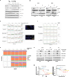
Fig. 2. Target engagement and cellular mechanism of action of AZD1390.
(A) AZD1390 cellular target engagement using phospho-Ser1981 ATM and downstream pathway modulation demonstrating dose-dependent (0 to 300 nM) target engagement (pATM) in LN18 GBM cells at 4-hour time points. Effect of AZD6738 (ATR inhibitor), AZD1775 (Wee1 inhibitor), and AZD2281 (PARP inhibitor, olaparib/Lynparza) selective clinical inhibitors. (B) AZD1390 cellular target engagement using phospho-KAP1 (pKAP1) and other downstream biomarkers demonstrating dose-dependent target engagement in NCI-H2228 lung cells after 1 and 6 hours of incubation with drug. A drug-washout time course (right) was used to monitor evidence of pathway reactivation after 6 hours from AZD1390 removal. (C) Number of pATM foci, γH2AX foci, and nuclear condensation/fragmentation detected in NCI-H2228 cells by immunofluorescence at various time points following various single doses of IR and effect of dosing AZD1390 before IR exposure in vitro. (D) Cell cycle phases as measured by DNA content of NCI-H2228 following 2- and 4-Gy IR at various doses of AZD1390. (E) Number of pATM, γH2AX foci, and micronuclei in cells gated by cell cycle phase at various time points after NCI-H2228 cells were treated with 2- or 4-Gy IR at various doses of AZD1390. (F) ATM pathway modulation by AZD1390 in three GBM cell lines indicated. Reduction in IR-induced cell cycle checkpoint (pChk2) by AZD1390 after 6 hours of incubation in the drug, confirmed in three p53 mutant GBM cell lines indicated. (G) Radiosensitization by AZD1390 (10 nM, orange curves) in p53 mutant and wild-type GBM cell lines in colony formation assays compared to DMSO control (black curves). AZD1390 was added to cells 1 hour before IR and incubated for 10 to 14 days before colonies were counted. n = 3, and error bars are SD. DEF37, dose enhancement factors at 37% survival.
Fig. 3
Fig. 3. Pharmacokinetics and pharmacodynamics of AZD1390.
(A) Correlation between free brain exposures of AZD1390 (PK) with PD modulation of pATM (Ser1981) in NCI-H2228 orthotopic lung-brain tumor model. Lower panel shows associated IHC PD images taken at various time points following IR ± AZD1390 (20 mg/kg). (B) pRad50 (Ser635) modulation following IR ± AZD1390 (20 mg/kg), with right panel showing representative IHC images (fig. S4B shows, in more detail, the scoring criteria for staining using this clinically validated pRAD50 IHC antibody). (C) Induction of apoptosis as measured by CC3 staining in the above tumor samples. Error bars indicate SD.
Fig. 4
Fig. 4. In vivo activity of AZD1390 in lung-brain metastatic models.
(A) Tumor growth was measured by bioluminescence, with inset images showing tumor growth rate measured using a Xenogen IVIS-200 imaging system from the start of treatment and assessed by the mean change in bioluminescence intensity. The first panel represents tumor growth relative to control background signal and associated survival plots shown using AZD1390 in combination with IR in the NCI-H2228 intracranial injection into the brain (ICB) model. The same animals used for tumor growth were assessed for survival in the second panel. (B) The first panel represents tumor growth effects as measured by bioluminescence relative to control background signal and associated survival plots using AZD1390 in combination with IR and TMZ in the NCI-H2228 ICB model. The same animals used for tumor growth were assessed for survival in the second panel. (C) The first panel represents tumor growth effects as measured by bioluminescence relative to control background signal and associated survival plots using AZD1390 in combination with IR in the NCI-H2228 ICA model. The same animals used for tumor growth were assessed for survival in the second panel.
Fig. 5
Fig. 5. Survival of a syngeneic mouse model of GBM treated with AZD1390.
(A) Survival of GL261 tumor-bearing mice dosed with AZD1390 (20 mg/kg) plus 2 × 5-Gy focal beam IR delivered by small-animal radiation research platform (SARRP). (B) Survival of GL261 tumor-bearing mice dosed with AZD1390 (5 mg/kg) plus 10 × 2-Gy focal beam IR (P = 0.0006 for AZD1390 + IR versus IR alone).
Fig. 6
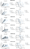
Fig. 6. In vivo subcutaneous efficacy studies using PDX models.
Mice were shielded from radiation except for the tumor region and exposed to 2 Gy × 5 days of IR [x-ray therapy (XRT)] and dosed QD with vehicle or AZD1390 1 hour before IR. Tumor growth (left column) and survival (right column) were indicated.

References

    1. Ajaz M., Jefferies S., Brazil L., Watts C., Chalmers A., Current and investigational drug strategies for glioblastoma. Clin. Oncol. R. Coll. Radiol. 26, 419–430 (2014). - PubMed
    1. Delgado-López P. D., Corrales-García E. M., Survival in glioblastoma: A review on the impact of treatment modalities. Clin. Transl. Oncol. 18, 1062–1071 (2016). - PubMed
    1. Lin X., DeAngelis L. M., Treatment of brain metastases. J. Clin. Oncol. 33, 3475–3484 (2015). - PMC - PubMed
    1. Smith G. C. M., Cary R. B., Lakin N. D., Hann B. C., Teo S.-H., Chen D. J., Jackson S. P., Purification and DNA binding properties of the ataxia-telangiectasia gene product ATM. Proc. Natl. Acad. Sci. U.S.A. 96, 11134–11139 (1999). - PMC - PubMed
    1. Bakkenist C. J., Beumer J. H., Schmitz J. C., ATM serine-1981 phosphorylation is a plausible biomarker. Cell Cycle 14, 3207–3208 (2015). - PMC - PubMed

Publication types

MeSH terms

Substances