. 2018 Jul 24;18(15):2146-2155.
doi: 10.1039/c8lc00035b.

Anti-thrombotic strategies for microfluidic blood processing

Affiliations

Anti-thrombotic strategies for microfluidic blood processing

Keith H K Wong et al. Lab Chip. .

Abstract

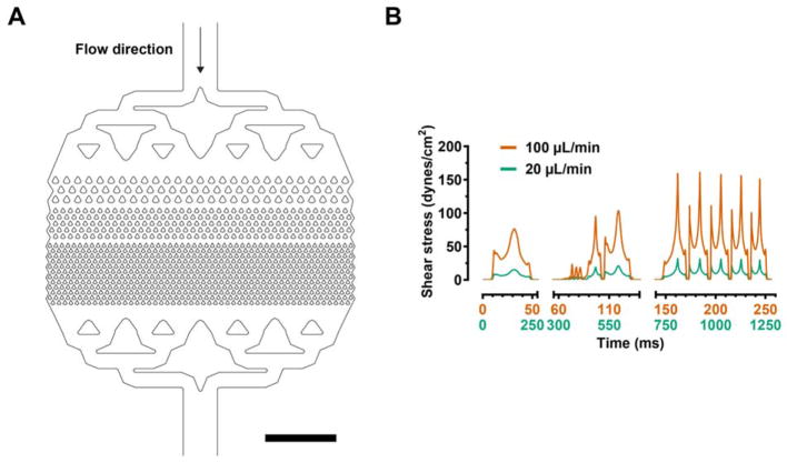
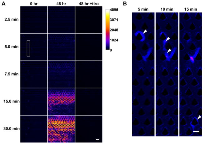
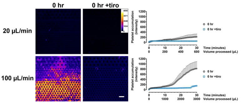
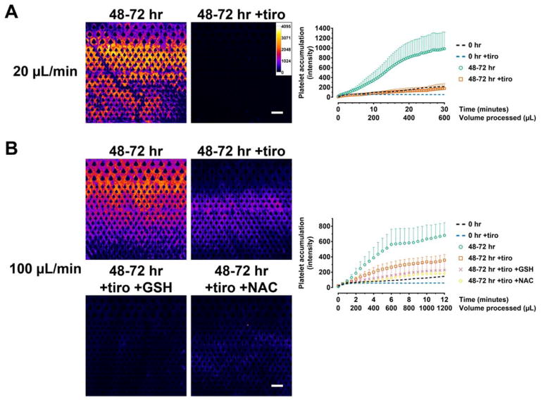
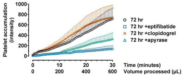
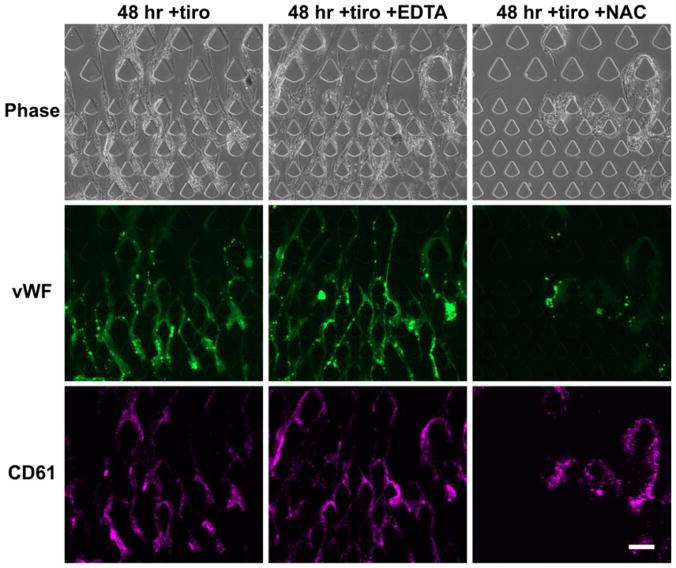
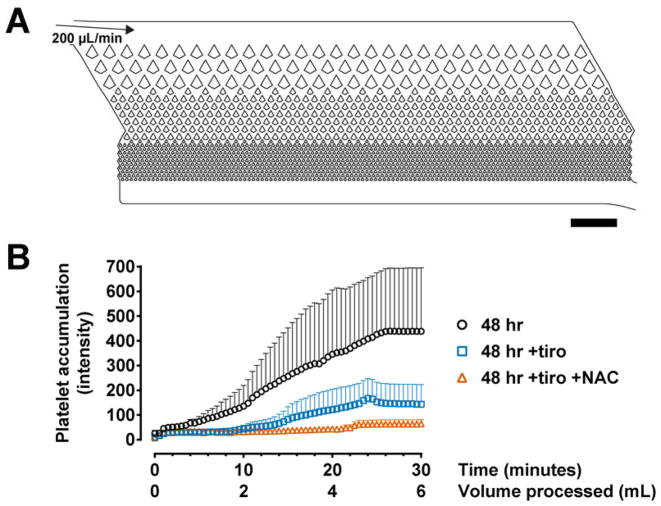
The redundant mechanisms involved in blood coagulation are crucial for rapid hemostasis. Yet they also create challenges in blood processing in medical devices and lab-on-a-chip systems. In this work, we investigate the effects of both shear stress and hypothermic blood storage on thrombus formation in microfluidic processing. For fresh blood, thrombosis occurs only at high shear, and the glycoprotein IIb/IIIa inhibitor tirofiban is highly effective in preventing thrombus formation. Blood storage generally activates platelets and primes them towards thrombosis via multiple mechanisms. Thrombus formation of stored blood at low shear can be adequately inhibited by glycoprotein IIb/IIIa inhibitors. At high shear, von Willebrand factor-mediated thrombosis contributes significantly and requires additional treatments with thiol-containing antioxidants-such as N acetylcysteine and reduced glutathione-that interfere with von Willebrand factor polymerization. We further demonstrate the effectiveness of these anti-thrombotic strategies in microfluidic devices made of cyclic olefin copolymer, a popular material used in the healthcare industry. This work identifies effective anti-thrombotic strategies that are applicable in a wide range of blood- and organ-on-a-chip applications.


Figures

Figure 1
Figure 1
(A) Computer-Aided Design (CAD) representation of the microfluidic filter device and (B) the fluid shear stress computed from the CFD model at the imposed bulk flow rates (20 and 100 μL/min). The blood flows through 3 stages of kite-shaped pillars with progressively narrower gap sizes (30 μm at the narrowest stage). Each plotted shear stress curve was calculated along a streamline that originated exactly one micron from the pillar side-wall in its narrowest gap, at the mid-height of the channel. One such streamline was seeded in the middle of the first, second, and third levels of the filter in (A). The surface normal for shear stress computation was then defined as the normal to the streamline with zero span-wise component due to symmetry at the midplane. The sections on the time axis correspond to the time needed for the fluid particle to travel across each stage. Scale bar represents 1 mm.
Figure 2
Figure 2
Time-lapse assay for quantifying platelet accumulation. Whole blood was labeled with DiOC6 prior to microfluidic processing and fluorescence images were acquired in 30-second intervals. The fluorescence intensity was pseudocolored for easy visualization. (A) Fresh (0 hr) blood could be processed without platelet accumulation under low shear (20 μL/min) but cold-stored (48 hr) blood resulted in stable aggregates. The presence of tirofiban (tiro) completely inhibited platelet accumulation. (B) Close-up images (with enhanced contrast) of the boxed region in (A) demonstrating that adhered platelets (from fresh blood) can be easily dislodged under flow. The arrowheads point to an aggregate (5 min) that was displaced and carried downstream over time (10 min and 15 min). Scale bar in (A) and (B) represents 100 μm and 50 μm, respectively.
Figure 3
Figure 3
Tirofiban effectively inhibits clot formation in fresh blood (0 hr) under both low- (20 μL/min) and high-shear (100 μL/min) flow conditions. Pseudocolored images show the extent of platelet accumulation at the end of the experiment (30 min). The intensity plots represent area-averaged fluorescence intensity (20 μL/min: 0 hr, n = 4; 0 hr +tiro, n = 5. 100 μL/min: 0 hr, n = 6; 0 hr +tiro, n = 5).
Figure 4
Figure 4
Anti-thrombotic strategies for sensitized (cold-stored) whole blood. (A) Hypothermic blood storage results in platelet activation that generally potentiates clot formation. Stored blood forms clots rapidly even under low shear. The presence of tirofiban in blood storage effectively inhibited clot formation. n = 5 each for 48–72 hr and 48–72 hr +tiro. (B) Under high shear, tirofiban reduced clot formation but the amount of clot was still significant. The addition of GSH and NAC prior to blood processing further reduced clot formation. 48–72 hr, n = 6; 48–72 hr +tiro, n = 17; 48–72 hr +tiro +GSH, n = 9; 48–72 hr +tiro +NAC, n = 9. Data from fresh blood (0 hr data from Figure 3) are included as dotted lines as a baseline reference but is not used for statistical comparisons. Scale bars represent 100 μm.
Figure 5
Figure 5
Mechanisms of storage-induced activation. Blood was stored with the indicated anti-platelet agents for 72 hours and processed at low shear. Eptifibatide, another clinically used GPIIb/IIIa inhibitor, effectively inhibited clot formation. Apyrase, an ADP scavenger, also resulted in clot reduction but was not as effective as eptifibatide. Clopidogrel, a P2Y12 inhibitor, did not reduce clot formation. 72 hr, n = 7; 72 hr +eptifibatide, n = 5; 72 hr +clopidogrel, n = 7; 72 hr +apyrase, n = 7.
Figure 6
Figure 6
High-shear processing of stored blood results in vWF-mediated thrombosis. Immunofluorescence staining identified vWF fibers that co-localize with platelet accumulation. Treatment with EDTA, which chelates diavlent ions required in the coagulation cascade, did not inhibit vWF fiber formation or platelet accumulation. Instead, the vWF fibers appeared longer and more continuous. The addition of NAC during blood processing effectively inhibited vWF fiber formation. Scale bar represents 100 μm.
Figure 7
Figure 7
Anti-thrombotic strategies are effective in microfluidic devices made of materials other than PDMS. (A) CAD representation of the microfluidic filter in the CTC-iChip manufactured using injection-molded COC plastic. The channel depth is 52 μm and the narrowest constrictions have a gap size of 30 μm. (B) Treatments with tirofiban and NAC produced similar anti-thrombotic effects at high shear as observed in PDMS devices. 48 hr, n = 6; 48 hr +tiro, n = 7; 48 hr +tiro +NAC, n = 7. Scale bar represents 1 mm.

References

    1. Toner M, Irimia D. Blood-on-a-chip. Annu Rev Biomed Eng. 2005;7:77–103. - PMC - PubMed
    1. Davis JA, et al. Deterministic hydrodynamics: taking blood apart. Proc Natl Acad Sci U S A. 2006;103:14779–84. - PMC - PubMed
    1. Yu ZTF, Aw Yong KM, Fu J. Microfluidic blood cell sorting: now and beyond. Small. 2014;10:1687–703. - PMC - PubMed
    1. Kang JH, et al. An extracorporeal blood-cleansing device for sepsis therapy. Nat Med. 2014;20:1211–6. - PubMed
    1. Cheng X, et al. A microfluidic device for practical label-free CD4(+) T cell counting of HIV-infected subjects. Lab Chip. 2007;7:170–8. - PMC - PubMed

Publication types