

An important step towards a prevascularized islet macroencapsulation device-effect of micropatterned membranes on development of endothelial cell network
- PMID: 29938334
- PMCID: PMC6018599
- DOI: 10.1007/s10856-018-6102-0
An important step towards a prevascularized islet macroencapsulation device-effect of micropatterned membranes on development of endothelial cell network
Abstract
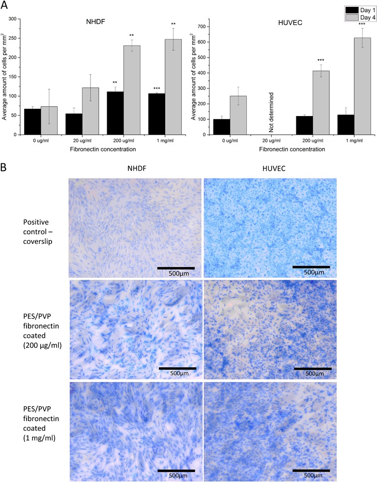
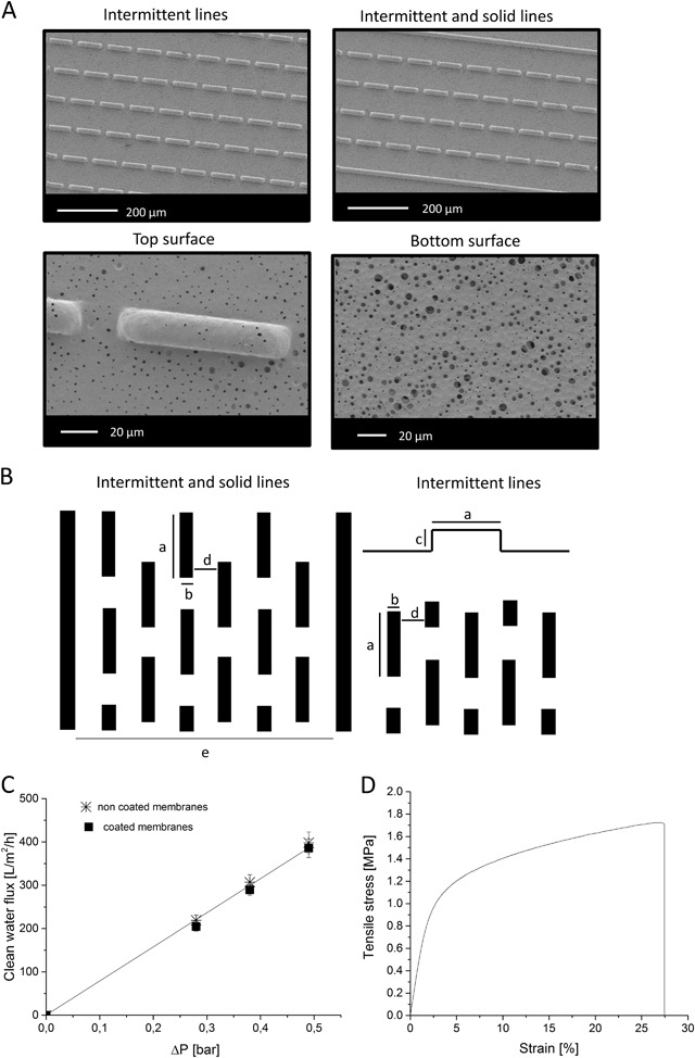
The development of immune protective islet encapsulation devices could allow for islet transplantation in the absence of immunosuppression. However, the immune protective membrane / barrier introduced there could also impose limitations in transport of oxygen and nutrients to the encapsulated cells resulting to limited islet viability. In the last years, it is well understood that achieving prevascularization of the device in vitro could facilitate its connection to the host vasculature after implantation, and therefore could provide sufficient blood supply and oxygenation to the encapsulated islets. However, the microvascular networks created in vitro need to mimic well the highly organized vasculature of the native tissue. In earlier study, we developed a functional macroencapsulation device consisting of two polyethersulfone/polyvinylpyrrolidone (PES/PVP) membranes, where a bottom microwell membrane provides good separation of encapsulated islets and the top flat membrane acts as a lid. In this work, we investigate the possibility of creating early microvascular networks on the lid of this device by combining novel membrane microfabrication with co-culture of human umbilical vein endothelial cell (HUVEC) and fibroblasts. We create thin porous microstructured PES/PVP membranes with solid and intermittent line-patterns and investigate the effect of cell alignment and cell interconnectivity as a first step towards the development of a stable prevascularized layer in vitro. Our results show that, in contrast to non-patterned membranes where HUVECs form unorganized HUVEC branch-like structures, for the micropatterned membranes, we can achieve cell alignment and the co-culture of HUVECs on a monolayer of fibroblasts attached on the membranes with intermittent line-pattern allows for the creation of HUVEC branch-like structures over the membrane surface. This important step towards creating early microvascular networks was achieved without the addition of hydrogels, often used in angiogenesis assays, as gels could block the pores of the membrane and limit the transport properties of the islet encapsulation device.
Conflict of interest statement
The authors declare that they have no conflict of interest.
Figures


References
MeSH terms
Substances
Grants and funding
LinkOut - more resources
Full Text Sources
Other Literature Sources
Medical
