. 2018 Nov 15;12(6):664-673.
doi: 10.5009/gnl18072.

Regulatory Dendritic Cells Induced by Mesenchymal Stem Cells Ameliorate Dextran Sodium Sulfate-Induced Chronic Colitis in Mice

Affiliations

Regulatory Dendritic Cells Induced by Mesenchymal Stem Cells Ameliorate Dextran Sodium Sulfate-Induced Chronic Colitis in Mice

Hannah Jo et al. Gut Liver. .

Abstract

Background/aims: Regulatory dendritic cells (rDCs), which can be induced by mesenchymal stem cells (MSCs), play an important role in inducing and maintaining homeostasis of regulatory T cells and exhibit anti-inflammatory functions. In this study, we investigated whether MSCs could differentiate DCs into rDCs and compared the therapeutic effects of rDCs and MSCs on dextran sodium sulfate (DSS)-induced chronic colitis mice.

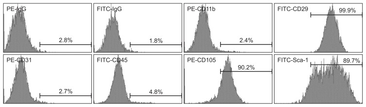
Methods: Immature DCs (imDCs) and lipopolysaccharide (LPS)-treated mature DCs (mDCs) were co-cultured with MSCs for 48 hours, and then the profiles of surface markers and cytokines and regulatory roles of these DCs for primary splenocytes were analyzed. In addition, the therapeutic effects of MSCs and DCs co-cultured with MSCs were compared in chronic colitis mice.

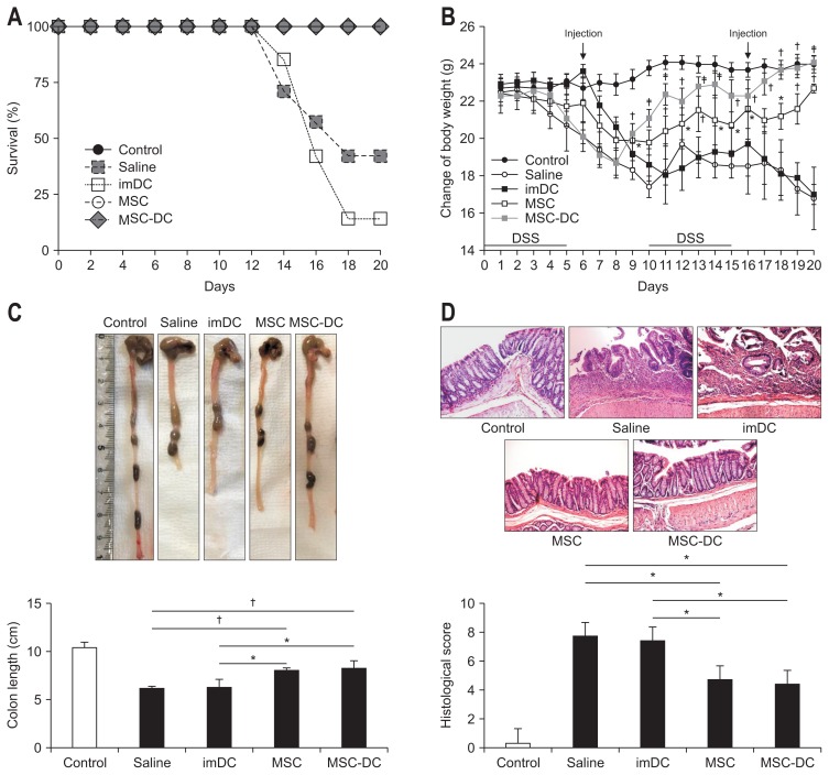
Results: After co-culture of imDCs (MSC-DCs) or LPS-treated mDCs (LPS+MSC-DCs) with MSCs, the expression of CD11c, CD80, CD86, interleukin 6 (IL-6), tumor necrosis factor-α (TNF-α), and interferon-γ (IFN-γ), was decreased, but that of CD11b, IL-10, and transforming growth factor-β (TGF-β) was increased. Furthermore, MSC-DCs and LPS+MSC-DCs induced the expression of CD4, CD25, and Foxp3 in primary splenocytes isolated from mice. In DSS-induced colitis mice, MSCs and MSC-DCs increased colon length, body weight, and survival rate and induced histological improvement. Moreover, in the colon tissues, the expression of IL-6, TNF-α, and IFN-γ decreased, but that of IL-10, TGF-β, and Foxp3 increased in the MSC- and MSC-DC-injected groups.

Conclusions: Our data suggest that MSCs differentiate DCs into rDCs, which ameliorate chronic colitis. Thus, rDCs stimulated by MSCs may be therapeutically useful for the treatment of chronic inflammatory diseases.

Keywords: Anti-inflammatory effect; Dendritic cells; Inflammatory bowel diseases; Mesenchymal stromal cells; T-lymphocytes, regulatory.


Conflict of interest statement

CONFLICTS OF INTEREST

No potential conflict of interest relevant to this article was reported.

Figures

Fig. 1
Fig. 1
The surface markers of mesenchymal stem cells (MSCs) were analyzed by flow cytometry. MSCs were negative for CD45 and CD11b (hematopoietic cell markers) and CD31 (endothelial cell marker). MSCs were positive for Sca-1 (progenitor cell marker) and CD105 and CD29. PE, phycoerythrin; FITC, fluorescein isothiocyanate; Sca, stem cell antigen.
Fig. 2
Fig. 2
Mesenchymal stem cells (MSCs) induce immature dendritic cells (imDCs) and lipopolysaccharide (LPS)-DCs into a distinct DC subset. LPS-DCs and imDCs were co-cultured with MSCs with or without LPS for 2 days. (A) Surface markers (i.e., CD11b, CD11c, CD80, and CD86) were analyzed by flow cytometry. (B) The expression of pro- (IL-6, TNF-α, and IFN-γ) and anti-inflammatory (IL-10 and TGF-β) cytokines was detected in four kinds of DCs (i.e., imDCs, LPS-DCs, MSC-DCs, and LPS+MSC-DCs) by reverse transcription-polymerase chain reaction, and then, relative mRNA expression was analyzed by densitometry to compare the expression levels (LPS-DCs vs MSC-DCs groups and LPS-DCs vs LPS+MSC-DCs groups). Statistical analysis was performed using a one-way analysis of variance. β-Actin was used as a loading control. Data are expressed as the mean±standard error of the mean in triplicate experiments. IL, interleukin; TNF, tumor necrosis factor; IFN, interferon; TGF, transforming growth factor. *p<0.05, p<0.001, and p<0.0001.
Fig. 3
Fig. 3
MSC-DCs induce differentiation of splenocytes into regulatory T cells (Tregs). The spleens were isolated from healthy and young C57BL/6 female mice. Naïve T-cell-enriched splenocytes were co-cultured with each group for 48 hours (imDCs, LPS-DCs, MSC-DCs, or LPS+MSC-DCs:splenocytes=1:5). CD4, CD25, and Foxp3 were used as markers of Tregs, and their mRNA levels were detected by reverse transcription-polymerase chain reaction (A). The levels of Foxp3 protein were assessed by Western blotting (B). MSCs, mesenchymal stem cells; DCs, dendritic cells; imDC, immature DCs; LPS, lipopolysaccharide. *p<0.001, and p<0.0001.
Fig. 4
Fig. 4
MSC-DCs ameliorate dextran sodium sulfate (DSS)-induced chronic colitis in mice. Saline or 1×10⁶ cells (imDCs, MSCs, and MSC-DCs) were administered by two intraperitoneal injections on days 6 and 16. (A) Survival rates of mice were analyzed (n=7). Injection with MSC-DCs significantly improved the survival rate. (B) The body weights of mice were recorded during the experiment (n=5). Treatment with MSCs and MSC-DCs significantly decreased the weight loss caused by DSS-induced colitis. (C) Colon length for each group (n=5). MSC-DCs prevented the shortening of the colon length (p=0.003). (D) Representative images of the colon tissue sections for histological examination (H&E, ×20). The saline group had the highest inflammatory score; in contrast, the MSC-DCs group had the lowest score (p=0.03). Statistical analysis was performed using a one-way analysis of variance. MSCs, mesenchymal stem cells; DCs, dendritic cells; imDC, immature DCs. *p<0.05, p<0.001.
Fig. 5
Fig. 5
MSC-DCs produce anti-inflammatory cytokines in dextran sodium sulfate (DSS)-induced chronic colitis mice. (A) Reverse transcription-polymerase chain reaction was performed to assess the mRNA levels of interleukin (IL)-6, tumor necrosis factor (TNF)-α, interferon (IFN)-γ, IL-10, and transforming growth factor (TGF)-β. (B) Western blotting was performed to analyze the expression levels of total STAT3, phospho-STAT3, TGF-β, and IL-10. MSCs, mesenchymal stem cells; DCs, dendritic cells. *p<0.05, p<0.001, and p<0.0001.
Fig. 6
Fig. 6
Both mesenchymal stem cells (MSCs) and MSC-DCs induce regulatory T cell (Treg) differentiation in vivo. (A) Immunohistochemical staining (×100) of Foxp3+ cells in the colon tissues. Representative images from one experiment are shown. Foxp3+ cells were counted from three random fields, with over 200 cells counted per field. (B) The number of Foxp3+ cells was expressed as a percentage of the total cell number. Foxp3 in the colon tissues was detected by Western blotting. Statistical analysis was performed using a one-way analysis of variance. DC, dendritic cells; imDC, immature DCs. *p<0.05, p<0.001, and p<0.0001.

Similar articles

Cited by

References

    1. Zhang YZ, Li YY. Inflammatory bowel disease: pathogenesis. World J Gastroenterol. 2014;20:91–99. doi: 10.3748/wjg.v20.i1.91. - DOI - PMC - PubMed
    1. Bouma G, Strober W. The immunological and genetic basis of inflammatory bowel disease. Nat Rev Immunol. 2003;3:521–533. doi: 10.1038/nri1132. - DOI - PubMed
    1. Klinker MW, Wei CH. Mesenchymal stem cells in the treatment of inflammatory and autoimmune diseases in experimental animal models. World J Stem Cells. 2015;7:556–567. doi: 10.4252/wjsc.v7.i3.556. - DOI - PMC - PubMed
    1. Duijvestein M, van den Brink GR, Hommes DW. Stem cells as potential novel therapeutic strategy for inflammatory bowel disease. J Crohns Colitis. 2008;2:99–106. doi: 10.1016/j.crohns.2007.12.002. - DOI - PubMed
    1. Dalal J, Gandy K, Domen J. Role of mesenchymal stem cell therapy in Crohn’s disease. Pediatr Res. 2012;71(4 Pt 2):445–451. doi: 10.1038/pr.2011.56. - DOI - PubMed

LinkOut - more resources