. 2018 Aug 1;128(8):3319-3332.
doi: 10.1172/JCI98194. Epub 2018 Jun 25.

The COPII cargo adapter SEC24C is essential for neuronal homeostasis

Affiliations

The COPII cargo adapter SEC24C is essential for neuronal homeostasis

Bo Wang et al. J Clin Invest. .

Abstract

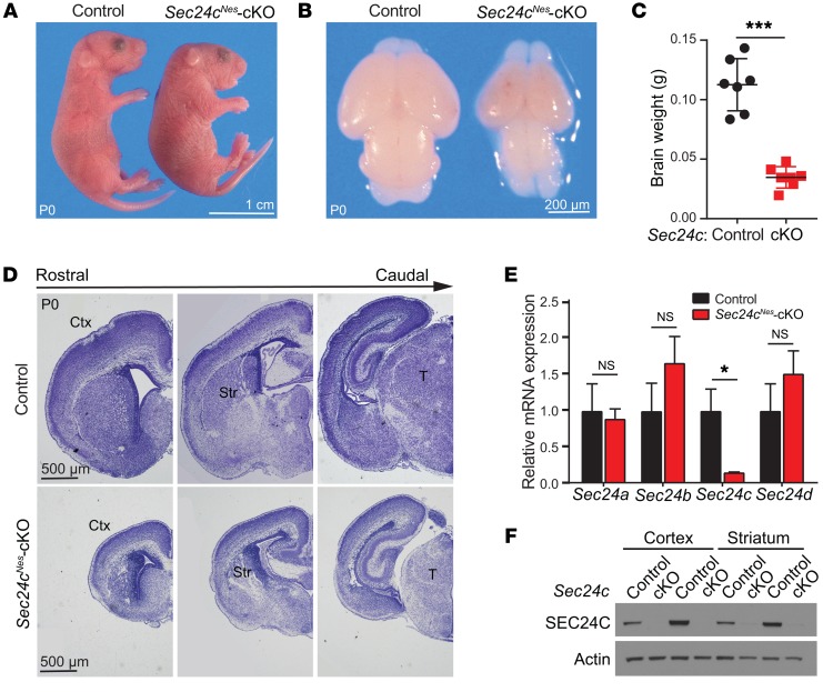
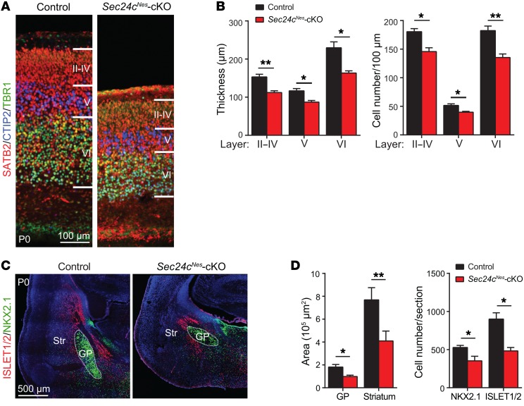
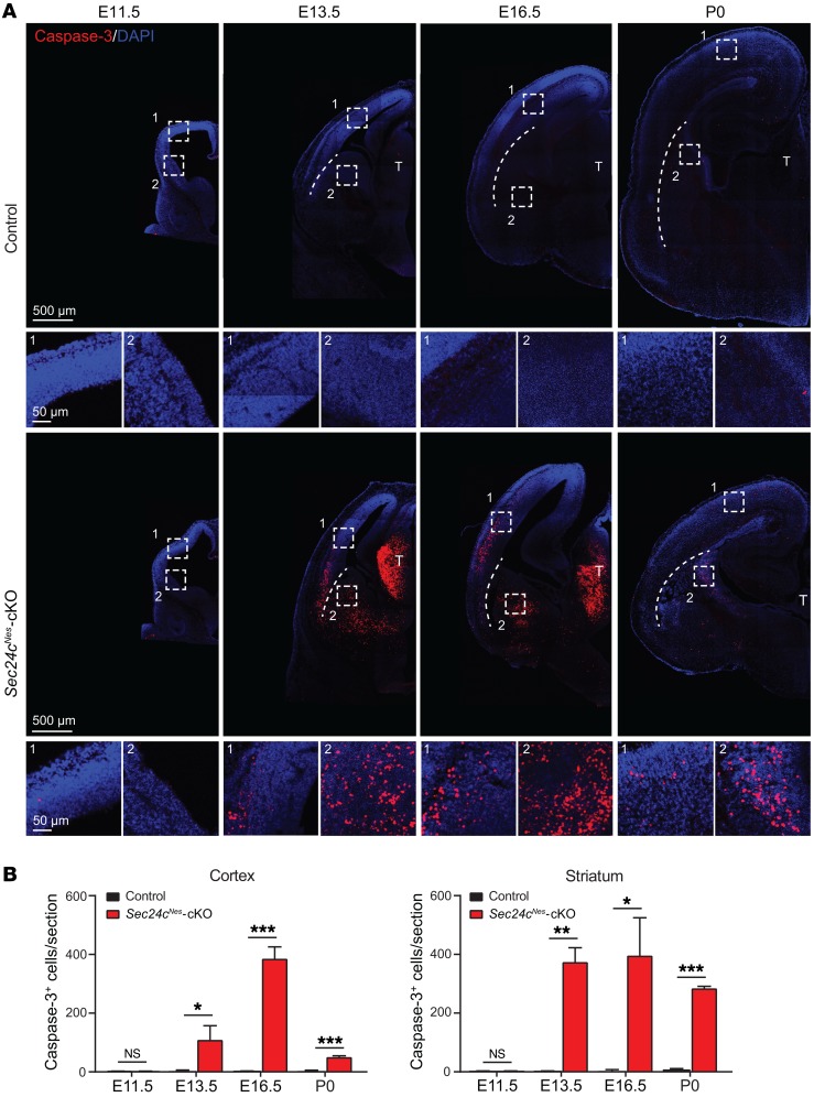
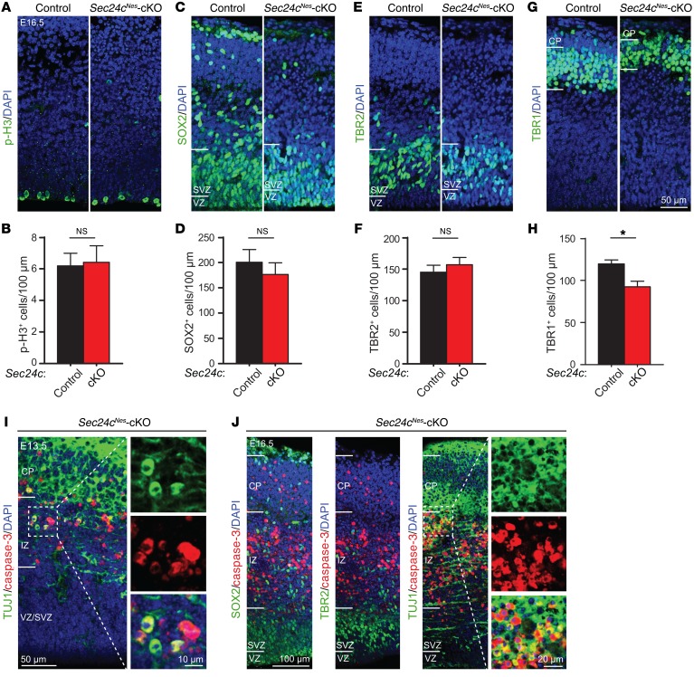
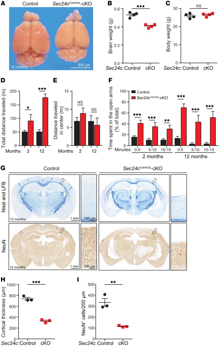
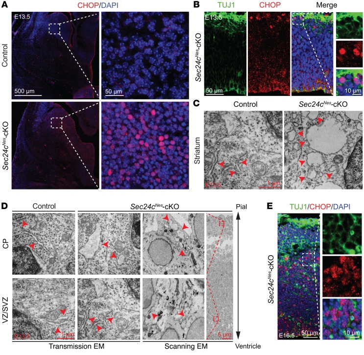
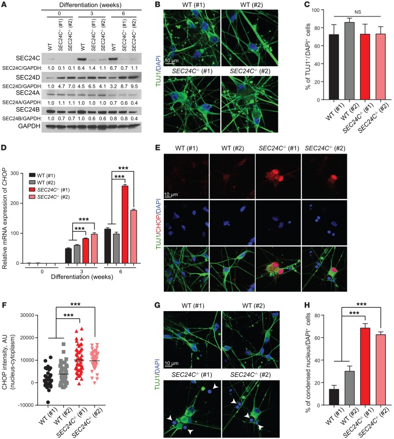
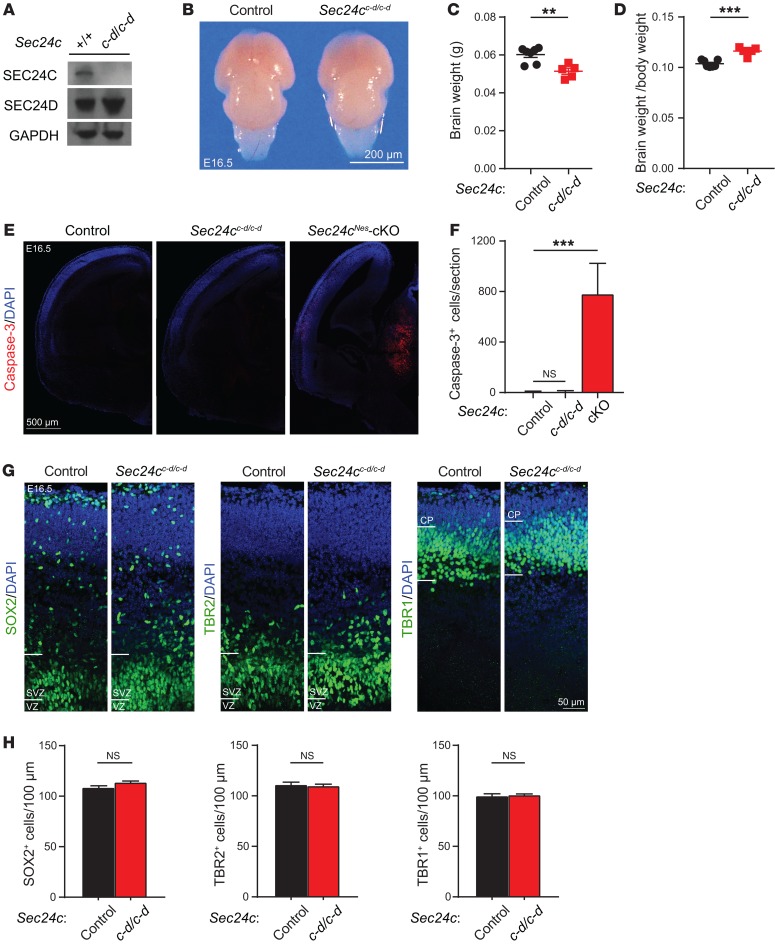
SEC24 family members are components of the coat protein complex II (COPII) machinery that interact directly with cargo or with other adapters to ensure proper sorting of secretory cargo into COPII vesicles. SEC24C is 1 of 4 mammalian SEC24 paralogs (SEC24A-D), which segregate into 2 subfamilies on the basis of sequence homology (SEC24A/SEC24B and SEC24C/SEC24D). Here, we demonstrate that postmitotic neurons, unlike professional secretory cells in other tissues, are exquisitely sensitive to loss of SEC24C. Conditional KO of Sec24c in neural progenitors during embryogenesis caused perinatal mortality and microcephaly, with activation of the unfolded protein response and apoptotic cell death of postmitotic neurons in the murine cerebral cortex. The cell-autonomous function of SEC24C in postmitotic neurons was further highlighted by the loss of cell viability caused by disrupting Sec24c expression in forebrain neurons of mice postnatally and in differentiated neurons derived from human induced pluripotent stem cells. The neuronal cell death associated with Sec24c deficiency was rescued in knockin mice expressing Sec24d in place of Sec24c. These data suggest that SEC24C is a major cargo adapter for COPII-dependent transport in postmitotic neurons in developing and adult brains and that its functions overlap at least partially with those of SEC24D in mammals.

Keywords: Cell stress; Development; Neurodegeneration; Neurodevelopment; Neuroscience.


Conflict of interest statement

Conflict of interest: The authors have declared that no conflict of interest exists.

Figures

Figure 1
Figure 1. CNS-specific deletion of Sec24c leads to microcephaly.
(A) Representative photographs of control and Sec24cNes-cKO mice at P0. (B) Representative images of dissected brains from control and Sec24cNes-cKO mice. (C) Brain weights (mean ± SEM) of Sec24cNes-cKO mice (n = 7) and control mice (n = 7) at P0. (D) Nissl staining of brain sections along the rostrocaudal axis in control and Sec24cNes-cKO mice at P0. (E) Sec24a, Sec24b, Sec24c, and Sec24d mRNA levels (mean ± SEM) in the cortices of Sec24cNes-cKO (n = 5) and control (n = 5) mice. (F) Immunoblot analyses of cortical and striatal extracts confirming the loss of SEC24C protein in Sec24cNes-cKO mice. n = 2 for each genotype. Scale bars: 1 cm (A), 200 μm (B), and 500 μm (D). cKO, conditional knockout; Ctx, cortex; Str, striatum; T, thalamus. *P < 0.05 and ***P < 0.001, by Student’s t test.
Figure 2
Figure 2. The brains of Sec24c-deficient mice contain fewer neurons than brains of control mice.
(A) Representative images of layers II–VI in control (n = 3) and Sec24cNes-cKO (n = 3) mice at P0, as delineated by expression of the callosal neuron marker SATB2, the layer V marker CTIP2, and the layer VI marker TBR1. Scale bar: 100 μm. (B) Thickness of designated cortical layers (mean ± SEM) and numbers of neurons within those layers (mean ± SEM) from Sec24cNes-cKO (n = 3) and control (n = 3) mice. (C) Representative images of immunostained brain sections showing ISLET1/2+ neurons in the striatum and NKX2.1+ neurons in the globus pallidus of control (n = 3) and Sec24cNes-cKO (n = 3) mice at P0. Scale bar: 500 μm. (D) Mean area (± SEM) covered by and mean number (± SEM) of ISLET1/2+ and NKX2.1+ cells in the striatum and globus pallidus of Sec24cNes-cKO (n = 3) and control (n = 3) mice. GP, globus pallidus. *P < 0.05 and **P < 0.01, by Student’s t test.
Figure 3
Figure 3. Sec24c deficiency leads to widespread apoptotic cell death in embryonic brains.
(A) Representative images of cleaved caspase-3 staining in brains from control and Sec24cNes-cKO mice at E11.5, E13.5, E16.5, and P0. Magnified areas from cortex (boxed area 1) and striatum (boxed area 2) are also shown. Scale bars: 500 μm (A) and 50 μm (A, insets). (B) Number (mean ± SEM) of caspase-3+ cells in the cortex and striatum in brains from control and Sec24cNes-cKO mice at E11.5, E13.5, E16.5, and P0. n = 3 for each genotype at each age. *P < 0.05, **P < 0.01, and ***P < 0.001, by Student’s t test.
Figure 4
Figure 4. Sec24c deficiency triggers cell death in postmitotic neurons.
(AF) Representative images and mean number (± SEM) of neurons in the VZ/SVZ region of the cortex from sections stained using antibodies against p-H3 (Ser10) (A and B), the neural stem cell marker SOX2 (C and D), or the intermediate progenitor cell marker TBR2 (E and F) (n = 3 E16.5 mice per genotype). (G and H) Representative images of anti-TBR1–stained brain sections and mean number (± SEM) of mature TBR1+ neurons (mean ± SEM) in the cortical plate region (n = 3 E16.5 mice per genotype). Scale bar: 50 μm (A, C, E, and G). (I) Representative image of a brain section from a E13.5 Sec24cNes-cKO mouse (n = 2) costained using antibodies against cleaved caspase-3 and the postmitotic neuronal cell marker TUJ1. Scale bars: 50 μm and 10 μm (insets). (J) Representative images of cortices from E16.5 Sec24cNes-cKO mice (n = 2) costained using antibodies against cleaved caspase-3 and SOX2, TBR2, or TUJ1. Scale bars: 100 μm and 20 μm (insets). *P < 0.05, by Student’s t test.
Figure 5
Figure 5. Postnatal deletion of Sec24c in the forebrain leads to hyperactivity and altered anatomy in the forebrain.
(A) Representative image of fixed brains dissected from a 12-month-old Sec24cCamk2a-cKO mouse and littermate control. Scale bar: 500 μm. (B and C) Brain (B) and body (C) weights (mean ± SEM) of 12-month-old Sec24cCamk2a-cKO and control mice (n = 4 mice/genotype). (DF) Behavioral tests were performed using 2-month-old Sec24cCamk2a-cKO mice (n = 8) and age-matched controls (n = 17) and 12-month-old Sec24cCamk2a-cKO mice (n = 4) and age-matched controls (n = 5). Total distance traveled (mean ± SEM) (D) and distance traveled in the center (mean ± SEM) (E) by mice subjected to the open field test. (F) Mean time (± SEM) spent in the open arms during the indicated intervals by mice subjected to the elevated plus maze test. (GI) Brain sections from 12-month-old Sec24cCamk2a-cKO and littermate control mice (n = 3 mice/genotype) were stained with Nissl and Luxol fast blue or immunostained with anti-NeuN antibody. Representative images (G), mean cortical thickness (± SEM) (H), and mean neuronal numbers (± SEM) (I) are shown. (G) Scale bars: 1 mm and 100 μm (insets). LFB, Luxol fast blue. *P < 0.05, **P < 0.01, and ***P < 0.001, by Student’s t test.
Figure 6
Figure 6. Sec24c deficiency causes ER stress.
(A) Representative images of immunostaining against CHOP in the striatum of control (n = 3) and Sec24cNes-cKO (n = 3) mice at E13.5. Scale bars: 500 μm and 50 μm (insets). (B) Representative images of CHOP staining in the cortex of Sec24cNes-cKO (n = 3) mice at E13.5. Scale bars: 50 μm and 10 μm (insets). (C) Representative ultrastructural images of E13.5 striatal neurons showing a swollen ER (denoted by red arrowheads) in Sec24c-deficient brain (n = 1). Scale bar: 1 μm. (D) Scanning and transmission EM analyses of the ultrastructure in cells located in the CP and the VZ/SVZ in Sec24cNes-cKO mice (n = 1). The ER structure is highlighted by arrowheads. Scale bars: 1 μm and 5 μm (inset). (E) Representative images of CHOP staining in the cortex of Sec24cNes-cKO mice at E16.5 (n = 3). Scale bars: 50 μm and 10 μm (insets).
Figure 7
Figure 7. Deletion of SEC24C leads to elevated cellular stress and the demise of mature neurons derived from hiPSCs.
(A) Immunoblot analysis of cell extracts from WT and SEC24C-KO hiPSCs (clones 1 and 2) at 0, 3, and 6 weeks of neuronal differentiation. (B) Representative images of TUJ1 immunostaining of WT and SEC24C-KO hiPSCs at 6 weeks of differentiation. Scale bar: 10 μm. (C) Percentage of TUJ1+ cells per total cells (DAPI+) (mean ± SEM) shows normal differentiation of the SEC24C-KO hiPSCs compared with WT clones 1 and 2. (D) mRNA levels of CHOP in WT and SEC24C-KO hiPSCs at 0, 3, and 6 weeks of differentiation were determined by real-time RT-PCR. Data are presented as the mean ± SEM. (E) Representative images of immunostaining show pronounced nuclear localization of CHOP in SEC24C-KO hiPSCs at 6 weeks of differentiation. Scale bar: 10 μm. (F) Quantification of nucleus minus cytoplasm CHOP intensity shows significant enrichment of nuclear CHOP in SEC24C-KO hiPSCs. (G) Representative nuclear staining with DAPI and (H) quantification indicate a significant increase in the percentage of cells with condensed nuclei (mean ± SEM) in SEC24C-KO hiPSCs. Scale bar: 10 μm. Data were collected from 3 independent experiments. ***P < 0.001, by 2-way ANOVA.
Figure 8
Figure 8. SEC24D can replace SEC24C during development of the embryonic mouse brain.
(A) Western blot of brain extract prepared from Sec24c+/+ (n = 1) and Sec24cc-d/c-d mice (n = 1) shows almost complete abolishment of SEC24C and elevated expression of SEC24D. (B and C) Representative photograph (B) and mean (± SEM) fixed brain weight (C) of control (n = 7) and Sec24cc-d/c-d (n = 5) mice at E16.5. Scale bar: 200 μm. (D) Average ratio (± SEM) of brain weight to body weight in Sec24cc-d/c-d mice (n = 5) compared with that of control mice (n = 7). (E and F) Representative images of cleaved caspase-3 staining (E) and mean number (± SEM) of cleaved caspase-3+ cells revealed no obvious cell death in the brains of Sec24cc-d/c-d mice. Brain sections from Sec24cNes-cKO mice at the same age were used as a positive control (n = 3 mice/genotype). Scale bar: 500 μm. (G and H) Representative images and the number of SOX2+, TBR2+, and TBR1+ cells (mean ± SEM) were normal in the Sec24cc-d/c-d mouse brain (n = 3) compared with control mice (n = 3). Scale bar: 50 μm. **P < 0.01 and ***P < 0.001, by Student’s t test.

Similar articles

Cited by

References

    1. Venditti R, Wilson C, De Matteis MA. Exiting the ER: what we know and what we don’t. Trends Cell Biol. 2014;24(1):9–18. doi: 10.1016/j.tcb.2013.08.005. - DOI - PubMed
    1. Hunn BH, Cragg SJ, Bolam JP, Spillantini MG, Wade-Martins R. Impaired intracellular trafficking defines early Parkinson’s disease. Trends Neurosci. 2015;38(3):178–188. doi: 10.1016/j.tins.2014.12.009. - DOI - PMC - PubMed
    1. Zanetti G, Pahuja KB, Studer S, Shim S, Schekman R. COPII and the regulation of protein sorting in mammals. Nat Cell Biol. 2011;14(1):20–28. - PubMed
    1. Lee MC, Miller EA, Goldberg J, Orci L, Schekman R. Bi-directional protein transport between the ER and Golgi. Annu Rev Cell Dev Biol. 2004;20:87–123. doi: 10.1146/annurev.cellbio.20.010403.105307. - DOI - PubMed
    1. Jensen D, Schekman R. COPII-mediated vesicle formation at a glance. J Cell Sci. 2011;124(Pt 1):1–4. - PubMed

Publication types

Substances