. 2018 Dec;32(12):6796-6807.
doi: 10.1096/fj.201800374. Epub 2018 Jun 25.

Lipin-1 regulates Bnip3-mediated mitophagy in glycolytic muscle

Affiliations

Lipin-1 regulates Bnip3-mediated mitophagy in glycolytic muscle

Abdullah A Alshudukhi et al. FASEB J. 2018 Dec.

Abstract

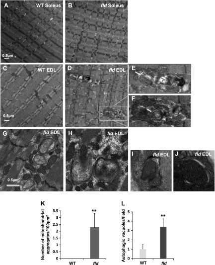
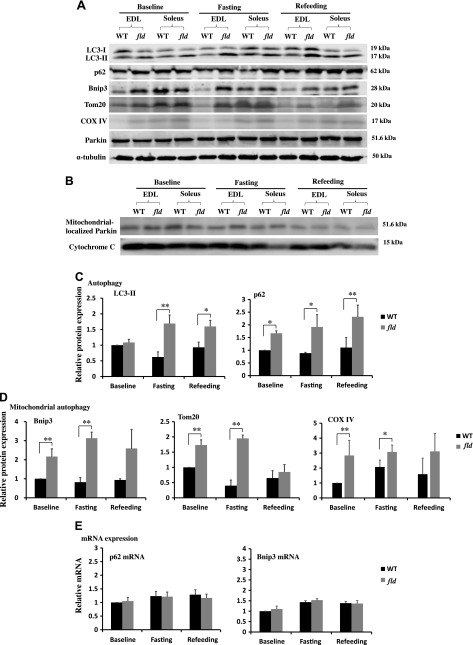
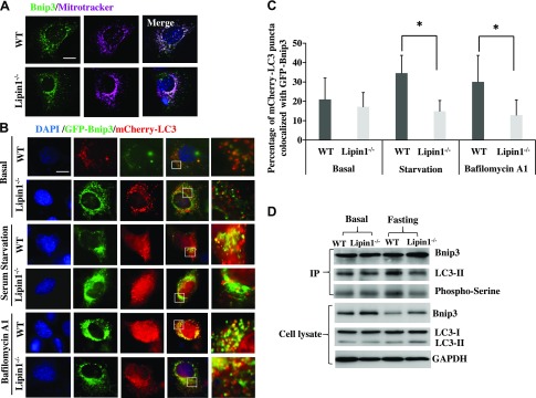
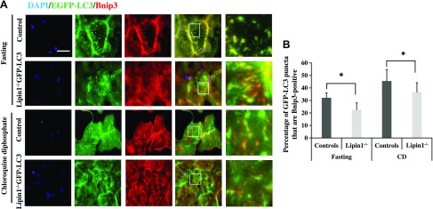
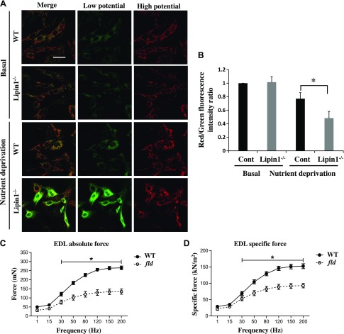
Autophagy of mitochondria (mitophagy) is essential for maintaining muscle mass and healthy skeletal muscle. Patients with heritable phosphatidic acid phosphatase lipin-1-null mutations present with severe rhabdomyolysis and muscle atrophy in glycolytic muscle fibers, which are accompanied with mitochondrial aggregates and reduced mitochondrial cytochrome c oxidase activity. However, the underlying mechanisms leading to muscle atrophy as a result of lipin-1 deficiency are still not clear. In this study, we found that lipin-1 deficiency in mice is associated with a marked accumulation of abnormal mitochondria and autophagic vacuoles in glycolytic muscle fibers. Our studies using lipin-1-deficient myoblasts suggest that lipin-1 participates in B-cell leukemia (BCL)-2 adenovirus E1B 19 kDa protein-interacting protein 3 (Bnip3)-regulated mitophagy by interacting with microtubule-associated protein 1A/1B-light chain (LC)3, which is an important step in the recruitment of mitochondria to nascent autophagosomes. The requirement of lipin-1 for Bnip3-mediated mitophagy was further verified in vivo in lipin-1-deficient green fluorescent protein-LC3 transgenic mice (lipin-1-/--GFP-LC3). Finally, we showed that lipin-1 deficiency in mice resulted in defective mitochondrial adaptation to starvation-induced metabolic stress and impaired contractile muscle force in glycolytic muscle fibers. In summary, our study suggests that deregulated mitophagy arising from lipin-1 deficiency is associated with impaired muscle function and may contribute to muscle rhabdomyolysis in humans.-Alshudukhi, A. A., Zhu, J., Huang, D., Jama, A., Smith, J. D., Wang, Q. J., Esser, K. A., Ren, H. Lipin-1 regulates Bnip3-mediated mitophagy in glycolytic muscle.

Keywords: LC3; contractile force; fld; mitochondrial autophagy; rhabdomyolysis.


Conflict of interest statement

The authors thank Dr. Noboro Mizushima (University of Tokyo, Tokyo, Japan) for the GFP-LC3 mice, Dr. Wen-Xing Ding (University of Kansas Medical School, Kansas City, KS, USA) for the GFP-Bnip3 plasmid, and Dr. Feng Zhang (McGovern Institute for Brian Research, Massachusetts Institute of Technology, Cambridge, MA, USA) for pSpCas9 (BB)-2A-Puro (PX495) v.2.0 (plasmid 62988; Addgene, Cambridge, MA, USA). This work was supported by startup funds from Wright State University (to H.R.), U.S. National Institutes of Health Center of Biomedical Research Excellence on Obesity and Cardiovascular Diseases Grant P20 GM103527-06, and Beginning Grant-in-Aid 11BGIA7710059 and Scientist Development Grant 12SDG12050697 from the American Heart Association (to H.R.). The authors declare no conflicts of interest.

Figures

Figure 1
Figure 1
Lipin-1 deficiency results in mitochondrial aggregation and an increase in autophagic vacuoles. Representative electron micrographs of soleus of WT (A) and fld (B), and EDL of WT (C) and fld (DJ) mice. The morphology of some of the vacuoles resembles autophagosomes (E, F, yellow arrows) and autolysosomes (E, blue arrows). GJ) Representative micrographs showing mitochondrion-containing autophagic vacuoles. K) Quantification of the number of mitochondrial aggregates per 100 μm2. L) Quantification of the number of autophagic vacuoles per view field. **P < 0.01.
Figure 2
Figure 2
Lipin-1 deficiency inhibits mitophagy. A) Representative immunoblots of key proteins involved in autophagy and mitophagy from EDL and soleus muscles of WT and fld mice fed with chow diet, deprived of food for 16 h (fasting) or food deprived for 16 h followed by refeeding for 5 h (refeeding). B) Western blot of Parkin in mitochondria-enriched fractions prepared from EDL and soleus muscles of WT and fld mice in basal, unfed, and refeeding states. The expression levels of cytochrome c in mitochondrial fraction were used as the loading control. C) Densitometry of LC3-II and p62 immunoblots of EDL muscles in WT and fld mice in the basal, unfed, and refeeding states. D) Densitometry of Bnip3, Tom20, and COX-IV immunoblots of EDL muscles in WT and fld mice in the basal, unfed, and refeeding states. E) Real-time RT-PCR of mRNA levels of p62 and Bnip3 in EDL muscle of WT and fld mice in the basal, unfed, and refeeding states. Data represent means ± se (n = 3/group). *P < 0.05, **P < 0.01. Experiments were repeated for at least 3 times.
Figure 3
Figure 3
Lipin-1 contributes to LC3-Bnip3–mediated autophagosome recruitment. A) Primary myoblasts isolated from WT and fld mice were transfected with GFP-Bnip3. Twenty-four hours after transfection, mitochondria were stained by Mitotracker dye. Subcellular colocalization of Bnip3 and mitochondria in the presence (WT) and absence (Lipin-1−/−) of lipin-1 were identified. Scale bar, 50 μm. B) WT and Lipin-1−/− myoblasts were transfected with GFP-Bnip3 and mCherry-LC3 plasmids. Twenty-four hours after transfection, cells were not treated, serum starved for 2 h, or serum starved for 2 h followed by 2 h of bafilomycin A1 (200 nM) treatment. Higher magnification images represent the regions enclosed in white squares. C) Quantification of the average percentage of mCherry-LC3 puncta colocalized with GFP-Bnip3. Data are means ± se. *P < 0.05. D) Lipin-1 deficiency suppressed the interaction of Bnip3 and LC3, likely through inhibition of Bnip3 phosphorylation. Lipin-1–deficient (Lipin-1−/−) and control (WT) myoblasts, transfected with GFP-Bnip3 and mcherry-LC3, were either not treated or treated with serum-free medium for 4 h. The cell lysates were subjected to immunoprecipitation with mouse anti-Bnip3 mAb. The immunoprecipitates were immunoblotted with rabbit anti-LC3 to evaluate the Bnip3-LC3 interaction or rabbit anti-phosphoserine antibody to assess Bnip3 phosphorylation.
Figure 4
Figure 4
Lipin-1 is essential in Bnip3–mediated mitophagy in vivo. A) Immunofluorescence detection of Bnip3 and GFP-LC3 in GFP-LC3 control and lipin-1−/−-GFP-LC3 transgenic mice that were food deprived for 16 h, or were treated with chloroquine diphosphate (50 mg/kg body weight, intraperitoneal injection for 10 d) followed by food deprivation for 16 h (n = 3 for each group). Higher magnification images represent the regions enclosed in white squares. EDL muscles were cryosectioned and stained with anti-Bnip3 antibody. Scale bar, 50 μm. Subcellular localization of Bnip3 (red) and GFP-LC3 (green) in GFP-LC3 control (Control) and lipin-1−/−-GFP-LC3 (Lipin-1−/−) were quantified in (B) as the percentage of GFP-LC3 puncta that are Bnip3+. Significant differences between groups were determined with Student’s t test. *P < 0.05.
Figure 5
Figure 5
Lipin-1 deficiency leads to reduced mitochondrial energy potential and impaired muscle force. A) Lipin-1 deficiency in primary myoblasts isolated from fld mice (compared with WT mice) led to reduced mitochondrial energy potential, as probed by JC-1 dye. Primary myoblasts isolated from WT and fld mice were either untreated or starved with an HBSS medium containing 10 mM HEPES for 6 h at 37°C followed by incubation with 5 μg/ml JC-1 dye at 37°C for 20 min. After extensive washing with HBSS, the medium was switched to HBSS before immediate live imaging. All images were acquired under the same instrument settings. Scale bar, 50 μm. B) The ratio of JC-1 red to green staining was subjected to image analysis to measure the mitochondrial membrane potential. Data are means ± se. *P < 0.05. Absolute force (C) and force per CSA (specific force) (D) in EDL of WT (n = 4) and fld (n = 4) mice. Tetanic contractions were elicited at 1 min intervals with stimulus frequencies of 1, 15, 30, 50, 80, 120, 150, and 200 Hz. *P < 0.05, by 1-way repeated-measures ANOVA and Bonferroni post hoc comparison.

References

    1. Schiaffino S., Reggiani C. (2011) Fiber types in mammalian skeletal muscles. Physiol. Rev. 91, 1447–1531 - PubMed
    1. Goodman C. A., Kotecki J. A., Jacobs B. L., Hornberger T. A. (2012) Muscle fiber type-dependent differences in the regulation of protein synthesis. PLoS One 7, e37890 - PMC - PubMed
    1. Sandri M. (2013) Protein breakdown in muscle wasting: role of autophagy-lysosome and ubiquitin-proteasome. Int. J. Biochem. Cell Biol. 45, 2121–2129 - PMC - PubMed
    1. He C., Klionsky D. J. (2009) Regulation mechanisms and signaling pathways of autophagy. Annu. Rev. Genet. 43, 67–93 - PMC - PubMed
    1. Choi A. M., Ryter S. W., Levine B. (2013) Autophagy in human health and disease. N. Engl. J. Med. 368, 1845–1846 - PubMed