

A high precision method for length-based separation of carbon nanotubes using bio-conjugation, SDS-PAGE and silver staining
- PMID: 29939999
- PMCID: PMC6016930
- DOI: 10.1371/journal.pone.0197972
A high precision method for length-based separation of carbon nanotubes using bio-conjugation, SDS-PAGE and silver staining
Abstract
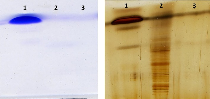
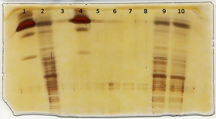
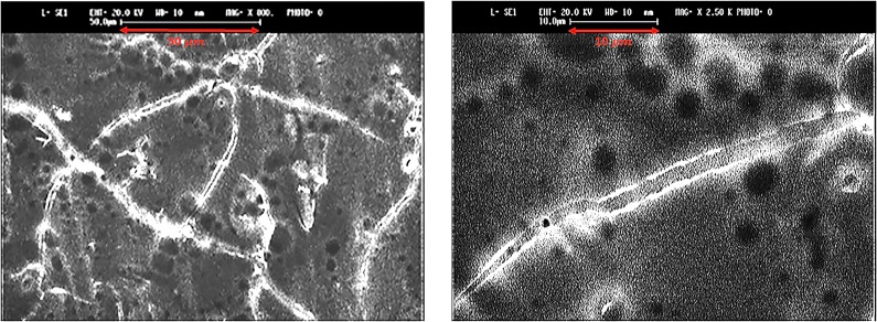
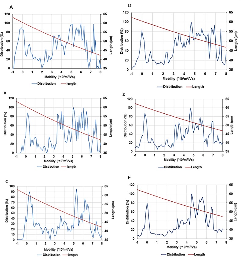
Parametric separation of carbon nanotubes, especially based on their length is a challenge for a number of nano-tech researchers. We demonstrate a method to combine bio-conjugation, SDS-PAGE, and silver staining in order to separate carbon nanotubes on the basis of length. Egg-white lysozyme, conjugated covalently onto the single-walled carbon nanotubes surfaces using carbodiimide method. The proposed conjugation of a biomolecule onto the carbon nanotubes surfaces is a novel idea and a significant step forward for creating an indicator for length-based carbon nanotubes separation. The conjugation step was followed by SDS-PAGE and the nanotube fragments were precisely visualized using silver staining. This high precision, inexpensive, rapid and simple separation method obviates the need for centrifugation, additional chemical analyses, and expensive spectroscopic techniques such as Raman spectroscopy to visualize carbon nanotube bands. In this method, we measured the length of nanotubes using different image analysis techniques which is based on a simplified hydrodynamic model. The method has high precision and resolution and is effective in separating the nanotubes by length which would be a valuable quality control tool for the manufacture of carbon nanotubes of specific lengths in bulk quantities. To this end, we were also able to measure the carbon nanotubes of different length, produced from different sonication time intervals.
Conflict of interest statement
The authors have declared that no competing interests exist.
Figures
Similar articles
-
A high precision length-based carbon nanotube ladder.RSC Adv. 2018 Oct 23;8(63):36049-36055. doi: 10.1039/c8ra05482g. eCollection 2018 Oct 22. RSC Adv. 2018. PMID: 35558502 Free PMC article.
-
Detecting Sonolysis of Polyethylene Glycol Upon Functionalizing Carbon Nanotubes.Methods Mol Biol. 2017;1530:147-164. doi: 10.1007/978-1-4939-6646-2_10. Methods Mol Biol. 2017. PMID: 28150202 Free PMC article.
-
Use of gel electrophoresis and Raman spectroscopy to characterize the effect of the electronic structure of single-walled carbon nanotubes on cellular uptake.Anal Chem. 2014 Mar 18;86(6):2882-7. doi: 10.1021/ac403827m. Epub 2014 Mar 7. Anal Chem. 2014. PMID: 24564772 Free PMC article.
-
Employing Raman spectroscopy to qualitatively evaluate the purity of carbon single-wall nanotube materials.J Nanosci Nanotechnol. 2004 Sep;4(7):691-703. doi: 10.1166/jnn.2004.116. J Nanosci Nanotechnol. 2004. PMID: 15570946 Review.
-
Controlled growth of carbon nanotubes.Philos Trans A Math Phys Eng Sci. 2004 Oct 15;362(1823):2143-60. doi: 10.1098/rsta.2004.1433. Philos Trans A Math Phys Eng Sci. 2004. PMID: 15370475 Review.
Cited by
-
Surface Modification of Curcumin Microemulsions by Coupling of KLVFF Peptide: A Prototype for Targeted Bifunctional Microemulsions.Polymers (Basel). 2022 Jan 22;14(3):443. doi: 10.3390/polym14030443. Polymers (Basel). 2022. PMID: 35160433 Free PMC article.
-
Advances and Perspectives in the Use of Carbon Nanotubes in Vaccine Development.Int J Nanomedicine. 2021 Aug 12;16:5411-5435. doi: 10.2147/IJN.S314308. eCollection 2021. Int J Nanomedicine. 2021. PMID: 34408416 Free PMC article. Review.
-
Construction of RNA nanotubes.Nano Res. 2019 Aug;12(8):1952-1958. doi: 10.1007/s12274-019-2463-z. Epub 2019 Jul 11. Nano Res. 2019. PMID: 32153728 Free PMC article.
-
Genetic diversity of Venturia inaequalis isolates from the scabs in apple trees in Gansu Province, China, using AFLP markers.PeerJ. 2022 Dec 16;10:e14512. doi: 10.7717/peerj.14512. eCollection 2022. PeerJ. 2022. PMID: 36545382 Free PMC article.
-
A Novel Method for Carbon Nanotube Functionalization Using Immobilized Candida antarctica Lipase.Nanomaterials (Basel). 2022 Apr 26;12(9):1465. doi: 10.3390/nano12091465. Nanomaterials (Basel). 2022. PMID: 35564174 Free PMC article.
References
-
- Baughman RH, Zakhidov AA, de Heer WA. Carbon nanotubes—the route toward applications. science. 2002;297(5582):787–92. doi: 10.1126/science.1060928 - DOI - PubMed
-
- Kong J, Franklin NR, Zhou C, Chapline MG, Peng S, Cho K, et al. Nanotube molecular wires as chemical sensors. science. 2000;287(5453):622–5. - PubMed
-
- Jones A, Bekkedahl T, Kiang C. Storage of hydrogen in single-walled carbon nanotubes. Nature. 1997;386:377.
-
- Chen P, Wu X, Lin J, Tan K. High H2 uptake by alkali-doped carbon nanotubes under ambient pressure and moderate temperatures. Science. 1999;285(5424):91–3. - PubMed
-
- Liu C, Fan Y, Liu M, Cong H, Cheng H, Dresselhaus MS. Hydrogen storage in single-walled carbon nanotubes at room temperature. Science. 1999;286(5442):1127–9. - PubMed
MeSH terms
Substances
LinkOut - more resources
Full Text Sources
Other Literature Sources
