

The structural basis for filovirus neutralization by monoclonal antibodies
- PMID: 29940415
- PMCID: PMC6141344
- DOI: 10.1016/j.coi.2018.05.001
The structural basis for filovirus neutralization by monoclonal antibodies
Abstract
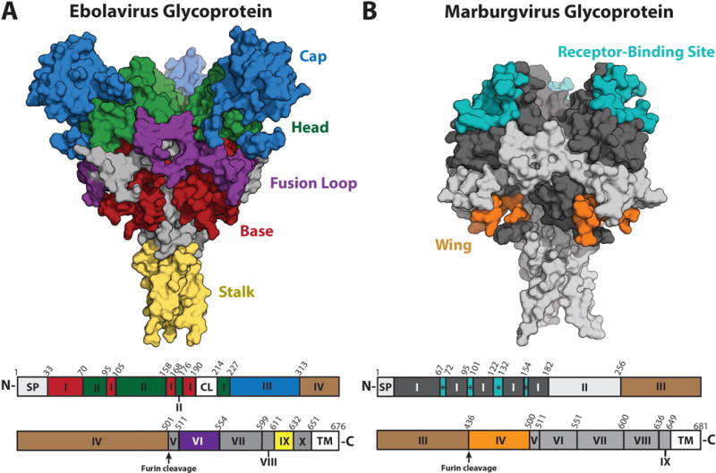
Filoviruses, including ebolaviruses and marburgviruses, are the causative agents of highly lethal disease outbreaks. The 2013-2016 Ebola virus outbreak was responsible for >28000 infections and >11000 deaths. Although there are currently no licensed vaccines or therapeutics for any filovirus-induced disease, monoclonal antibodies (mAbs) are among the most promising options for therapeutic development. Hundreds of mAbs have been isolated from human survivors of filovirus infections that target the viral spike glycoprotein (GP). The binding, neutralization, and cross-reactivity of many of these mAbs has been determined. Several mAbs have been characterized structurally, and this information has been crucial for strategizing therapeutic and vaccine design. Here we present an overview of the structural features of the neutralizing/protective epitopes on filovirus glycoproteins.
Copyright © 2018 Elsevier Ltd. All rights reserved.
Figures
Similar articles
-
Achieving cross-reactivity with pan-ebolavirus antibodies.Curr Opin Virol. 2019 Feb;34:140-148. doi: 10.1016/j.coviro.2019.01.003. Epub 2019 Mar 15. Curr Opin Virol. 2019. PMID: 30884329 Free PMC article. Review.
-
Macaque Monoclonal Antibodies Targeting Novel Conserved Epitopes within Filovirus Glycoprotein.J Virol. 2015 Oct 14;90(1):279-91. doi: 10.1128/JVI.02172-15. Print 2016 Jan 1. J Virol. 2015. PMID: 26468532 Free PMC article.
-
Pan-ebolavirus and Pan-filovirus Mouse Monoclonal Antibodies: Protection against Ebola and Sudan Viruses.J Virol. 2015 Oct 14;90(1):266-78. doi: 10.1128/JVI.02171-15. Print 2016 Jan 1. J Virol. 2015. PMID: 26468533 Free PMC article.
-
Generation and Characterization of Anti-Filovirus Nucleoprotein Monoclonal Antibodies.Viruses. 2019 Mar 14;11(3):259. doi: 10.3390/v11030259. Viruses. 2019. PMID: 30875741 Free PMC article.
-
Characterization of a putative filovirus vaccine: virus-like particles.Virol Sin. 2013 Apr;28(2):65-70. doi: 10.1007/s12250-013-3306-9. Epub 2013 Feb 6. Virol Sin. 2013. PMID: 23385315 Free PMC article. Review.
Cited by
-
Achieving cross-reactivity with pan-ebolavirus antibodies.Curr Opin Virol. 2019 Feb;34:140-148. doi: 10.1016/j.coviro.2019.01.003. Epub 2019 Mar 15. Curr Opin Virol. 2019. PMID: 30884329 Free PMC article. Review.
-
Soluble CD4 inhibits Ebola virus infection by targeting endosomal receptor-binding site.iScience. 2025 May 2;28(6):112573. doi: 10.1016/j.isci.2025.112573. eCollection 2025 Jun 20. iScience. 2025. PMID: 40491485 Free PMC article.
-
Rational design of next-generation filovirus vaccines with glycoprotein stabilization, nanoparticle display, and glycan modification.bioRxiv [Preprint]. 2025 Mar 2:2025.03.02.641072. doi: 10.1101/2025.03.02.641072. bioRxiv. 2025. PMID: 40060701 Free PMC article. Preprint.
-
Therapeutic strategies to target the Ebola virus life cycle.Nat Rev Microbiol. 2019 Oct;17(10):593-606. doi: 10.1038/s41579-019-0233-2. Epub 2019 Jul 24. Nat Rev Microbiol. 2019. PMID: 31341272 Review.
-
Proteo-Genomic Analysis Identifies Two Major Sites of Vulnerability on Ebolavirus Glycoprotein for Neutralizing Antibodies in Convalescent Human Plasma.Front Immunol. 2021 Jul 16;12:706757. doi: 10.3389/fimmu.2021.706757. eCollection 2021. Front Immunol. 2021. PMID: 34335620 Free PMC article.
References
-
-
Outbreaks Chronology: Ebola Virus Disease | Ebola Hemorrhagic Fever | CDC. 2017,
-
-
-
Outbreak Table | Marburg Hemorrhagic Fever | CDC. 2014,
-
-
- Kondratowicz AS, Lennemann NJ, Sinn PL, Davey RA, Hunt CL, Moller-Tank S, Meyerholz DK, Rennert P, Mullins RF, Brindley M, et al. T-cell immunoglobulin and mucin domain 1 (TIM-1) is a receptor for Zaire Ebolavirus and Lake Victoria Marburgvirus. Proc Natl Acad Sci U S A. 2011;108:8426–8431. - PMC - PubMed
Publication types
MeSH terms
Substances
Grants and funding
LinkOut - more resources
Full Text Sources
Other Literature Sources
