. 2018;114(3):242-250.
doi: 10.1159/000489387. Epub 2018 Jun 25.

Survival of Immunoglobulins from Human Milk to Preterm Infant Gastric Samples at 1, 2, and 3 h Postprandial

Affiliations

Survival of Immunoglobulins from Human Milk to Preterm Infant Gastric Samples at 1, 2, and 3 h Postprandial

Veronique Demers-Mathieu et al. Neonatology. 2018.

Abstract

Background: Human milk immunoglobulins (Ig) are an important support for the naïve infant immune system; yet the extent to which these proteins survive within the infant digestive tract, particularly for preterm infants, is poorly studied.

Objectives: Our objective was to evaluate the survival of human milk Igs in the preterm stomach across postprandial time.

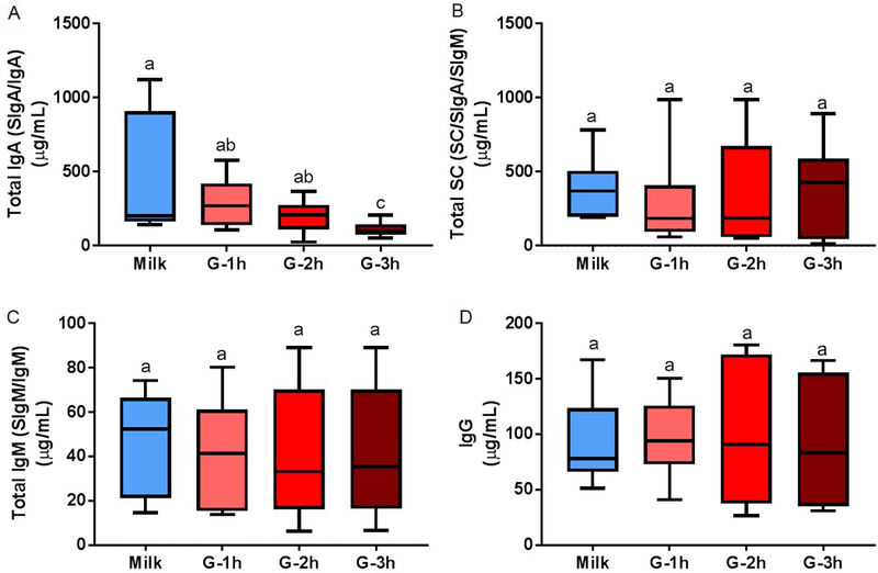
Methods: Human milk and infant gastric samples were collected from 11 preterm (23-32 weeks gestational age) mother-infant pairs within 7-98 days postnatal age. Preterm gastric samples were collected 1, 2, and 3 h after the beginning of the feeding. Samples were analyzed for concentration of total IgA (secretory IgA [SIgA]/IgA), total secretory component (SC/SIgA/SIgM), total IgM (SIgM/IgM), and IgG via enzyme-linked immunosorbent assay. Ig-chain fragment peptides were determined using peptidomic analysis. One-way analysis of variance with repeated measures followed by Tukey's multiple comparison tests was applied.

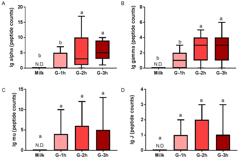
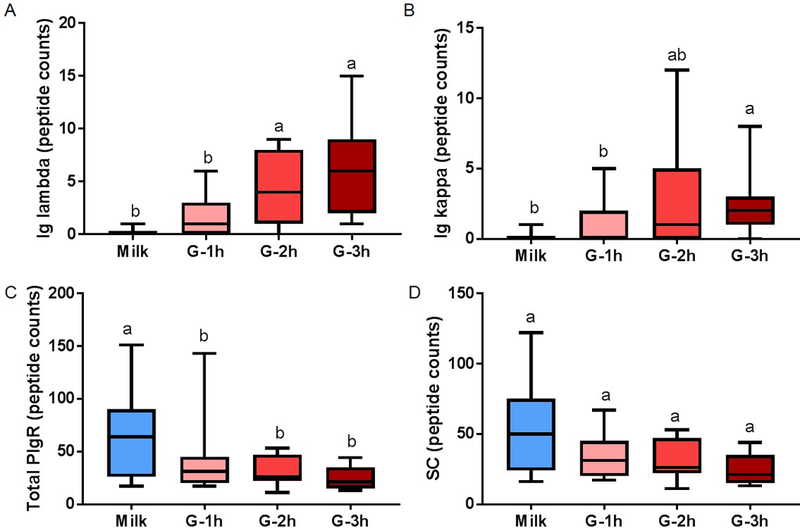
Results: Concentrations of total IgA were lower in the gastric contents at 3 h postprandial compared with human milk and gastric contents at 1 and 2 h. Human milk SC/SIgA/SIgM, IgG, and total IgM concentrations remained stable in the preterm stomach across postprandial time. Peptide counts from the Ig alpha-chain and the Ig gamma-chain increased in gastric contents from 1 to 2 h postprandial. Peptide counts from the human milk Ig-chain, Ig-chain, and SC were stable across postprandial time. These peptides from Ig-chains were not present in human milk but were released in the stomach due to their partial degradation.

Conclusions: Human milk total SC (SIgA/SC/SIgM), total IgM, and IgG survived mostly intact through the preterm infant stomach, while total IgA was -partially digested.

Keywords: Breast milk; Digestion; Gastric contents; Passive immunity; Peptidomic antibodies; Polymeric immunoglobulin receptor; Premature infants.


Conflict of interest statement

Disclosure Statement

There are no financial relationships or conflicts of interest to disclose.

Figures

Fig. 1.
Fig. 1.
Form of immunoglobulins (Ig) secreted into human milk by plasma cells. IgA is mainly secreted as a dimer IgA joined by the J-chain polypeptide and contains alpha heavy constant chains; IgG is secreted as a monomer (without a J-chain) and possesses gamma heavy constant chains; IgM is secreted as a pentamer joined by the J-chain and contains mu heavy constant chains. All Igs contain at least two light constant chains (kappa-chain or lambda-chain), two light variable region chains and two heavy variable region chains, which form the specific antigen binding site.
Fig. 2.
Fig. 2.
Schematization of the origin of IgG, secretory IgA (SIgA), IgA, secretory IgM (SIgM), and IgM in human milk. (A) Monomeric IgG is likely transported from maternal blood after binding to neonatal Fc receptors (FcRn) on the basolateral membrane of mammary epithelial cells and transported via vesicles to the alveolar lumen. (B) Plasma cells produce dimeric IgA and pentamer IgM in the interstitial spaces of the mammary gland. IgA and IgM bind to the secretory component (SC) of polymeric immunoglobulin receptor (PIgR) on the basolateral membrane of the mammary epithelial cell and the IgA-PIgR complex and IgM-PIgR complex travel to the apical membrane. PIgR is cleaved by a protease (unknown), releasing SC, which covalently binds to IgA or IgM, creating the complexes secretory IgA (SIgA) and secretory IgM (SIgM), which are secreted across the apical membrane. (C) Milk plasma cells can produce monomeric IgG, dimeric IgA and polymeric IgM in human milk inside the alveolar lumen and contribute to production of IgG, as well as IgA and IgM (without the SC).
Fig. 3.
Fig. 3.
Concentration of immunoglobulin (Ig) in human milk and preterm infant gastric samples at 1 (G-1h), 2 (G-2h) and 3 h (G-3h) after the beginning of feeding. Concentrations of (A) total IgA (secretory IgA (SIgA)/IgA), (B) total secretory component (SC/SIgA/SIgM), (D) total IgM (secretory IgM (SIgM)/IgM) and IgG were determined by ELISA. Paired milk and gastric samples were collected in preterm infants (23–32 wk of gestational age (GA) and 7–98 days of postnatal age). Letters a, b and c show statistically significant differences between groups (p < 0.05) using one-way ANOVA with repeated measures followed by Tukey’s multiple comparison tests. Values are min, median and max, n = 11.
Fig. 4.
Fig. 4.
Peptide counts of immunoglobulin (Ig) fragments in human milk and preterm infant gastric samples at 1 (G-1h), 2 (G-2h) and 3 h (G-3h) after the beginning of feeding. Peptide counts of (A) total Ig alpha-chain (from IgA or SIgA), (B) total Ig gamma-chain (from IgG), (C) Ig mu-chain (from IgM or SIgM) and (D) Ig J-chain (from IgA, SIgA, IgM or SIgM). Paired milk and gastric samples were collected in preterm-delivering mother-infant pairs (23–32 wk of gestational age (GA) and 7–98 days of postnatal age). Letters a, b and c show statistically significant differences between groups (p < 0.05) using one-way ANOVA with repeated measures followed by Tukey’s multiple comparison tests. Values are min, median and max, n = 11.
Fig. 5.
Fig. 5.
Peptide counts of immunoglobulin (Ig) fragments in human milk (blue boxplot) and preterm infant gastric samples at 1 (G-1h), 2 (G-2h) and 3 h (G-3h) after the beginning of feeding. Peptide counts of (A) Ig lambda (from IgA, SIgA, IgM, SIgM or IgG), (B) Ig kappa (from IgA, SIgA, IgM, SIgM or IgG), (C) total polymeric immunoglobulin receptor (PIgR) (f19–764) and (D) secretory component (SC, f19–603 of PIgR). Paired milk and gastric samples were collected in preterm-delivering mother-infant pairs (23–32 wk of gestational age (GA) and 7–98 days of postnatal age). Letters a, b and c show statistically significant differences between groups (p < 0.05) using one-way ANOVA with repeated measures followed by Tukey’s multiple comparison tests. Values are min, median and max, n = 11.

Similar articles

Cited by

References

    1. Oddy WH: Breastfeeding protects against illness and infection in infants and children: a review of the evidence. Breastfeed Rev 2001;9:11. - PubMed
    1. Van de Perre P: Transfer of antibody via mother’s milk. Vaccine 2003;21:3374–3376. - PubMed
    1. Goldman AS, Garza C, Nichols BL, Goldblum RM: Immunologic factors in human milk during the first year of lactation. J Pediatr 1982;100:563–567. - PubMed
    1. Chandra R: Immunoglobulin and protein levels in breast milk produced by mothers of preterm infants. Nutr Res 1982;2:27–30.
    1. Koenig Á, Diniz EMdA, Barbosa SFC, Vaz FAC: Immunologic factors in human milk: the effects of gestational age and pasteurization. J Hum Lact 2005;21:439–443. - PubMed