. 2018;14(9):1562-1573.
doi: 10.1080/15548627.2018.1462426. Epub 2018 Aug 17.

IRE1B degrades RNAs encoding proteins that interfere with the induction of autophagy by ER stress in Arabidopsis thaliana

Affiliations

IRE1B degrades RNAs encoding proteins that interfere with the induction of autophagy by ER stress in Arabidopsis thaliana

Yan Bao et al. Autophagy. 2018.

Abstract

Macroautophagy/autophagy is a conserved process in eukaryotes that contributes to cell survival in response to stress. Previously, we found that endoplasmic reticulum (ER) stress induces autophagy in plants via a pathway dependent upon AT5G24360/IRE1B (INOSITOL REQUIRING 1-1), an ER membrane-anchored factor involved in the splicing of AT1G42990/BZIP60 (basic leucine zipper protein 60) mRNA. IRE1B is a dual protein kinase and ribonuclease, and here we determined the involvement of the protein kinase catalytic domain, nucleotide binding and RNase domains of IRE1B in activating autophagy. We found that the nucleotide binding and RNase activity of IRE1B, but not its protein kinase activity or splicing target BZIP60, are required for ER stress-mediated autophagy. Upon ER stress, the RNase activity of IRE1B engages in regulated IRE1-dependent decay of messenger RNA (RIDD), in which mRNAs of secreted proteins are degraded by IRE1 upon ER stress. Twelve genes most highly targeted by RIDD were tested for their role in inhibiting ER stress-induced autophagy, and 3 of their encoded proteins, AT1G66270/BGLU21 (β-glucosidase 21), AT2G16005/ROSY1/ML (MD2-related lipid recognition protein) and AT5G01870/PR-14 (pathogenesis-related protein 14), were found to inhibit autophagy upon overexpression. From these findings, IRE1B is posited to be a 'licensing factor' linking ER stress to autophagy by degrading the RNA transcripts of factors that interfere with the induction of autophagy.

Abbreviations: ACT2: actin 2; ATG: autophagy-related; BGLU21: β-glucosidase 21; BIP3: binding protein 3; BZIP: basic leucine zipper; DAPI: 4', 6-diamidino-2-phenylindole; DTT: dithiothreitol; ER: endoplasmic reticulum; ERN1: endoplasmic reticulum to nucleus signaling 1; IRE1: inositol requiring 1; GFP: green fluorescent protein; MAP3K5/ASK1: mitogen-activated protein kinase kinase kinase 5; MAPK8/JNK1: mitogen-activated protein kinase 8/c-Jun N-terminal kinase 1; MDC: monodansylcadaverine; PR-14: pathogenesis-related protein 14; RIDD: Regulated IRE1-Dependent Decay of Messenger RNA; ROSY1/ML: interactor of synaptotagmin1/MD2-related lipid recognition protein; Tm: tunicamycin; UPR: unfolded protein response; WT: wild-type.

Keywords: Arabidopsis; ER stress; IRE1; RIDD; autophagosome; autophagy; mRNA degradation.


Figures

Figure 1.
Figure 1.
ER stress-induced autophagy in Arabidopsis seedling roots is independent of BZIP60. (a) Possible mechanisms by which IRE1B links ER stress to autophagy, either by BZIP60 splicing, protein phosphorylation, IRE1B clustering or Regulated IRE1-dependent Decay (RIDD). (b) Schematic diagram of T-DNA insertions in bzip60 mutants. Exons and intron are depicted to scale by boxes and lines, respectively. The positions of T-DNA insertions are indicated by triangles, the numbers below show the insertion sites or start and stop codons in base pair units, and the arrows below indicate primer-binding sites. (c) RT-PCR analysis of BZIP60 gene expression in wild-type (WT) and the 3 bzip60 mutants using the primers depicted in (b). Total RNA was isolated from 7-day-old seedlings. AT3G18780/ACT2 (actin 2) was used as a loading control. (d) Seven-day-old WT and 3 bzip60 mutants were transferred to 1/2 MS liquid medium plus DMSO as control, or supplemented with 5 μg/mL Tm for 6 h to induce ER stress. Autophagosomes were visualized by MDC staining and confocal microscopy. Bar: 50 μm. (e) The number of autophagosomes per root section was assessed following Tm treatment and staining by MDC. Error bars represent SE, n > 20 for 3 biological replicates. Asterisks indicate significant differences (P < 0.05) using the Student t test compared with WT under control conditions.
Figure 2.
Figure 2.
Complementation of autophagy defect in ire1a ire1b null mutant protoplasts by IRE1B mutant constructs. (a) Illustration of the disposition of IRE1B in the ER membrane. The cytoplasmic domain bears both RNase and protein kinase subdomains. Point mutations used in this study that disrupt the nucleotide binding site, kinase catalytic and RNase domains are indicated. (b) RT-PCR analysis of the expression of spliced BZIP60 mRNA, BIP3 and IRE1B after 6-h treatment ± 5 μg/mL Tm in WT and ire1 ire1b protoplasts, or in ire1a ire1b protoplasts transfected with various IRE1B mutants. (c) Protoplasts were cotransformed with GFP-ATG8E and the indicated constructs and treated as in (b), and the number of successfully transformed protoplasts with active autophagy, defined as 3 or more autophagosomes per protoplast, was assessed using epifluorescence microscopy. Three replicates with 100 protoplasts per replicate were analyzed. Error bars represent SE. Asterisks indicate significant differences (P < 0.05) using the Student t test compared with WT under control conditions. (d) Leaf protoplasts from WT and the ire1a ire1b mutant expressing GFP-ATG8E alone, or from the ire1a ire1b mutant background coexpressing GFP-ATG8E and IRE1B constructs bearing mutations as described in (a), were treated with 5 μg/mL Tm for 6 h in the dark, then imaged by confocal microscopy. Bar: 10 μm.
Figure 3.
Figure 3.
Complementation of defects in ER stress-induced autophagy induction in transgenic ire1a ire1b mutant plants bearing IRE1B mutant constructs. (a) Seven-day-old WT or ire1a ire1b seedlings expressing various IRE1B mutant constructs were treated in liquid 1/2 MS medium with 5 μg/mL Tm for 6 h and then stained with MDC. Autophagosomes were visualized by confocal microscopy, bar: 50 μm. (b) The number of autophagosomes in root sections was assessed by fluorescence microscopy. 15 sections per sample were analyzed, with 3 biological replicates. Error bars represent SE. Asterisks indicate significant differences (P < 0.05) using the Student t test compared with WT under control conditions. (c) Expression of IRE1B and BZIP60 splicing (BZIP60s) in the treated seedlings was analyzed by RT-PCR. ACT2 was employed as a loading control.
Figure 4.
Figure 4.
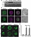
Clustering of IRE1B is induced upon ER stress. (a) BZIP60 is spliced by IRE1B-YFP but not by the RNase dead IRE1B fusion in Tm-treated ire1a ire1b protoplasts. Leaf protoplasts from WT and ire1a ire1b plants, or protoplasts transfected with a plasmid encoding IRE1B-YFP or IRE1BN820A-YFP in an ire1a ire1b mutant background, were incubated in the dark for 12 h, treated with DMSO (as the control) or 5 μg/mL Tm for 6 h and RNA was extracted. RT-PCR was carried out to detect BZIP60 mRNA splicing (BZIP60s), and ACT2 was used as a loading control. (b) Colocalization of IRE1B with an ER marker. Protoplasts isolated from leaves of WT plants were cotransfected with IRE1B-YFP-expressing constructs and mCherry-tagged ER or Golgi markers, incubated in the dark for 12 h and observed using confocal microscopy. Bar: 10 μm. (c) Confocal microscopy analysis of the clustering of IRE1B-YFP and RNase-dead IRE1BN820A-YFP. Leaf protoplasts from WT seedlings were transfected with a plasmid expressing an ER-YFP marker, IRE1B-YFP or RNase dead IRE1BN820A-YFP, incubated in the dark for 12 h, and then treated with DMSO or 5 μg/mL Tm for 6 h before imaging by confocal microscopy. Bar: 10 μm. (d) Quantitation of the number of clusters per cell for at least 9 protoplasts/treatment derived from images such as those shown in (c), with 3 biological replicates. Error bars = SD.
Figure 5.
Figure 5.
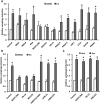
Some RIDD target genes repress ER stress induced autophagy. (a) Suppression of autophagy induction by transfecting WT leaf protoplasts with RIDD target genes. The top 12 RIDD target genes expressed from a 35S promoter were introduced into protoplasts together with GFP-ATG8E. After incubation in the dark for 12 h, samples were treated with DMSO (as the control) or 5 μg/mL Tm for 6 h. Autophagosomes were quantified and normalized to WT protoplasts transfected with the empty vector (Control) with DMSO treatment. The average of 3 biological replicates is shown, with 100 protoplasts per replicate, and error bars represent SE. (b) Inhibition of RIDD target gene translation results in failure of autophagy suppression. ROSY1/ML, PR-14 and BGLU21 and mutant forms of each (mROSY1/mML, mPR-14 and mBGLU21) with a mutated start codon were expressed from a 35S promoter in protoplasts with GFP-ATG8E. Protoplasts were treated and examined as in (a). (c) Overexpression of RIDD target genes has no effect on sucrose starvation-induced autophagy. RIDD target genes were coexpressed in protoplasts with GFP-ATG8E and incubated in the presence or absence of 0.5% sucrose for 2 days in the dark. Autophagosomes were quantified and normalized to WT protoplasts transfected with the empty vector (Control). The average of 3 biological replicates is shown, with 100 protoplasts per replicate, and error bars represent SE. For all experiments, asterisks indicate significant differences (P < 0.05) using the Student t test compared with vector alone under control conditions.

References

    1. Sah SK, Reddy KR, Li J.. Abscisic acid and abiotic stress tolerance in crop plants. Front Plant Sci. 2016;7:571 Epub 2016/ 05/21. - PMC - PubMed
    1. Boyer J. Plant productivity and environment. Science. 1982;218:443–448. - PubMed
    1. Cramer GR, Urano K, Delrot S, et al. Effects of abiotic stress on plants: a systems biology perspective. BMC Plant Biol. 2011;11:163 Epub 2011/ 11/19. - PMC - PubMed
    1. Yang X, Bassham DC. New insight into the mechanism and function of autophagy in plant cells. Int Rev Cell Mol Biol. 2015;320:1–40. Epub 2015/ 11/29. - PubMed
    1. Michaeli S, Galili G, Genschik P, et al. Autophagy in plants–what’s new on the menu? Trends Plant Sci. 2016;21:134–144. Epub 2015/ 11/26. - PubMed

Publication types