

Interference with KCTD9 inhibits NK cell activation and ameliorates fulminant liver failure in mice
- PMID: 29940856
- PMCID: PMC6019787
- DOI: 10.1186/s12865-018-0256-x
Interference with KCTD9 inhibits NK cell activation and ameliorates fulminant liver failure in mice
Abstract
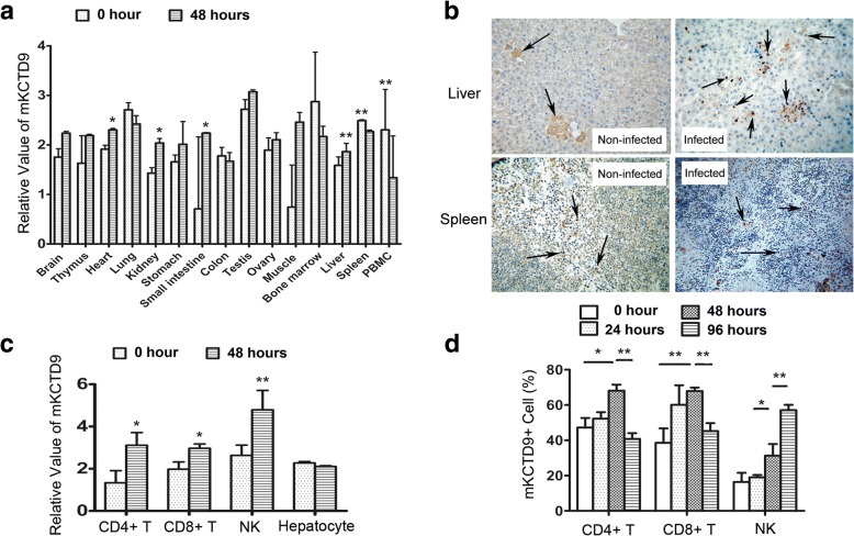
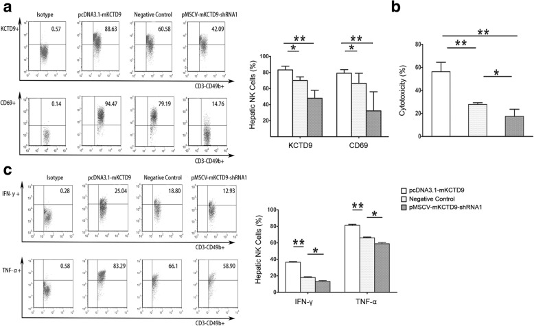
Background: Potassium channel tetramerisation domain containing 9 (KCTD9), a member of KCTD family with a DNA-like pentapeptide repeat domain, was found to be increased particularly in NK cells of patients with HBV-induced acute-on-chronic liver failure (HBV-ACLF) and experimental viral fulminant hepatitis. Knockdown of KCTD9 in immortalized NK cells inhibits cytokines production and cytotoxicity. As NK cell activation was shown to exacerbate liver damage in viral fulminant hepatitis, we propose that target inhibition of KCTD9 may prohibit NK cells activity and thus ameliorate liver damage in viral fulminant hepatitis.
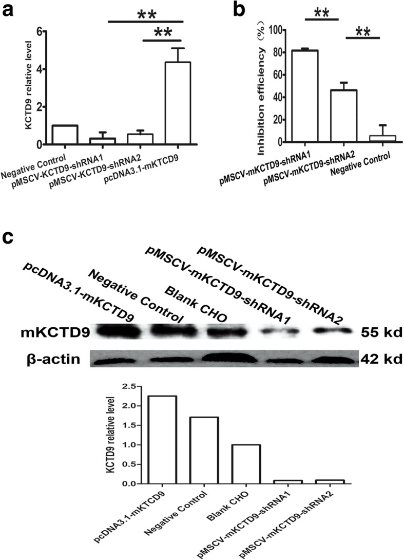
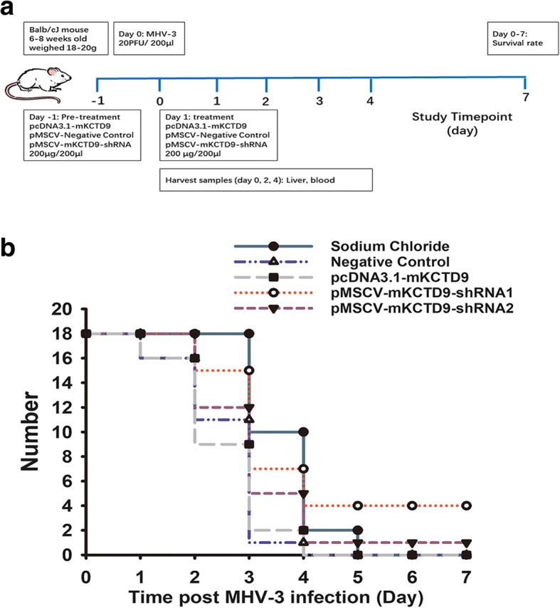
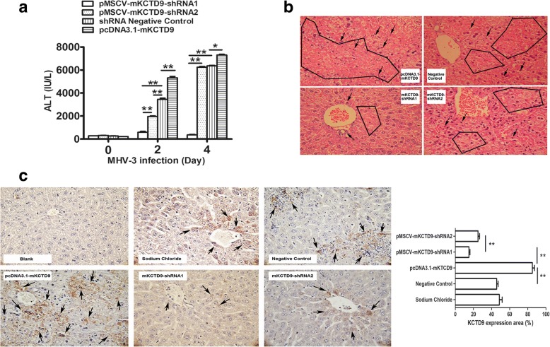
Result: Hydrodynamic delivery of plasmid expressing short-hairpin RNA against KCTD9 resulted in impaired NK cells function as demonstrated by reduced cytokine production and cytotoxicity, and ameliorated liver injury as manifested by improved liver histology and survival rate. In contrast, delivery of plasmid expressing KCTD9 led to deteriorated disease progression.
Conclusion: Interference with KCTD9 expression exert beneficial effect in viral fulminant hepatitis therapy. Such effect may be mediated by impairment of NK cell activation.
Keywords: KCTD9; Liver failure; MHV-3; NK cell; Short hairpin RNA.
Conflict of interest statement
Ethics approval and consent to participate
All experiments were approved by and conducted in compliance with the guidelines of the Care and Use of Laboratory Animals and Committees on Animal Experimentation, Tongji Hospital, Tongji Medical College, Huazhong University of Science and Technology.
Consent for publication
Not applicable.
Competing interests
The authors declare that they have no competing interests.
Publisher’s Note
Springer Nature remains neutral with regard to jurisdictional claims in published maps and institutional affiliations.
Figures
Similar articles
-
Kctd9 Deficiency Impairs Natural Killer Cell Development and Effector Function.Front Immunol. 2019 Apr 10;10:744. doi: 10.3389/fimmu.2019.00744. eCollection 2019. Front Immunol. 2019. PMID: 31024568 Free PMC article.
-
KCTD9 contributes to liver injury through NK cell activation during hepatitis B virus-induced acute-on-chronic liver failure.Clin Immunol. 2013 Mar;146(3):207-16. doi: 10.1016/j.clim.2012.12.013. Epub 2013 Jan 7. Clin Immunol. 2013. PMID: 23376586
-
[KCTD9, a novel potassium channel related gene, was highly expressed in hepatic NK cells and T cells of fulminant hepatitis mice induced by MHV-3].Zhonghua Gan Zang Bing Za Zhi. 2011 Nov;19(11):833-7. doi: 10.3760/cma.j.issn.1007-3418.2011.11.010. Zhonghua Gan Zang Bing Za Zhi. 2011. PMID: 22433305 Chinese.
-
Hepatitis B virus infection: Defective surface antigen expression and pathogenesis.World J Gastroenterol. 2018 Aug 21;24(31):3488-3499. doi: 10.3748/wjg.v24.i31.3488. World J Gastroenterol. 2018. PMID: 30131655 Free PMC article. Review.
-
Interplay between the Hepatitis B Virus and Innate Immunity: From an Understanding to the Development of Therapeutic Concepts.Viruses. 2017 Apr 28;9(5):95. doi: 10.3390/v9050095. Viruses. 2017. PMID: 28452930 Free PMC article. Review.
Cited by
-
Comprehensive analysis of circRNAs from cashmere goat skin by next generation RNA sequencing (RNA-seq).Sci Rep. 2020 Jan 16;10(1):516. doi: 10.1038/s41598-019-57404-9. Sci Rep. 2020. PMID: 31949277 Free PMC article.
-
C-C chemokine receptor 5 is essential for conventional NK cell trafficking and liver injury in a murine hepatitis virus-induced fulminant hepatic failure model.J Transl Med. 2023 Nov 29;21(1):865. doi: 10.1186/s12967-023-04665-8. J Transl Med. 2023. PMID: 38017505 Free PMC article.
-
Systematic analysis on multiple Gene Expression Omnibus data sets reveals fierce immune response in hepatitis B virus-related acute liver failure.J Cell Mol Med. 2020 Sep;24(17):9798-9809. doi: 10.1111/jcmm.15561. Epub 2020 Jul 19. J Cell Mol Med. 2020. PMID: 32686296 Free PMC article.
-
Potential therapies for acute-on-chronic liver failure.Liver Int. 2025 Mar;45(3):e15545. doi: 10.1111/liv.15545. Epub 2023 Mar 8. Liver Int. 2025. PMID: 36800487 Free PMC article. Review.
-
KCTD15 Protein Expression in Peripheral Blood and Acute Myeloid Leukemia.Diagnostics (Basel). 2020 Jun 4;10(6):371. doi: 10.3390/diagnostics10060371. Diagnostics (Basel). 2020. PMID: 32512747 Free PMC article.
References
Publication types
MeSH terms
Substances
LinkOut - more resources
Full Text Sources
Other Literature Sources
Molecular Biology Databases
Research Materials
