. 2018 Jun 26;18(1):153.
doi: 10.1186/s12886-018-0817-z.

Comparison of 5-year progression of retinitis pigmentosa involving the posterior pole among siblings by means of SD-OCT: a retrospective study

Affiliations

Comparison of 5-year progression of retinitis pigmentosa involving the posterior pole among siblings by means of SD-OCT: a retrospective study

Leonardo Colombo et al. BMC Ophthalmol. .

Abstract

Background: The aim of this study is to analyze and compare the progression of photoreceptor atrophy among siblings affected by retinitis pigmentosa by means of spectral SD-OCT.

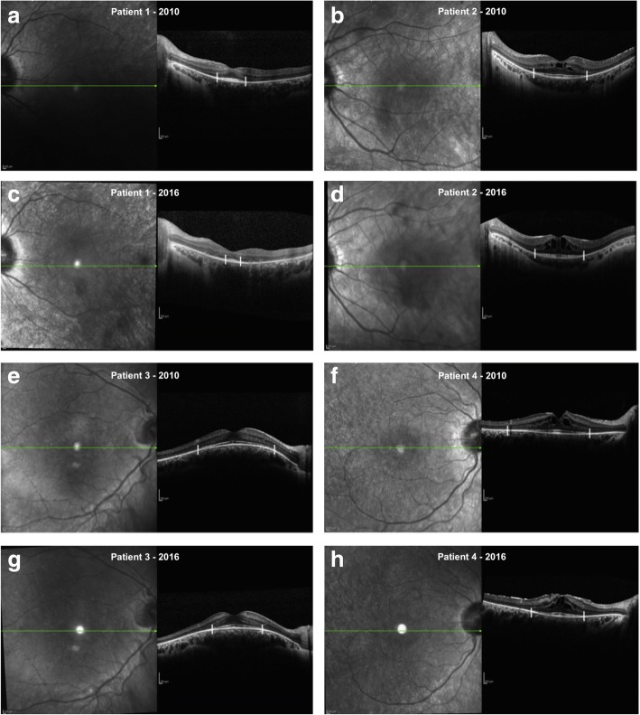
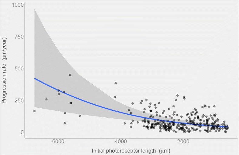
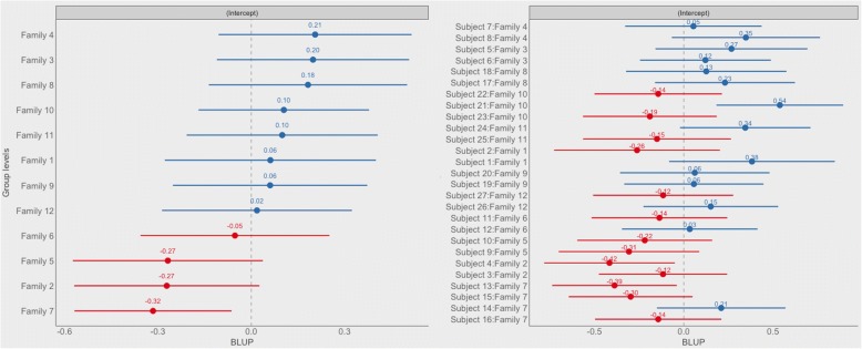
Methods: Fifty three eyes of 27 patients belonging to 12 family clusters were analyzed. To assess the annual progression rate of photoreceptor atrophy, the ellipsoid zone (EZ) line was measured in OCT sections through the fovea. We used multivariate generalized mixed effects to model the rate of progression and its relation to the initial ellipsoid zone line width.

Results: During our 4.84 years (± 1.44) mean follow up time (range 3-7) 53 eyes were examined. The ellipsoid zone line width declined with a yearly average rate of 76.4 μm (4.16% / year) (p-value < 0.0001). Progression rates were poorly correlated within family clusters (p-value = 0.23) and showed statistical difference between affected siblings (p-value = 0.007). There was no correlation between inter-familiar progression rate and mode of inheritance (p-value = 0.98) as well as between age and ellipsoid zone line width among siblings (p-value = 0.91).

Conclusion: RP could be extremely heterogeneous even among siblings: an accurate and sensitive method to follow the progression of the disease is fundamental for future development of clinical trials and therapy strategies.

Keywords: Disease progression; Ellipsoid zone; Retinitis pigmentosa; SD-OCT; Siblings.


Conflict of interest statement

Ethics approval and consent to participate

The study was approved by the internal review boards of San Paolo Hospital – University of Milan. Written informed consent was obtained from each participant.

Consent for publication

Not applicable.

Competing interests

Prof Luca Rossetti is a section editor for Glaucoma of BMC Ophthalmology.

Other authors declare no competing interests.

Publisher’s Note

Springer Nature remains neutral with regard to jurisdictional claims in published maps and institutional affiliations.

Figures

Fig. 1
Fig. 1
EZ band measurement: EZ limit was considered where the hyperreflective band decline to zero. In a, b and e, f horizontal scan passing through the fovea of two couples of siblings affected by RP acquired in 2010, in c, d and g, h same scan of same patients acquired 6 years later (2016). Patient in a was 17 years old and in 2010 and 23 years old in 2016 (c). His brother (b) was 18 years old in 2010 and 24 years old in 2016 (d). Patient in e was 14 years old and in 2010 and 20 years old in 2016 (g). His brother (f) was 20 years old in 2010 and 26 years old in 2016 (h)
Fig. 2
Fig. 2
The blue line shows the progression rate (in microns/year) of the photoreceptor length decrease at different baseline lengths (in microns). The curved relationship is a direct consequence of the generalized linear model when displayed on the response scale. The grey band represents the 95% point wise confidence intervals. Single observations are overlaid as semitransparent black dots
Fig. 3
Fig. 3
Forest plot of the random effects via BULP (Best Unbiased Linear Predictions) derived from the fitted model of the progression rate. BULP are the best prediction of the group mean (Family or subject), given the observations, from mixed models. The estimated intercept is indicated filled dot and numeric values are reported above each dot. Horizontal lines represent the 95% confidence intervals of the estimates. Reported estimates refer to the intercepts on the log-link function scale of the model and are ordered based on the estimated intercept of the family. Notice how subjects belonging to the same family show very variable estimated intercepts

References

    1. Hartong DT, Berson EL, Dryja TP. Retinitis pigmentosa. Lancet. 2006;368(9549):1795–1809. doi: 10.1016/S0140-6736(06)69740-7. - DOI - PubMed
    1. Bundey S, Crews SJ. A study of retinitis pigmentosa in the city of Birmingham. I prevalence. J Med Genet. 1984;21(6):417–420. doi: 10.1136/jmg.21.6.417. - DOI - PMC - PubMed
    1. Haim M, Holm NV, Rosenberg T. Prevalence of retinitis pigmentosa and allied disorders in Denmark. I Main results. Acta Ophthalmol (Copenh) 1992;70(2):178–186. doi: 10.1111/j.1755-3768.1992.tb04121.x. - DOI - PubMed
    1. Bunker C, Berson E, Bromley W, et al. Prevalence of retinitis Pigmentosa in Maine. Am J Ophthalmol. 1984;97(3):357–365. doi: 10.1016/0002-9394(84)90636-6. - DOI - PubMed
    1. Farber DB, Heckenlively JR, Sparkes RS, Bateman JB. Molecular genetics of retinitis pigmentosa. West J Med. 1991;155(4):388–399. - PMC - PubMed

LinkOut - more resources