. 2018 Jun 26;19(1):127.
doi: 10.1186/s12931-018-0831-y.

Changes in pulmonary endothelial cell properties during bleomycin-induced pulmonary fibrosis

Affiliations

Changes in pulmonary endothelial cell properties during bleomycin-induced pulmonary fibrosis

Shinpei Kato et al. Respir Res. .

Abstract

Background: Pulmonary fibrosis is a progressive and lethal disease characterized by damage to the lung parenchyma with excess extracellular matrix deposition. The involvement of endothelial cells in fibrosis development is unclear.

Methods: We isolated pulmonary endothelial cells, using a magnetic-activated cell sorting system, from mice with pulmonary fibrosis induced by intratracheal bleomycin. We characterized endothelial cells isolated at various times in the course of pulmonary fibrosis development.

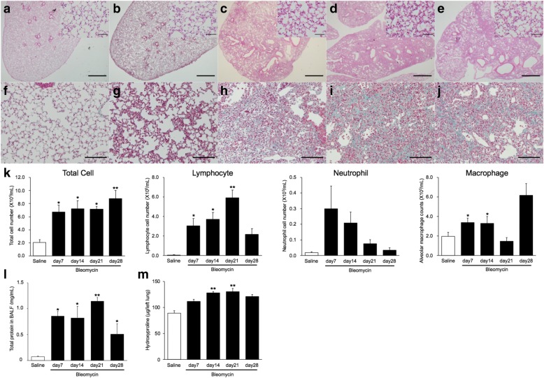
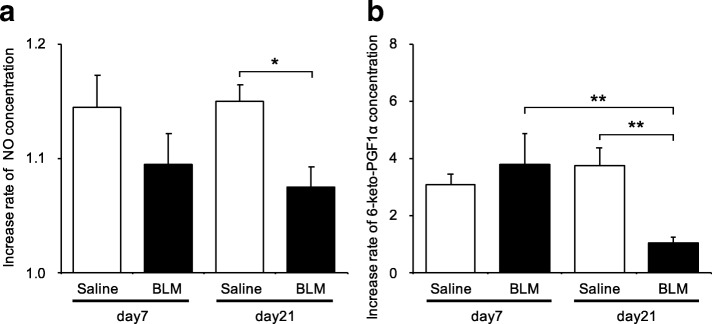
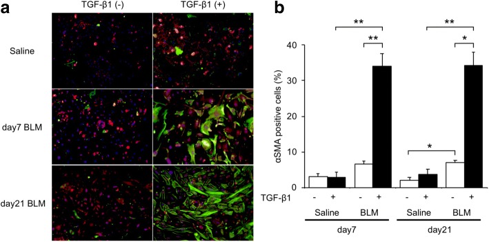
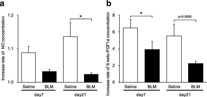
Results: Inflammatory cell infiltration was observed at 7 days after bleomycin administration, and fibrotic changes with increased collagen content were observed on day 21. Endothelial cells were isolated at these two timepoints. Levels of von Willebrand factor, plasminogen activator inhibitor-1 and matrix metalloproteinase-12 were elevated in lung endothelial cells isolated from bleomycin-treated mice at days 7 and 21. This indicated that intratracheal bleomycin administration induced endothelium injury. Expression of fibrogenic mediators, transforming growth factor (TGF)-β, connective tissue growth factor and platelet-derived growth factor-C was elevated in the cells from bleomycin-treated, compared with untreated, lungs. When endothelial cells were treated with TGF-β, α-smooth muscle actin (SMA) expression and collagen production were increased only in those cells from bleomycin-treated mouse lungs. Thapsigargin-induced prostaglandin I2 and nitric oxide production, decreased in endothelial cells from bleomycin-treated mouse lungs, compared with controls, was further suppressed by TGF-β.

Conclusion: Bleomycin administration induced functional changes in lung endothelial cells, indicating potential involvement of endothelium in pulmonary fibrogenesis.

Keywords: Bleomycin; Endothelial cell; Fibrosis; Nitric oxide; Prostaglandin I2; TGF-β; α-SMA.


Conflict of interest statement

Ethics approval and consent to participate

This study was approved by the Animal Care and Use Committee of Hamamatsu University School of Medicine and all experiments were performed according to guidelines of this Committee.

Consent for publication

Not applicable.

Competing interests

The authors declare that they have no competing interests.

Publisher’s Note

Springer Nature remains neutral with regard to jurisdictional claims in published maps and institutional affiliations.

Figures

Fig. 1
Fig. 1
Intratracheal bleomycin-induced pulmonary fibrosis. Representative optical microscopy images of mouse lungs. Hematoxylin-eosin stain for saline (a) and bleomycin-treated lungs at day 7 (b), day 14 (c), day 21 (d) and day 28 (e). Scale bars indicate 500 μm (100 μm in inset). Masson’s trichrome stained saline (f) and bleomycin-treated lungs at day 7 (g), day 14 (h), day 21 (i) and day 28 (j). Scale bars indicate 200 μm. k Total and differential cell counts in bronchoalveolar lavage (BAL) fluid from saline and bleomycin-treated mice. BAL fluid was collected at days 7, 14, 21 and 28 after intratracheal bleomycin administration. Total cell numbers and lymphocyte counts in BAL fluid from bleomycin-treated lungs were significantly higher than in controls. Neutrophils in BAL fluid from bleomycin-treated lungs tended to be increased on day 7. l Total protein concentration in BAL fluid from saline and bleomycin-treated mice. BAL fluid was collected at days 7, 14, 21 and 28 after intratracheal bleomycin administration. Total protein concentrations in BAL fluid from bleomycin-treated lungs were significantly higher compared with that in controls. m Hydroxyproline content in lungs from saline and bleomycin-treated mice. Lung homogenates from saline and bleomycin-treated mice (n = 3 per group) were analyzed for hydroxyproline with a colorimetric assay. Hydroxyproline content was higher in the bleomycin-treated lungs. Results are means ± standard error, from three to seven mice. *p < 0.05, compared with saline-treated mice; **p < 0.05, compared with bleomycin-treated mice on day 7
Fig. 2
Fig. 2
Functional performance of endothelial cells from bleomycin-treated lungs, assessed by response to thapsigargin. a Intracellular nitric oxide concentrations in endothelial cells were measured using DAF–FM/DA. Thapsigargin (1 μM) was added and the DAF–FM fluorescence intensity in whole cells was measured at 515 nm. The fluorescence intensity ratio indicates the relative fluorescence intensity of intracellular nitric oxide in thapsigargin-treated endothelial cells over that in untreated endothelial cells. The fluorescence intensity ratio of intracellular nitric oxide was significantly attenuated in endothelial cells from bleomycin-treated lungs at day 21, compared with in saline-treated lungs. b 6-Keto PGF released from endothelial cells. The concentration of 6-keto PGF in the culture medium was measured by ELISA after incubation of endothelial cells with 10 μM thapsigargin. The relative concentration ratio indicates the concentration of 6-keto PGF in thapsigargin-treated endothelial cells over that in untreated cells and these ratios were compared for cells from saline and bleomycin-treated mice. Relative rates of 6-keto PGF production were significantly attenuated in thapsigargin-stimulated endothelial cells from bleomycin-treated lungs, compared with in those from saline-treated lungs, both isolated on day 21. These levels were also attenuated relative to those in endothelial cells from bleomycin-treated lungs that were not stimulated by thapsigargin at the same day. Data are means ± standard error from three or four mice. *p < 0.05, **p < 0.01
Fig. 3
Fig. 3
Expression of mediators, determined by quantitative real-time PCR. Levels of mRNA for various mediators were compared in endothelial cells from saline and bleomycin-treated mouse lungs. Quantitative real-time PCR was performed using three or four independently prepared cDNA samples from endothelial cells harvested from saline or bleomycin-treated lungs on days 7 and 21. Gene expression was calculated asΔCt, the Ct of a gene of interest minus the Ct of GAPDH from the same sample. Results were normalized to expression levels in endothelial cells from untreated lungs at day 0 and are means from three experiments. Data are means ± standard error of the mean for three or four mice. *p < 0.05, **p < 0.01, compared with saline-treated mice
Fig. 4
Fig. 4
Fibrotic mediator proteins released from endothelial cells. Protein levels of TGF-β1 (a), CTGF (b) and PDGF-C (c) were quantified by ELISA. Concentrations of TGF-β1, CTGF and PDGF-C were significantly higher in the culture medium of endothelial cells from bleomycin-treated lungs, compared with those from saline-treated lungs. Data are means ± standard error of the mean for three to six mice. *p < 0.05, **p < 0.01, compared with saline-treated mice
Fig. 5
Fig. 5
Total collagen content in endothelial cells, with or without TGF-β1. Total soluble collagen content was measured by the Sircol assay. The collagen content in the culture medium from endothelial cells isolated from bleomycin-treated lungs was higher than samples from saline-treated lungs. TGF-β induced significantly increased collagen content in endothelial cells from bleomycin-treated lungs. However, in medium from endothelial cells isolated from saline-treated mice lungs, TGF-β did not affect total collagen content. Results are means ± standard error of the mean from three or four mice per group. *p < 0.05, **p < 0.01, compared with saline-treated mice
Fig. 6
Fig. 6
TGF-β1-induced increased α-SMA staining in endothelial cells from bleomycin-treated lungs. a Shown are representative fluorescence microscopy images of endothelial cells from bleomycin-treated lungs, cultured with or without TGF-β1. The α-SMA (green) and CD31 (red) stained images were merged with those showing a nuclear stain (Hoechst, blue). b Comparison of the percentage of α-SMA positive endothelial cells from saline and bleomycin-treated lungs, cultured with or without TGF-β1. Cells were imaged and images captured with IX83 and the percentage of α-SMA positive cells expressed as the percentage of total cell number, estimated from Hoechst 33,342 nuclear staining. The percentage of α-SMA positive cells was significantly elevated in cells from bleomycin-treated lungs and in any endothelial cells treated with TGF-β1, compared with in the corresponding controls. TGF-β1 treatment further enhanced the percent of α-SMA stained cells in lung endothelial cultures from bleomycin-treated mice. Data are means ± standard error, from three to four mice. * p < 0.05, ** p < 0.01
Fig. 7
Fig. 7
Effects of TGF-β on the functional properties of lung endothelial cells isolated from bleomycin-treated mice. a Intracellular nitric oxide response to TGF-β was significantly attenuated in cells isolated on day 21 after bleomycin treatment (p = 0.0138, compared with cells from saline-treated mice). b The increase in 6-keto-PGF levels caused by TGF-β was attenuated in cells isolated on day 7 or 21 from bleomycin-treated mice (p = 0.0206, p = 0.0506, respectively, compared with cells from saline-treated mice). Data are means ± standard error, from three to four mice. *, p < 0.05

Similar articles

Cited by

References

    1. American Thoracic S, European Respiratory S. American Thoracic Society/European Respiratory Society international multidisciplinary consensus classification of the idiopathic interstitial pneumonias. This joint statement of the American Thoracic Society (ATS), and the European Respiratory Society (ERS) was adopted by the ATS board of directors, June 2001 and by the ERS executive committee, June 2001. Am J Respir Crit Care Med. 2002;165:277–304. doi: 10.1164/ajrccm.165.2.ats01. - DOI - PubMed
    1. Wolters PJ, Collard HR, Jones KD. Pathogenesis of idiopathic pulmonary fibrosis. Annu Rev Pathol. 2014;9:157–179. doi: 10.1146/annurev-pathol-012513-104706. - DOI - PMC - PubMed
    1. King TE, Jr, Pardo A, Selman M. Idiopathic pulmonary fibrosis. Lancet. 2011;378:1949–1961. doi: 10.1016/S0140-6736(11)60052-4. - DOI - PubMed
    1. Bantsimba-Malanda C, Marchal-Somme J, Goven D, Freynet O, Michel L, Crestani B, Soler P. A role for dendritic cells in bleomycin-induced pulmonary fibrosis in mice? Am J Respir Crit Care Med. 2010;182:385–395. doi: 10.1164/rccm.200907-1164OC. - DOI - PubMed
    1. Raghu G, Collard HR, Egan JJ, Martinez FJ, Behr J, Brown KK, Colby TV, Cordier JF, Flaherty KR, Lasky JA, et al. An official ATS/ERS/JRS/ALAT statement: idiopathic pulmonary fibrosis: evidence-based guidelines for diagnosis and management. Am J Respir Crit Care Med. 2011;183:788–824. doi: 10.1164/rccm.2009-040GL. - DOI - PMC - PubMed

Publication types