

Pathological changes and bacteriological assessments in the urinary tract of pregnant goats experimentally infected with Brucella melitensis
- PMID: 29940976
- PMCID: PMC6019509
- DOI: 10.1186/s12917-018-1533-x
Pathological changes and bacteriological assessments in the urinary tract of pregnant goats experimentally infected with Brucella melitensis
Abstract
Background: This study was conducted to investigate the pathological changes and distribution of B. melitensis in the urinary tract of pregnant goats following acute experimental infection. Six Jamnapari crossbred does in their third trimester of pregnancy were randomly assigned into two groups; Group 1 was uninfected control and Group 2 was inoculated conjunctival with 0.1 mL of the inoculums containing 109 cfu/mL of live B. melitensis. All does were sacrificed 30 days post-inoculation before the kidney, ureter, urinary bladder, urethra and vaginal swab were collected for isolation of B. melitensis. The same tissue samples were fixed in 10% neutral buffered formalin for hematoxylin and eosin, and immunoperoxidase staining.
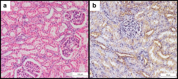
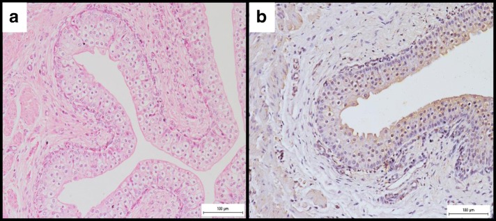
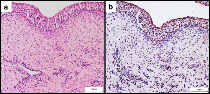
Results: None of the goats showed clinical signs or gross lesions. The most consistent histopathology finding was the infiltration of mononuclear cells, chiefly the macrophages with few lymphocytes and occasionally neutrophils in all organs along the urinary tract of the infected goats of Group 2. Other histopathology findings included mild necrosis of the epithelial cells of the renal tubules, congestion and occasional haemorrhages in the various tissues. Kidneys showed the most severe lesions. Immunoperoxidase staining revealed the presence of B. melitensis within the infiltrating macrophages and the epithelium of renal tubules, ureter, urethra and urinary bladder. Most extensive distribution was observed in the urinary bladder. Brucella melitensis was successfully isolated at low concentration (3.4 × 10³ cfu/g) in the various organs of the urinary tract and at high concentration (2.4 × 10⁸ cfu/mL) in the vaginal swabs of all infected goats. Although B. melitensis was successfully isolated from the various organs of the urinary tract, it was not isolated from the urine samples that were collected from the urinary bladder at necropsy.
Conclusion: This study demonstrates the presence of low concentrations of B. melitensis in the organs of urinary tract of pregnant does, resulting in mild histopathology lesions. However, B. melitensis was not isolated from the urine that was collected from the urinary bladder.
Keywords: Brucella melitensis; Goats; Histopathology; Immunoperoxidase; Urinary tract.
Conflict of interest statement
Ethics approval
The experiment was approved by the Institutional Animal Care and Use Committee, Universiti Putra Malaysia (IACUC no. R019/2014).
Consent for publication
Not applicable.
Competing interests
The authors declare that they have no competing interests.
Publisher’s Note
Springer Nature remains neutral with regard to jurisdictional claims in published maps and institutional affiliations.
Figures
References
-
- Xavier MN, Paixao TA, den Hartigh AB, Tsolis RM, Santos RL. Pathogenesis of Brucella spp. Open Vet Sci J. 2010;4:109–118. doi: 10.2174/1874318801004010109. - DOI
MeSH terms
LinkOut - more resources
Full Text Sources
Other Literature Sources
Medical
