

Increased extracellular matrix deposition during chondrogenic differentiation of dental pulp stem cells from individuals with neurofibromatosis type 1: an in vitro 2D and 3D study
- PMID: 29941005
- PMCID: PMC6020206
- DOI: 10.1186/s13023-018-0843-1
Increased extracellular matrix deposition during chondrogenic differentiation of dental pulp stem cells from individuals with neurofibromatosis type 1: an in vitro 2D and 3D study
Abstract
Background: Neurofibromatosis 1 (NF1) presents a wide range of clinical manifestations, including bone alterations. Studies that seek to understand cellular and molecular mechanisms underlying NF1 orthopedic problems are of great importance to better understand the pathogenesis and the development of new therapies. Dental pulp stem cells (DPSCs) are being used as an in vitro model for several diseases and appear as a suitable model for NF1. The aim of this study was to evaluate in vitro chondrogenic differentiation of DPSCs from individuals with NF1 using two-dimensional (2D) and three-dimensional (3D) cultures.
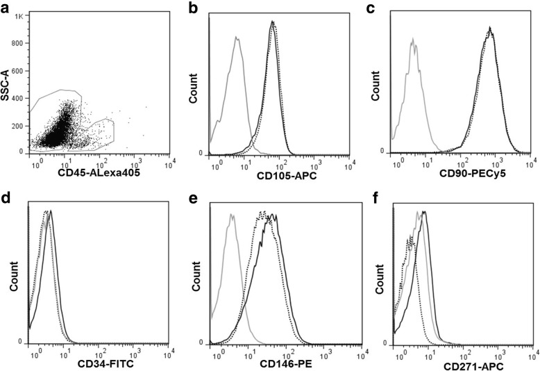
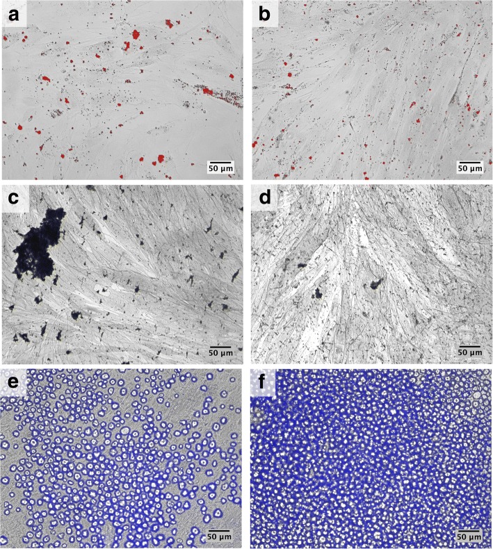
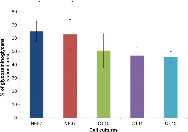
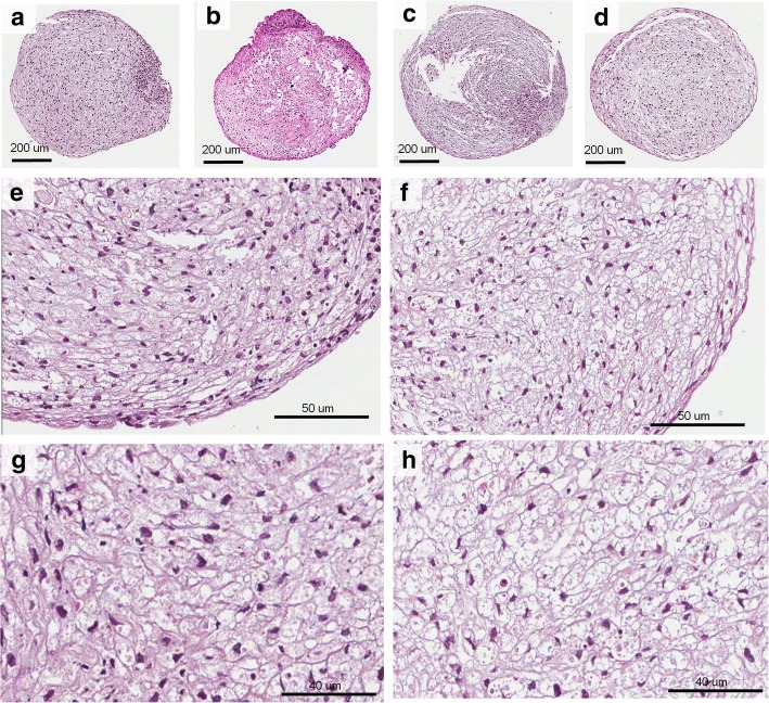
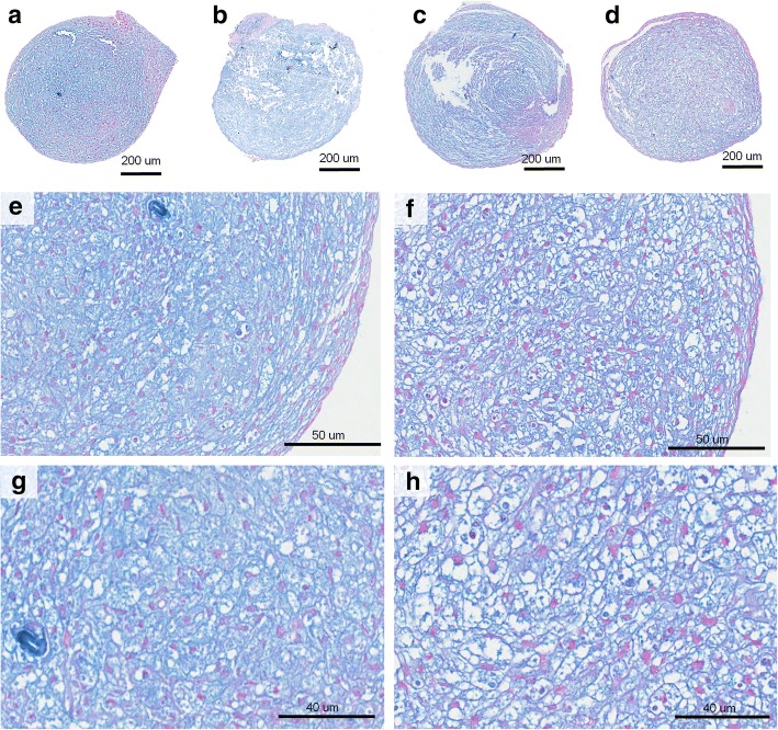
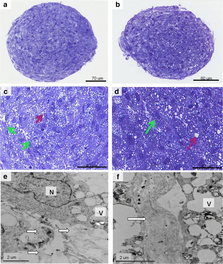
Results: To fulfill the criteria of the International Society for Cellular Therapy, DPSCs were characterized by surface antigen expression and by their multipotentiality, being induced to differentiate towards adipogenic, osteogenic, and chondrogenic lineages in 2D cultures. Both DPSCs from individuals with NF1 (NF1 DPSCs) and control cultures were positive for CD90, CD105, CD146 and negative for CD13, CD14, CD45 and CD271, and successfully differentiated after the protocols. Chondrogenic differentiation was evaluated in 2D and in 3D (pellet) cultures, which were further evaluated by optical microscopy and transmission electron microscopy (TEM). 2D cultures showed greater extracellular matrix deposition in NF1 DPSCs comparing with controls during chondrogenic differentiation. In semithin sections, control pellets hadhomogenous-sized intra and extracelullar matrix vesicles, whereas NF1 cultures had matrix vesicles of different sizes. TEM analysis showed higher amount of collagen fibers in NF1 cultures compared with control cultures.
Conclusion: NF1 DPSCs presented increased extracellular matrix deposition during chondrogenic differentiation, which could be related to skeletal changes in individuals with NF1.
Keywords: Cell culture; Cell differentiation; Chondrogenesis; Neurofibromatosis 1.
Conflict of interest statement
Ethics approval and consent to participate
This study was approved by the institution’s Ethics Committee (No. 519.858/2014). All participants signed a consent form.
Consent for publication
Not applicable.
Competing interests
The authors declare that they have no competing interests.
Publisher’s Note
Springer Nature remains neutral with regard to jurisdictional claims in published maps and institutional affiliations.
Figures
Similar articles
-
Multilineage Differentiation Potential of Human Dental Pulp Stem Cells-Impact of 3D and Hypoxic Environment on Osteogenesis In Vitro.Int J Mol Sci. 2020 Aug 26;21(17):6172. doi: 10.3390/ijms21176172. Int J Mol Sci. 2020. PMID: 32859105 Free PMC article.
-
Cryopreservation Method for the Effective Collection of Dental Pulp Stem Cells.Tissue Eng Part C Methods. 2017 May;23(5):251-261. doi: 10.1089/ten.TEC.2016.0519. Tissue Eng Part C Methods. 2017. PMID: 28314378
-
Characterization of pulp and follicle stem cells from impacted supernumerary maxillary incisors.Pediatr Dent. 2014 May-Jun;36(3):79-84. Pediatr Dent. 2014. PMID: 24960375
-
Isolation and characterization of dental pulp stem cells from a patient with Papillon-Lefèvre syndrome.J Endod. 2013 Jan;39(1):31-8. doi: 10.1016/j.joen.2012.09.024. Epub 2012 Oct 24. J Endod. 2013. PMID: 23228254
-
The chondrogenic differentiation potential of dental pulp stem cells.Eur Cell Mater. 2020 Feb 21;39:121-135. doi: 10.22203/eCM.v039a08. Eur Cell Mater. 2020. PMID: 32083715
Cited by
-
Epigenetic Regulation of Dental Pulp Stem Cell Fate.Stem Cells Int. 2020 Oct 13;2020:8876265. doi: 10.1155/2020/8876265. eCollection 2020. Stem Cells Int. 2020. PMID: 33149742 Free PMC article. Review.
-
Characterization of Dental Pulp Stem Cell Populations in the Teeth of Patients With Neurofibromatosis Type 1 - Therapeutic Potential for Bone Tissue Engineering.In Vivo. 2023 Mar-Apr;37(2):548-558. doi: 10.21873/invivo.13113. In Vivo. 2023. PMID: 36881087 Free PMC article.
-
An update on human periapical cyst-mesenchymal stem cells and their potential applications in regenerative medicine.Mol Biol Rep. 2020 Mar;47(3):2381-2389. doi: 10.1007/s11033-020-05298-6. Epub 2020 Feb 6. Mol Biol Rep. 2020. PMID: 32026284 Review.
References
-
- Abramowicz A, Gos M. Neurofibromin in neurofibromatosis type 1 - mutations in NF1gene as a cause of disease. Dev Period Med. 2014;18:297–306. - PubMed
Publication types
MeSH terms
Grants and funding
LinkOut - more resources
Full Text Sources
Other Literature Sources
Medical
Research Materials
Miscellaneous
