. 2018 Jun 26;9(1):173.
doi: 10.1186/s13287-018-0903-4.

Early gestational mesenchymal stem cell secretome attenuates experimental bronchopulmonary dysplasia in part via exosome-associated factor TSG-6

Affiliations

Early gestational mesenchymal stem cell secretome attenuates experimental bronchopulmonary dysplasia in part via exosome-associated factor TSG-6

Sushma Chaubey et al. Stem Cell Res Ther. .

Abstract

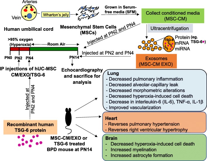
Background: Mesenchymal stem cells (MSCs) are promising tools for the treatment of human lung disease and other pathologies relevant to newborn medicine. Recent studies have established MSC exosomes (EXO), as one of the main therapeutic vectors of MSCs in mouse models of multifactorial chronic lung disease of preterm infants, bronchopulmonary dysplasia (BPD). However, the mechanisms underlying MSC-EXO therapeutic action are not completely understood. Using a neonatal mouse model of human BPD, we evaluated the therapeutic efficiency of early gestational age (GA) human umbilical cord (hUC)-derived MSC EXO fraction and its exosomal factor, tumor necrosis factor alpha-stimulated gene-6 (TSG-6).

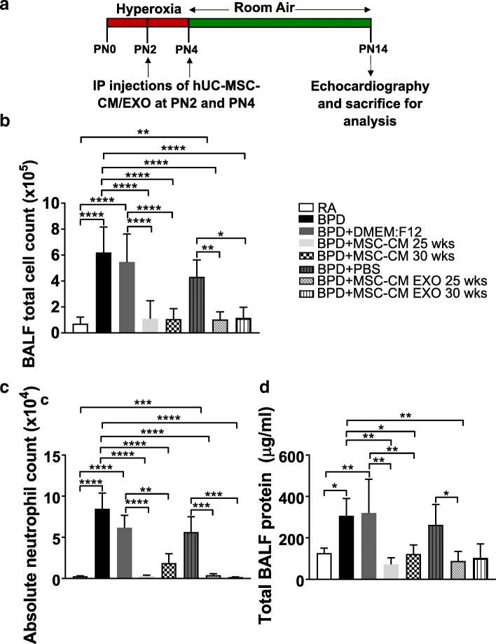
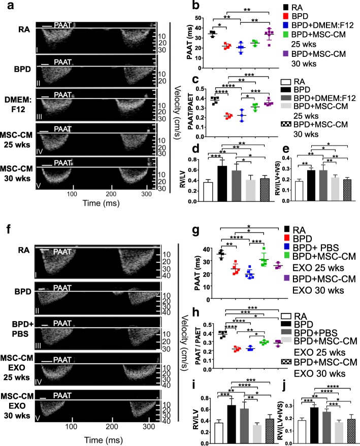
Methods: Conditioned media (CM) and EXO fractions were isolated from 25 and 30 weeks GA hUC-MSC cultures grown in serum-free media (SFM) for 24 h. Newborn mice were exposed to hyperoxia (> 95% oxygen) and were given intraperitoneal injections of MSC-CM or MSC-CM EXO fractions at postnatal (PN) day 2 and PN4. They were then returned to room air until PN14 (in a mouse model of severe BPD). The treatment regime was followed with (rh)TSG-6, TSG-6-neutralizing antibody (NAb), TSG-6 (si)RNA-transfected MSC-CM EXO and their appropriate controls. Echocardiography was done at PN14 followed by harvesting of lung, heart and brain for assessment of pathology parameters.

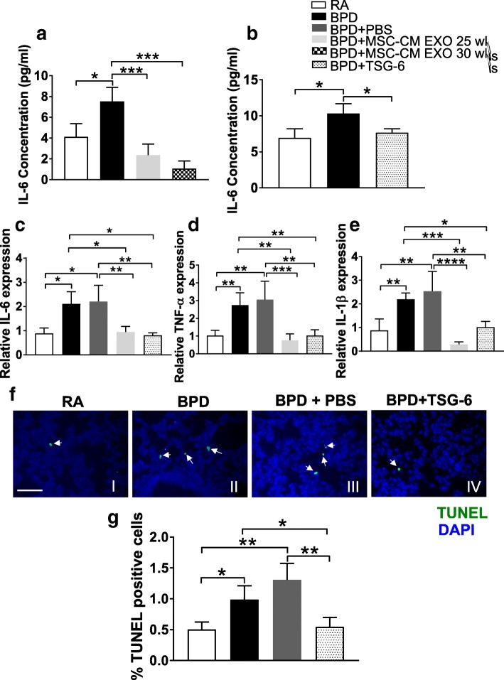
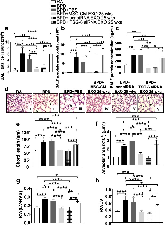
Results: Systemic administration of CM or EXO in the neonatal BPD mouse model resulted in robust improvement in lung, cardiac and brain pathology. Hyperoxia-exposed BPD mice exhibited pulmonary inflammation accompanied by alveolar-capillary leakage, increased chord length, and alveolar simplification, which was ameliorated by MSC CM/EXO treatment. Pulmonary hypertension and right ventricular hypertrophy was also corrected. Cell death in brain was decreased and the hypomyelination reversed. Importantly, we detected TSG-6, an immunomodulatory glycoprotein, in EXO. Administration of TSG-6 attenuated BPD and its associated pathologies, in lung, heart and brain. Knockdown of TSG-6 by NAb or by siRNA in EXO abrogated the therapeutic effects of EXO, suggesting TSG-6 as an important therapeutic molecule.

Conclusions: Preterm hUC-derived MSC-CM EXO alleviates hyperoxia-induced BPD and its associated pathologies, in part, via exosomal factor TSG-6. The work indicates early systemic intervention with TSG-6 as a robust option for cell-free therapy, particularly for treating BPD.

Keywords: Bronchopulmonary dysplasia; Exosomes; Hyperoxia-induced lung injury; Mesenchymal stem cells; Newborn; Pulmonary hypertension; Secretome; TSG-6; Wharton’s jelly.


Conflict of interest statement

Ethics approval

The collection of umbilical cord was approved by the Drexel University Institutional Review Board with a waiver of consent, as umbilical cords are considered discarded material. All experiments conformed to the guidelines issued by the committee on animal research of Drexel University. The study has been approved by the ethics board of Jefferson Hospital University.

Competing interests

The authors declare that they have no competing interests.

Publisher’s Note

Springer Nature remains neutral with regard to jurisdictional claims in published maps and institutional affiliations.

Figures

Fig. 1
Fig. 1
hUC MSC secretome treatment reverses pulmonary inflammation and alveolar-capillary leak associated with hyperoxia-induced lung injury in the BPD mouse model. a Schematic representation of BPD mouse model and secretome injection regime. The mice were kept in 100% oxygen from birth to PN4 followed by RA exposure till PN14. The mice were used for echocardiography and sacrificed for analysis at PN14. IP injections of hUC MSC-CM or hUC MSC-CM EXO were given at PN2 and PN4. 10 μg of MSC-CM and 2.5 μg of MSC-CM EXO (GA 25 wks and 30 wks), resuspended in 100 μl of PBS respectively, was injected into each neonatal mouse at PN2 and PN4. b-d Histogram showing BALF total cell count (b), BALF absolute neutrophil count (c), total BALF protein (d), in RA, BPD and vehicle (DMEM:F12 or PBS)-injected, MSC-CM or EXO 25 wks-injected, MSC-CM or EXO 30 wks-injected BPD mice at PN14. All values are expressed as mean ± standard error of the mean (SEM); eight experiments, N = 3–9 mice per group; one-way ANOVA with Tukey’s post hoc correction; *p < 0.05; **p < 0.01; ***p < 0.001; ****p < 0.0001. BALF bronchoalveolar lavage fluid, BPD bronchopulmonary dysplasia, CM conditioned medium, EXO exosomes, hUC human umbilical cord, i.p. intraperitoneal, MSC mesenchymal stem cell, PBS phosphate-buffered saline, PN postnatal, RA room air
Fig. 2
Fig. 2
hUC MSC secretome treatment reverses altered lung morphology associated with hyperoxia-induced lung injury in the BPD mouse model. a Representative images of lung histology with H&E stain from the five experimental groups, RA (I), BPD (II), BPD + DMEM:F12 (III), BPD + MSC-CM 25 wks (IV), BPD + MSC-CM 30 wks (V). Asterisks depicts the increased alveolar simplification in the BPD and DMEM:F12-injected BPD mice as compared to RA. ×200 magnification, Scale bar: 50 μm. b Representative images of lung histology with H&E stain from the five experimental groups, RA (I), BPD (II), BPD + PBS (III), BPD + MSC-CM EXO 25 wks (IV), BPD + MSC-CM EXO 30 wks (V). Asterisks depict the increased alveolar simplification in the BPD and PBS-injected BPD mice as compared to RA. ×200 magnification, Scale bar: 50 μm. c-g Histogram depicting the mean chord length (c), septal thickness (d), alveolar area (e), number of branches (f), number of junctions (g) in lungs of RA, BPD, DMEM:F12 or PBS-injected, MSC-CM or EXO 25 wks-injected, MSC-CM or EXO 30 wks-injected BPD mice at PN14. All values are expressed as mean ± standard error of the mean (SEM); eight experiments, N = 3–7 mice per group; one-way ANOVA with Tukey’s post hoc correction; *p < 0.05; **p < 0.01; ***p < 0.001; ****p < 0.0001. BPD bronchopulmonary dysplasia, CM conditioned medium, EXO exosomes, MSC mesenchymal stem cell, PBS phosphate-buffered saline, PN postnatal, RA room air
Fig. 3
Fig. 3
hUC MSC secretome treatment reverses PH and RV hypertrophy in hyperoxia-induced lung injury. a Echocardiography shows reversal of cardiac dysfunction on MSC-CM treatment in the BPD mouse model. The white line in the echocardiogram depicts PAAT values for the specific group, where PAAT is the pulmonary artery acceleration time. b Graph demonstrating PAAT values from the five experimental groups at PN14. c Graph depicting PAAT/PAET ratio where PAET is the pulmonary artery ejection time. d Histogram showing the RV to LV ratio in the five experimental groups. e Fulton’s index [RV/(LV + IVS)], reflecting right ventricular hypertrophy, in the five experimental groups at PN14. f Echocardiography shows reversal of cardiac dysfunction on MSC-CM EXO treatment in the BPD mouse model. The white line in the echocardiogram depicts PAAT values for the specific group. g Graph demonstrating PAAT values from the five experimental groups at PN14. h Graph depicting PAAT/PAET ratio where PAET is the pulmonary artery ejection time. i Histogram showing the RV to LV ratio in the five experimental groups. j Fulton’s index [RV/(LV + IVS)], reflecting right ventricular hypertrophy, in the five experimental groups at PN14. All values are expressed as mean ± standard error of the mean (SEM); 5 experiments, N = 3–7 mice per group; one-way ANOVA with Tukey’s post hoc correction; *p < 0.05; **p < 0.01; ***p < 0.001; ****p < 0.0001. BPD bronchopulmonary dysplasia, CM conditioned medium, EXO exosomes, IVS interventricular septa, LV left ventricular, MSC mesenchymal stem cell, PAAT pulmonary artery acceleration time, PAET pulmonary artery ejection time, PBS phosphate-buffered saline, RA room air, RV right ventricular
Fig. 4
Fig. 4
EXO-depleted MSC-CM 25 wks injections does not reverse hyperoxia-induced pulmonary inflammation, altered morphology, PH and RVH in the BPD mouse brain. a-c Histograms showing BALF total cell count (a), BALF absolute neutrophil count (b), total BALF protein (c) in RA, BPD, DMEM:F12-injected, MSC-CM 25 wks and EXO-depleted MSC-CM 25 wks-injected BPD mice at PN14. d-f Histograms showing the mean chord length (d), septal thickness (e), alveolar area (f) in lungs of RA, BPD, DMEM:F12-injected, MSC-CM 25 wks and EXO-depleted MSC-CM 25 wks-injected BPD mice. g-h Graph demonstrating PAAT/PAET ratio (g), RV to LV ratio (h), reflecting RV hypertrophy, in the five experimental groups at PN14. MSC-CM 25 wks data set used earlier in this manuscript was used for comparison with the EXO- depleted MSC-CM 25 wks group. All values are expressed as mean ± SEM; five experiments, N = 3–9 mice per group; one-way ANOVA with Tukey’s post hoc correction; *p < 0.05; **p < 0.01; ***p < 0.001; ****p < 0.0001. BPD bronchopulmonary dysplasia, CM conditioned medium, EXO exosomes, LV left ventricular, MSC mesenchymal stem cell, PAAT pulmonary artery acceleration time, PAET pulmonary artery ejection time, PBS phosphate-buffered saline, RA room air, RV right ventricular
Fig. 5
Fig. 5
TSG-6 reverses hyperoxia-induced pulmonary inflammation and altered morphology in the BPD mice. a Schematic representation of BPD mouse model and TSG-6 injection regime. 5 μg of rhTSG-6 resuspended in total 100 μl of PBS was injected into each neonatal mouse at PN2 and PN4. b-d Histograms showing BALF total cell count (b), BALF absolute neutrophil count (c), total BALF protein (d) in RA, BPD, PBS-injected and TSG-6-injected BPD mice at PN14. e Representative images of lung histology with H&E stain from the four experimental groups, RA (I), BPD (II), BPD + PBS (III), BPD + TSG-6 (IV). Asterisks depict the increased alveolar simplification in the BPD and PBS-injected BPD mice as compared to RA. ×200 magnification, Scale bar: 50 μm. f-k Histograms showing the mean chord length (f), septal thickness (g), alveolar area (h), number of branches (i), junctions (j), junction voxels (k) in lungs of RA, BPD, PBS-injected, TSG-6-injected BPD mice. All values are expressed as mean ± SEM; four experiments, N = 3–8 mice per group; one-way ANOVA with Tukey’s post hoc correction; *p < 0.05; **p < 0.01; ***p < 0.001; ****p < 0.0001. BALF bronchoalveolar lavage fluid, BPD bronchopulmonary dysplasia, CM conditioned medium, EXO exosomes, i.p. intraperitoneal, MSC mesenchymal stem cell, PAAT pulmonary artery acceleration time, PAET pulmonary artery ejection time, PBS phosphate-buffered saline, PN postnatal, RA room air, TSG-6 tumor necrosis factor alpha-stimulated gene-6
Fig. 6
Fig. 6
TSG-6 treatment corrects PH and RVH and attenuates hypomyelination and GFAP decrease in BPD mouse brain. a Echocardiography shows reversal of cardiac dysfunction on TSG-6 treatment in the BPD mouse model. The white line in the echocardiogram depicts PAAT values for specific group. b Graph demonstrating PAAT values from the four experimental groups at PN14. c Graph depicting PAAT/PAET ratio from the four experimental groups at PN14. d Histogram showing the RV to LV ratio in the four experimental groups. e Fulton’s index, [RV/(LV + IVS)], reflecting RV hypertrophy, in the four experimental groups at PN14. All values are expressed as mean ± SEM; four experiments, N = 3–8 mice per group; one-way ANOVA with Tukey’s post hoc correction; *p < 0.05; **p < 0.01; ***p < 0.001; ****p < 0.0001. BALF bronchoalveolar lavage fluid, BPD bronchopulmonary dysplasia, CM conditioned medium, IVS interventricular septa, LV left ventricular, PBS phosphate-buffered saline, RA room air, RV right ventricular, TSG-6 tumor necrosis factor alpha-stimulated gene-6
Fig. 7
Fig. 7
TSG-6 treatment decreases proinflammatory cytokines and cell death in lungs of the BPD mouse model. a IL-6 levels measured using ELISA in lung tissue of RA, BPD, MSC-CM EXO 25 wks- and MSC-CM EXO 30 wks-injected BPD mice at PN14. b IL-6 levels measured using ELISA in lung tissue of RA, BPD and TSG-6-injected BPD mice at PN14. c-e Pro-inflammatory cytokines levels in the lung tissues of RA, BPD and BPD mice injected with PBS, MSC-CM EXO 25 wks and TSG-6. Histogram showing relative expression of IL-6 (c), TNF-α (d) and IL-1β (e) as determined using real-time PCR. f Representative TUNEL immunofluorescence images of lung from the four experimental groups, RA (I), BPD (II), BPD + PBS (III), BPD + TSG-6 (IV). TUNEL-positive cells are labeled with FITC (green). The nuclei are counterstained with DAPI. Arrows depict TUNEL-positive dead cells in the field. Scale bar: 50 μm. (g) Histogram depicting the quantitative analysis of TUNEL-positive cells in the lungs of RA, BPD, PBS-injected and TSG-6-injected BPD mice. TUNEL-positive cells are expressed as a percentage, as described in Additional file 1: Methods. All values are expressed as mean ± SEM; 3–4 experiments, N = 3–5 mice per group; one-way ANOVA with Tukey’s post hoc correction; *p < 0.05; **p < 0.01; ***p < 0.001; ****p < 0.0001. BPD bronchopulmonary dysplasia, CM conditioned medium, DAPI 4,6-diamidino-2-phenylindole, EXO exosomes, IL-6 interleukin-6, IL-1β interleukin 1 beta, MSC mesenchymal stem cell, PBS phosphate-buffered saline, RA room air, TNF-α tumor necrosis factor alpha, TSG-6 tumor necrosis factor alpha-stimulated gene-6, TUNEL terminal deoxynucleotidyl transferase dUTP nick-end labeling
Fig. 8
Fig. 8
Administration of TSG-6 neutralizing antibody abrogates the therapeutic effects of MSC-CM EXO 25 wks. a-c Histogram showing BALF total cell count (a), BALF absolute neutrophil count (b), total BALF protein (c), in RA, BPD and PBS-injected, MSC-CM EXO 25 wks-injected, Isotype IgG + EXO 25 wks and NAb TSG-6 + EXO 25 wks-injected BPD mice at PN14. d Representative images of lung histology (H&E staining) from the six experimental groups, RA (I), BPD (II), BPD + PBS (III), BPD + MSC-CM EXO 25 wks (IV), BPD + Isotype IgG + EXO 25 wks (V), BPD + NAb TSG-6 + EXO 25 wks (VI). Asterisks depict the increased alveolar simplification in the BPD, PBS and NAb TSG-6 + EXO-injected BPD mice as compared to RA. ×200 magnification, Scale bar: 50 μm. e-f Histograms showing the mean chord length (e), alveolar area (f) in lungs of RA, BPD, PBS-injected, Isotype IgG + EXO 25 wks- injected and NAb TSG-6 + EXO 25 wks-injected BPD mice. g-h Histogram showing the RV to LV ratio (g) and Fulton’s index [RV/(LV + IVS)] (h), reflecting right ventricular hypertrophy at PN14. 5 μg of NAb resuspended in total 100 μl of PBS was injected into the neonatal mice at PN2 and PN4. RA, BPD, BPD + PBS and BPD + MSC-CM EXO 25 wks samples used for comparison in this analysis are from Figs. 1, 2 and 3. All values are expressed as mean ± SEM; six experiments, N = 3–8 mice per group; one-way ANOVA with Tukey’s post hoc correction; *p < 0.05; **p < 0.01; ***p < 0.001; ****p < 0.0001. BALF bronchoalveolar lavage fluid, BPD bronchopulmonary dysplasia, CM conditioned medium, EXO exosomes, IVS interventricular septa, LV left ventricular, MSC mesenchymal stem cell, NAb neutralizing antibody, PBS phosphate-buffered saline, RA room air, RV right ventricular, TSG-6 tumor necrosis factor alpha-stimulated gene-6
Fig. 9
Fig. 9
Administration of EXO isolated from TSG-6 siRNA knockdown MSCs did not suppress hyperoxia-induced inflammation and associated BPD pathology. a-c Histogram showing BALF total cell count (a), BALF absolute neutrophil count (b), total BALF protein (c), in RA, BPD and PBS-injected, MSC-CM EXO 25 wks-injected, scr siRNA EXO 25 wks and TSG-6 siRNA EXO 25 wks-injected BPD mice at PN14. d Representative images of lung histology (H&E staining) from the six experimental groups, RA (I), BPD (II), BPD + PBS (III), BPD + MSC-CM EXO 25 wks (IV), BPD + scr siRNA EXO 25 wks (V), BPD + TSG-6 siRNA EXO 25 wks (VI). Asterisks depict the increased alveolar simplification in the BPD, PBS and TSG-6 siRNA EXO 25 wks-injected BPD mice as compared to RA. ×200 magnification, Scale bar: 50 μm. e-f Histograms showing the mean chord length (e), alveolar area (f) in lungs of RA, BPD, PBS-injected, scr siRNA EXO 25 wks-injected and TSG-6 siRNA EXO 25 wks-injected BPD mice. g-h Histogram showing the RV to LV ratio (g) and Fulton’s index [RV/(LV + IVS)] (h), reflecting right ventricular hypertrophy at PN14. RA, BPD, BPD + PBS and BPD + MSC-CM EXO 25 wks samples used for comparison in this analysis are from Figs. 1, 2 and 3. All values are expressed as mean ± SEM; six experiments, N = 3–8 mice per group; one-way ANOVA with Tukey’s post hoc correction; *p < 0.05; **p < 0.01; ***p < 0.001; ****p < 0.0001. BALF bronchoalveolar lavage fluid, BPD bronchopulmonary dysplasia, CM conditioned medium, EXO exosomes, IVS interventricular septa, LV left ventricular, MSC mesenchymal stem cell, PBS phosphate-buffered saline, RA room air, RV right ventricular, scr siRNA scrambled small interfering ribonucleic acid, TSG-6 tumor necrosis factor alpha-stimulated gene-6
Fig. 10
Fig. 10
Schematic representation of MSC-CM/EXO/TSG-6 treatment regimen and outcomes in the mouse model of BPD. BPD pathology is reversed in the pups injected with secretome of hUC-derived MSCs. This therapeutic effect is mediated, at least in part, by exosomal factor TSG-6. BPD bronchopulmonary dysplasia, CM conditioned medium, EXO exosomes, hUC human umbilical cord, i.p. intraperitoneal, MSC mesenchymal stem cell, PN postnatal, TSG-6 tumor necrosis factor alpha-stimulated gene-6

References

    1. Balany J, Bhandari V. Understanding the impact of infection, inflammation, and their persistence in the pathogenesis of bronchopulmonary dysplasia. Front Med. 2015;2:90. doi: 10.3389/fmed.2015.00090. - DOI - PMC - PubMed
    1. Kim DH, Kim HS, Choi CW, et al. Risk factors for pulmonary artery hypertension in preterm infants with moderate or severe bronchopulmonary dysplasia. Neonatology. 2012;101:40–46. doi: 10.1159/000327891. - DOI - PubMed
    1. Nakanishi H, Uchiyama A, Kusuda S. Impact of pulmonary hypertension on neurodevelopmental outcome in preterm infants with bronchopulmonary dysplasia: a cohort study. J Perinatol. 2016;36:890–896. doi: 10.1038/jp.2016.108. - DOI - PMC - PubMed
    1. Reich B, Hoeber D, Bendix I, et al. Hyperoxia and the immature brain. Dev Neurosci. 2016;38:311–30. - PubMed
    1. Pierro M, Ionescu L, Montemurro T, et al. Short-term, long-term and paracrine effect of human umbilical cord-derived stem cells in lung injury prevention and repair in experimental bronchopulmonary dysplasia. Thorax. 2013;68:475–484. doi: 10.1136/thoraxjnl-2012-202323. - DOI - PubMed

Publication types

Substances