. 2018 Jul 18;38(29):6586-6596.
doi: 10.1523/JNEUROSCI.1034-18.2018. Epub 2018 Jun 25.

Axonal Ensheathment in the Nervous System of Lamprey: Implications for the Evolution of Myelinating Glia

Affiliations

Axonal Ensheathment in the Nervous System of Lamprey: Implications for the Evolution of Myelinating Glia

Marie-Theres Weil et al. J Neurosci. .

Abstract

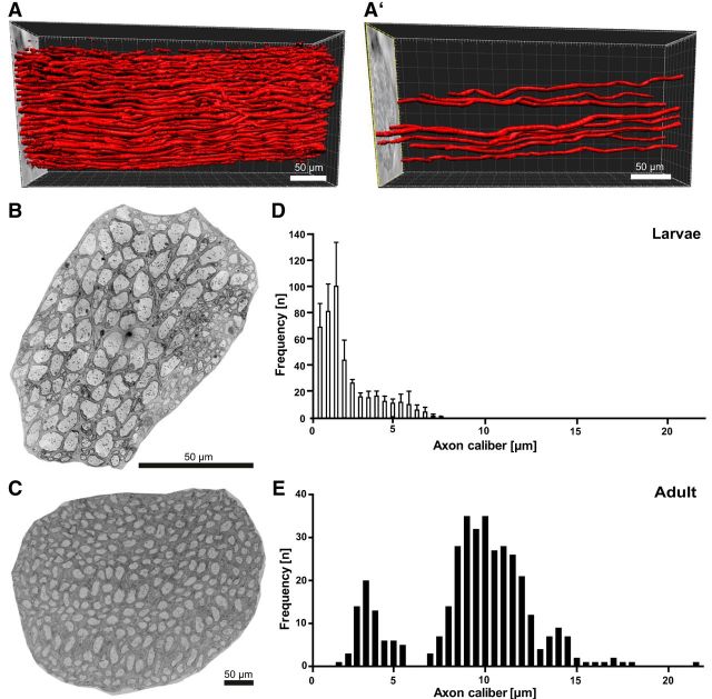
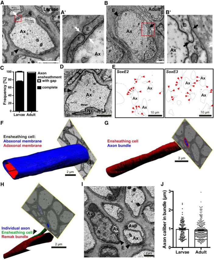
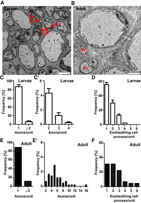
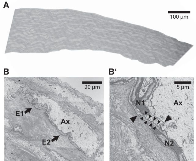
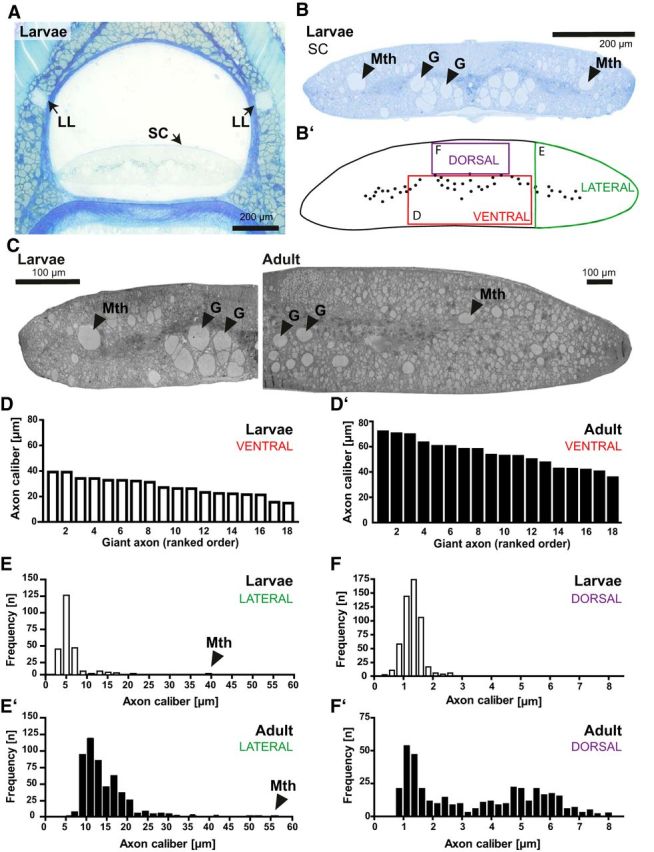
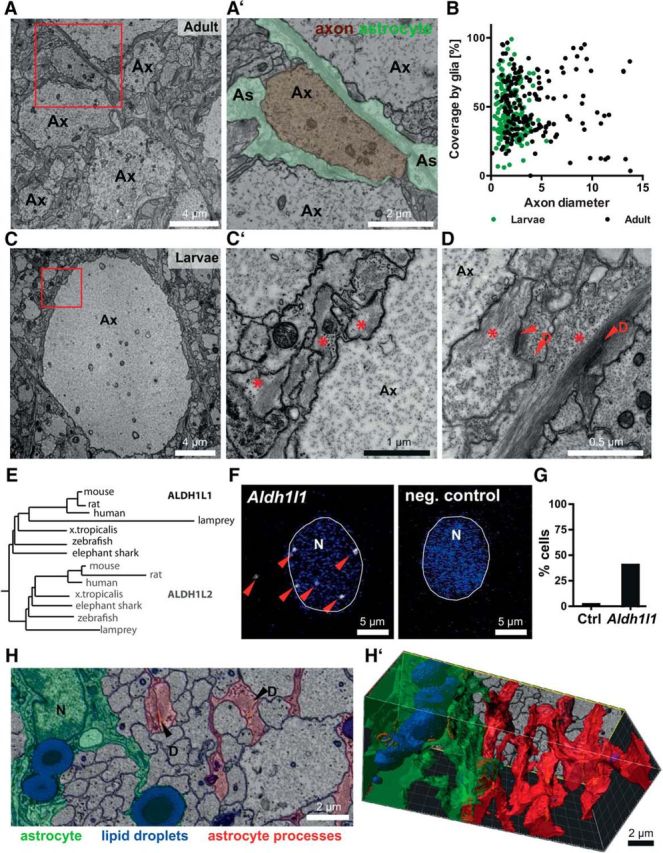
In the nervous system, myelination of axons enables rapid impulse conduction and is a specialized function of glial cells. Myelinating glia are the last cell type to emerge in the evolution of vertebrate nervous systems, presumably in ancient jawed vertebrates (gnathostomata) because jawless vertebrates (agnathans) lack myelin. We have hypothesized that, in these unmyelinated species, evolutionary progenitors of myelinating cells must have existed that should still be present in contemporary agnathan species. Here, we used advanced electron microscopic techniques to reveal axon-glia interactions in the sea lamprey Petromyzon marinus By quantitative assessment of the spinal cord and the peripheral lateral line nerve, we observed a marked maturation-dependent growth of axonal calibers. In peripheral nerves, all axons are ensheathed by glial cells either in bundles or, when larger than the threshold caliber of 3 μm, individually. The ensheathing glia are covered by a basal lamina and express SoxE-transcription factors, features of mammalian Remak-type Schwann cells. In larval lamprey, the ensheathment of peripheral axons leaves gaps that are closed in adults. CNS axons are also covered to a considerable extent by glial processes, which contain a high density of intermediate filaments, glycogen particles, large lipid droplets, and desmosomes, similar to mammalian astrocytes. Indeed, by in situ hybridization, these glial cells express the astrocyte marker Aldh1l1 Specimens were of unknown sex. Our observations imply that radial sorting, ensheathment, and presumably also metabolic support of axons are ancient functions of glial cells that predate the evolutionary emergence of myelin in jawed vertebrates.SIGNIFICANCE STATEMENT We used current electron microscopy techniques to examine axon-glia units in a nonmyelinated vertebrate species, the sea lamprey. In the PNS, lamprey axons are fully ensheathed either individually or in bundles by cells ortholog to Schwann cells. In the CNS, axons associate with astrocyte orthologs, which contain glycogen and lipid droplets. We suggest that ensheathment, radial sorting, and metabolic support of axons by glial cells predate the evolutionary emergence of myelin in ancient jawed vertebrates.

Keywords: Schwann cell; axon–glia interaction; electron microscopy; myelin; oligodendrocyte; radial sorting.


Figures

Figure 1.
Figure 1.
Evolutionary relationships of species groups and representative model species discussed in the present study. Consequential evolutionary innovations are indicated (red arrowheads). (Figure adapated with permission from Schweitzer et al., 2006; Grillner and Robertson, 2016; and Salzer and Zalc, 2016).
Figure 2.
Figure 2.
Spatial organization and maturation-dependent radial growth of peripheral lamprey axons. A, X-ray tomography of an adult lamprey LLN over a length of 370 μm. 3D reconstruction visualizes numerous large-caliber axons. A′, Representative axons selected from A. B, C, Scanning electron micrographs of cross-sectioned LLN in larval (B) and adult (C) lamprey. Note the different scale bar sizes. D, E, Calibers of individual axons in entire larval (D) and adult (E) LLNs indicating maturation-dependent radial growth. Data are shown as mean and SD. n = 1675 axons in 2 larvae; n = 373 axons in 1 adult.
Figure 3.
Figure 3.
Axonal ensheathment by Schwann cell orthologs in the lamprey PNS. A, B, Electron micrographs of a cross-sectioned LLN in larval (A) and adult (B) lamprey. Note the glial ensheathment (E) of axons (Ax). Red boxes indicate areas magnified in A′ and B′, respectively. A′, B′, Axonal ensheathment occasionally displayed gaps (A′) in larval but was closed (B′) in adult lamprey. C, Quantification of gapped and closed axonal ensheathment in entire cross-sectioned LLNs. Note the maturation-dependent closure of ensheathment gaps. Data are shown as mean and SD. n = 787 axons in 2 larvae; n = 343 axons in 1 adult. D, Electron micrograph of an axon (Ax) in the cross-sectioned adult LLN highlighting an ensheathing cell nucleus (N). (E) FISH of a cross-sectioned LLN detecting SoxE2 and SoxE3, the lamprey orthologs of Sox9 and Sox10, respectively. Note that SoxE2 and SoxE3-labeling (pseudocolor representation by black puncta marked by arrowheads) partially outlines axons (Ax, black lines). F–H, FIB-SEM micrographs and 3D reconstruction. F, Adaxonal (red) and abaxonal (blue) plasma membrane of a representative cell ensheathing an individual axon in the larval LLN. Note the structural homogeneity over at least 20 μm. See also Movie 1. G, Bundle of multiple axons (blue) ensheathed by a single ensheathing cell (red) showing homogeneity over at least 20 μm. See also Movie 2. H, As an anecdotal observation, an axon (blue) with its individual ensheathment (green) leaves a bundle of multiple axons (red). I, Electron micrograph of a LLN highlighting bundles of two or more axons (Ax). For quantification of axons per bundle, see Figure 4. E, Ensheathing cell; N, nucleus. J, Calibers of LLN axons ensheathed in bundles. Data are shown as mean and SEM. n = 120 axons in 2 larvae; n = 274 axons in 1 adult.
Figure 4.
Figure 4.
Numbers of axons and glial processes per bundle in the lamprey PNS. A, B, Electron micrographs of LLNs in larval (A) and adult (B) lamprey to exemplify axons in bundles (arrowheads) or with multiple ensheathing cell processes (Ax). C, C′, E, E′, Frequencies of axons per bundle in the entire LLN of larval (C,C′) and adult (E,E′) lamprey. C, E, Percentage of individually ensheathed axons; that is, axon/ensheathing cell units containing one axon versus units containing several axons. C′, E′, Subdivision of the numbers of axons in axon/ensheathing cell-units containing ≥2 axons. Note the maturation-dependent increase in the number of axons per bundle. D, F, Frequencies of ensheathing cell processes per bundle in the entire LLN of larval (D) and adult (F) lamprey. Data are shown as mean and SD. n = 1675 axons in 2 larvae; n = 411 axons in 1 adult.
Figure 5.
Figure 5.
Continuous longitudinal ensheathment of axons in the lamprey PNS. A, Scanning electron micrograph of a longitudinally sectioned LLN of an adult lamprey. B, Higher magnification to exemplify a segment of a longitudinally sectioned axon (Ax) with two adjacent ensheathing cells (arrows pointing at E1, E2). B′, Magnification from B to highlight the cell-to-cell contact between the ensheathing cells as identified by their nuclei (N1, N2). Large arrowheads point at the adaxonal and the abaxonal surfaces of the contact between the neighboring ensheathing cells. Note the direct apposition of their plasma membranes (small arrowheads). One representative of n = 27 contacts between longitudinally adjacent ensheathing glia in one adult lamprey is shown. Note that ensheathment gaps reminiscent of mammalian nodes of Ranvier were not observed, suggesting that glial ensheathment is largely continuous along peripheral lamprey axons.
Figure 6.
Figure 6.
Maturation-dependent radial growth of central lamprey axons. A, Micrograph of a semithin-sectioned larval body piece stained with methylene blue and Azure II. SCs and LLNs are indicated. B, B′, Magnification of the larval SC (B) and scheme (B′) to illustrate regions selected for quantification of axonal calibers in the ventral, lateral, and dorsal SC (red, green, and purple boxes, respectively). Arrowheads point at giant reticulospinal axons (G) and Mauthner axons (Mth). C, Scanning electron micrograph of the larval (left) and adult (right) SC illustrating maturation-dependent growth. Displayed is one half SC each. Note the different scale bar sizes. D, D′, Calibers of the 18 giant reticulospinal axons in the ventral SC of larval (D) and adult (D′) lamprey. n = 18 giant axons each in 1 larvae and 1 adult. E, E′, Axonal calibers in the lateral SC of larval (E) and adult (E′) lamprey. Arrowheads point at values for giant Mauthner axons (Mth). n = 239 axons in 2 larvae; n = 500 axons in 1 adult. F, F′, Axonal calibers in the dorsal SC of larval (F) and adult (F′) lamprey. n = 510 axons in 2 larvae; n = 449 axons in 1 adult. Note the maturation-dependent radial growth of axons in all analyzed SC regions.
Figure 7.
Figure 7.
Axonal coverage by astrocyte orthologs in the lamprey CNS. A, Electron micrograph of the dorsal SC in adult lamprey. The red box is magnified in A′. A′, Magnification of an axon (Ax) pseudocolored in brown to highlight its coverage by glial processes (As; green). B, Coverage of axonal surface with glial processes. Coverage of axonal surface with glial processes in the larval (green data points) and adult (black data points) SC plotted against axonal caliber. n = 199 axons in 2 larvae; n = 175 axons in 1 adult. Note that there is no evident correlation between axonal coverage and caliber. C, Electron micrograph of the dorsal SC in larval lamprey. The red box is magnified in C′. C′, Magnification of an axon (Ax) and glial processes to highlight that the glial cells display a high density of intermediate filaments and glycogen-particles (red stars). D, Desmosomes (D) were also frequent. Note that a high density of intermediate filaments, glycogen particles, and desmosomes are ultrastructural features of astrocytes. E, Sequence relationships of the astrocyte marker ALDH1L1 and the related ALDH1L2 in a phylogenetic tree. Note that lamprey ALDH1L1 clusters together with its orthologs in other species. F, FISH of lamprey SC with probes specific for lamprey Aldh1l1 or a negative control (E. coli Kd12). Maximum intensity projection images showing Aldh1l1 labeling (pseudocolor representation in white) in proximity to nuclei (N) (DAPI, blue). G, A considerable portion of cells displayed Aldh1l1 labeling in the adult SC. n = 198 DAPI-positive cells in 1 adult lamprey. H, H′, FIB-SEM micrograph (H) of the dorsal larval SC and 3D reconstruction (H′). A glial cell body and its primary processes are pseudocolored in green; large lipid droplets are pseudocolored in blue. Glial processes not connected to that cell are highlighted in red. D, Desmosome. See also Movie 3.

Similar articles

Cited by

References

    1. Banerjee S, Bhat MA (2008) Glial ensheathment of peripheral axons in Drosophila. J Neurosci Res 86:1189–1198. 10.1002/jnr.21574 - DOI - PMC - PubMed
    1. Bartels M, Krenkel M, Cloetens P, Möbius W, Salditt T (2015) Myelinated mouse nerves studied by X-ray phase contrast zoom tomography. J Struct Biol 192:561–568. 10.1016/j.jsb.2015.11.001 - DOI - PubMed
    1. Beirowski B, Babetto E, Golden JP, Chen YJ, Yang K, Gross RW, Patti GJ, Milbrandt J (2014) Metabolic regulator LKB1 is crucial for Schwann cell-mediated axon maintenance. Nat Neurosci 17:1351–1361. 10.1038/nn.3809 - DOI - PMC - PubMed
    1. Bertolini B. (1964) Ultrastructure of the spinal cord of the lamprey. J Ultrastruct Res 11:1–24. 10.1016/S0022-5320(64)80089-7 - DOI - PubMed
    1. Brodin L, Shupliakov O (2006) Giant reticulospinal synapse in lamprey: molecular links between active and periactive zones. Cell Tissue Res 326:301–310. 10.1007/s00441-006-0216-2 - DOI - PubMed

Publication types

LinkOut - more resources