. 2018 Aug 27;62(9):e00344-18.
doi: 10.1128/AAC.00344-18. Print 2018 Sep.

Validating a 14-Drug Microtiter Plate Containing Bedaquiline and Delamanid for Large-Scale Research Susceptibility Testing of Mycobacterium tuberculosis

Collaborators, Affiliations

Validating a 14-Drug Microtiter Plate Containing Bedaquiline and Delamanid for Large-Scale Research Susceptibility Testing of Mycobacterium tuberculosis

Paola M V Rancoita et al. Antimicrob Agents Chemother. .

Abstract

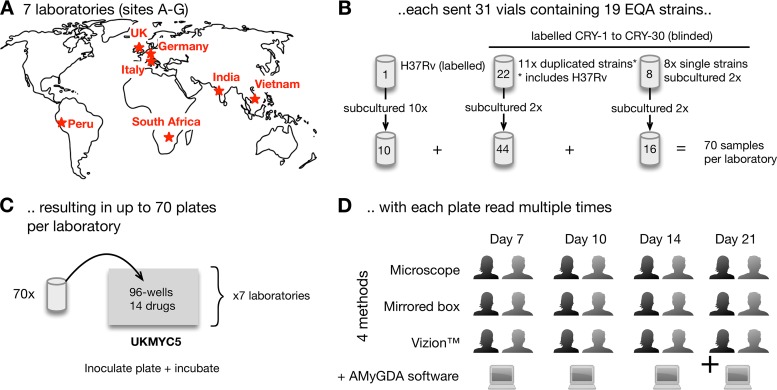
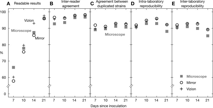
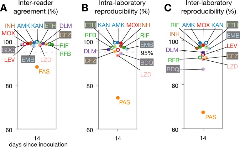
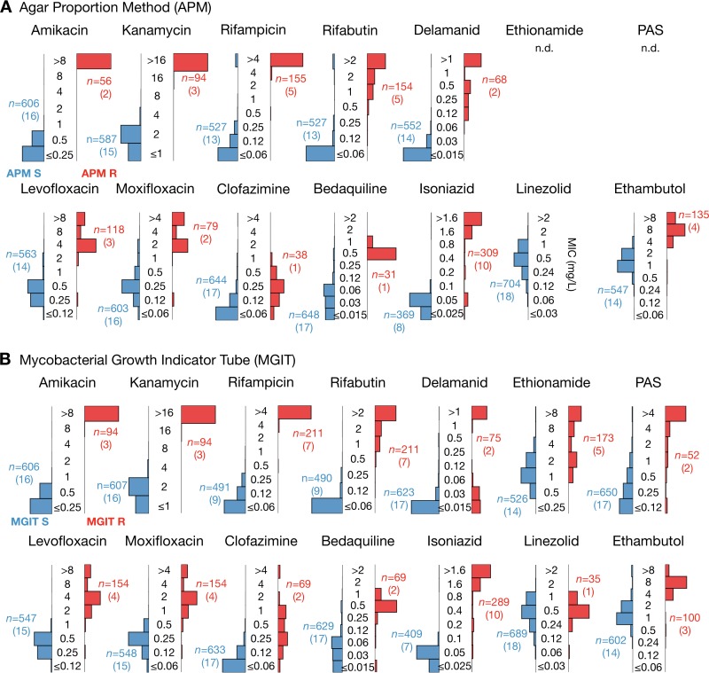
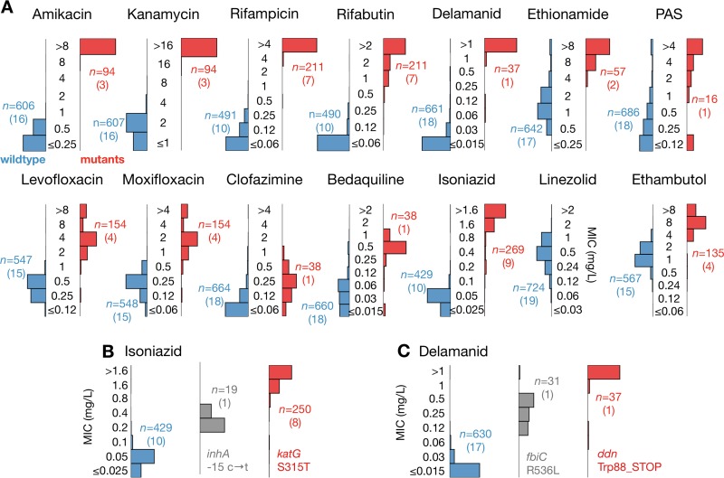
The UKMYC5 plate is a 96-well microtiter plate designed by the CRyPTIC Consortium (Comprehensive Resistance Prediction for Tuberculosis: an International Consortium) to enable the measurement of MICs of 14 different antituberculosis (anti-TB) compounds for >30,000 clinical Mycobacterium tuberculosis isolates. Unlike the MYCOTB plate, on which the UKMYC5 plate is based, the UKMYC5 plate includes two new (bedaquiline and delamanid) and two repurposed (clofazimine and linezolid) compounds. UKMYC5 plates were tested by seven laboratories on four continents by use of a panel of 19 external quality assessment (EQA) strains, including H37Rv. To assess the optimal combination of reading method and incubation time, MICs were measured from each plate by two readers, using three methods (mirrored box, microscope, and Vizion digital viewing system), after 7, 10, 14, and 21 days of incubation. In addition, all EQA strains were subjected to whole-genome sequencing and phenotypically characterized by the 7H10/7H11 agar proportion method (APM) and by use of MGIT960 mycobacterial growth indicator tubes. We concluded that the UKMYC5 plate is optimally read using the Vizion system after 14 days of incubation, achieving an interreader agreement of 97.9% and intra- and interlaboratory reproducibility rates of 95.6% and 93.1%, respectively. The mirrored box had a similar reproducibility. Strains classified as resistant by APM, MGIT960, or the presence of mutations known to confer resistance consistently showed elevated MICs compared to those for strains classified as susceptible. Finally, the UKMYC5 plate records intermediate MICs for one strain for which the APM measured MICs close to the applied critical concentration, providing early evidence that the UKMYC5 plate can quantitatively measure the magnitude of resistance to anti-TB compounds that is due to specific genetic variation.

Keywords: Mycobacterium tuberculosis; antibiotic resistance; antimicrobial agents; diagnostics.


Figures

FIG 1
FIG 1
Study design for validating the UKMYC5 plate. (A and B) Seven laboratories (A) were each sent 31 vials (B) containing 19 different genotypically characterized strains. H37Rv ATCC 27294 was subcultured and tested 10 times, while the other strains were subcultured in duplicate. (C) Each strain was inoculated onto a UKMYC5 96-well plate, starting from independent bacterial suspensions. Due to the large number of strains, the culturing and inoculation were performed over a period of weeks in each laboratory. (D) The MICs of each drug were read independently at 7, 10, 14, and 21 days postinoculation by two laboratory scientists, using three methods, as long as the positive-control wells showed acceptable and visible growth. Each plate was also photographed and the image analyzed using AMyGDA software (see Fig. S2 in the supplemental material).
FIG 2
FIG 2
For each reading method, the percentage of readable results (A), interreader agreement (B), agreement between two plates inoculated with different subcultures of the same strain in a single laboratory (C), and intralaboratory (D) and interlaboratory (E) reproducibilities were determined (see Table S2 in the supplemental material). These data include both on- and off-scale MICs. Data from site F were excluded from this analysis.
FIG 3
FIG 3
Most drugs performed well on the UKMYC5 plate; however, results for para-aminosalicylic acid (PAS) were consistently anomalous. (A) Interreader agreement for the 14 different anti-TB drugs for MICs measured after 14 days of incubation by use of the Vizion system. (B and C) Intralaboratory (B) and interlaboratory (C) reproducibilities. In each graph, a dashed line is drawn at 95%. Results for all reading days can be found in Fig. S3 and Table S7 in the supplemental material. RIF, rifampin; RFB, rifabutin; INH, isoniazid; EMB, ethambutol; LEV, levofloxacin; MOX, moxifloxacin; AMK, amikacin; KAN, kanamycin; ETH, ethionamide; CFZ, clofazimine; PAS, para-aminosalicylic acid; LZD, linezolid; DLM, delamanid; BDQ, bedaquiline.
FIG 4
FIG 4
MIC distributions for H37Rv for the 14 drugs on the UKMYC5 plate, as measured at day 14 by use of the Vizion system, are plotted as bar charts in gray. Each drug is annotated with the number of total measurements (n). These distributions are compared to MICs measured using three different methods. First, the published mode of the MIC for each drug (where available), measured using a frozen microtiter plate, is indicated by a black filled triangle (15, 16). The agreement with the MICs measured by use of the UKMYC5 plate is given for each drug. Next, the modal MICs measured by the agar proportion method (APM; teal triangles) and the resazurin microtiter assay (REMA; orange triangles) were plotted. n/d, not done.
FIG 5
FIG 5
Strains identified as resistant by the agar proportion method (APM) (A) or the mycobacterial growth indicator tube method (MGIT) (B) consistently recorded elevated MICs on the UKMYC5 plate, according to the Vizion instrument, after 14 days of incubation. The 19 EQA strains were categorized as either susceptible or resistant by using either APM or MGIT results. The distributions of MICs (milligrams per liter) measured by all laboratories in this study (excluding those from site F) for the susceptible and resistant strains were then plotted in blue and red, respectively. The critical concentrations used for APM are reported in Table S13 in the supplemental material. The number of UKMYC5 measurements (n) for each bar chart is given, with the number of EQA strains provided in parentheses.
FIG 6
FIG 6
The UKMYC5 plate not only registers higher MICs for strains containing known resistance-conferring genetic variation but also reports intermediate MICs for two strains containing mutations that confer intermediate resistance to delamanid and isoniazid. (A) UKMYC5 MIC distribution for each drug, split by whether the strain contains (red) or does not contain (blue) mutations that confer resistance. Drugs that share the same resistance genes are paired. The list of genetic variants used to make this classification can be found in Table S15 in the supplemental material. The number of UKMYC5 measurements (n) for each bar chart is given, with the number of EQA strains provided in parentheses. (B) Intermediate isoniazid MICs were consistently recorded on the UKMYC5 plate for one strain containing a mutation in the promoter of inhA. (C) Intermediate delamanid MICs were consistently recorded on the UKMYC5 plate for one strain containing the Arg563Leu mutation encoded within the fbiC gene.

Similar articles

Cited by

References

    1. World Health Organization. 2017. Global tuberculosis report. WHO, Geneva, Switzerland.
    1. World Health Organization. 2015. The End TB strategy. WHO, Geneva, Switzerland.
    1. Sotgiu G, Tiberi S, Centis R, D'Ambrosio L, Fuentes Z, Zumla A, Migliori GB. 2017. Applicability of the shorter ‘Bangladesh regimen’ in high multidrug-resistant tuberculosis settings. Int J Infect Dis 56:190–193. doi:10.1016/j.ijid.2016.10.021. - DOI - PubMed
    1. Schaberg T, Reichert B, Schülin T, Lode H, Mauch H. 1995. Rapid drug susceptibility testing of Mycobacterium tuberculosis using conventional solid media. Eur Respir J 8:1688–1693. doi:10.1183/09031936.95.08101688. - DOI - PubMed
    1. Franzblau SG, Witzig RS, McLaughlin JC, Torres P, Madico G, Hernandez A, Degnan MT, Cook MB, Quenzer VK, Ferguson RM, Gilman RH. 1998. Rapid, low-technology MIC determination with clinical Mycobacterium tuberculosis isolates by using the microplate Alamar Blue assay. J Clin Microbiol 36:362–366. - PMC - PubMed

Publication types

MeSH terms

LinkOut - more resources