A quarter century of EUS-FNA: Progress, milestones, and future directions
- PMID: 29941723
- PMCID: PMC6032705
- DOI: 10.4103/eus.eus_19_18
A quarter century of EUS-FNA: Progress, milestones, and future directions
Abstract
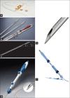
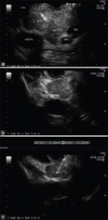
Tissue acquisition using EUS has considerably evolved since the first EUS-FNA was reported 25 years ago. Its introduction was an important breakthrough in the endoscopic field. EUS-FNA has now become a part of the diagnostic and staging algorithm for the evaluation of benign and malignant diseases of the gastrointestinal tract and of the organs in its proximity, including lung diseases. This review aims to present the history of EUS-FNA development and to provide a perspective on the recent developments in procedural techniques and needle technologies that have significantly extended the role of EUS and its clinical applications. There is a bright future ahead for EUS-FNA in the years to come as extensive research is conducted in this field and various technologies are continuously implemented into clinical practice.
Keywords: EUS; EUS-FNA; tissue acquisition.
Conflict of interest statement
There are no conflicts of interest
Figures


References
-
- Pauli EM, Ponsky JL. Principles of Flexible Endoscopy for Surgeons. New York: Springer; 2013. A history of flexible gastrointestinal endoscopy; pp. 1–10.
-
- Newman PG, Rozycki GS. The history of ultrasound. Surg Clin North Am. 1998;78:179–95. - PubMed
-
- Dimagno E, Regan P, Wilson D, et al. Ultrasonic endoscope. Lancet. 1980;315:629–31. - PubMed
-
- Strohm WD, Phillip J, Hagenmüller F, et al. Ultrasonic tomography by means of an ultrasonic fiberendoscope. Endoscopy. 1980;12:241–4. - PubMed
-
- Tio TL, Tytgat GN. Endoscopic ultrasonography in the assessment of intra-and transmural infiltration of tumours in the oesophagus, stomach and papilla of vater and in the detection of extraoesophageal lesions. Endoscopy. 1984;16:203–10. - PubMed
Publication types
LinkOut - more resources
Full Text Sources
Other Literature Sources

